

Abstract
Background
Pes planovalgus is one of the most common pediatric skeletal deformities. There have been no studies to analyze in detail the spatiotemporal variables of gait following arthroereisis.
Purpose of the study
The purpose of our study was to assess gait parameters in patients with symptomatic flexible flatfoot following treatment with the talus screw.
Methods
This was a prospective study assessing the 22 patients treated surgically due to symptomatic flexible flatfoot with the talus screw. Patients underwent gait assessment with a G-Sensor. We analyzed the following gait parameters: gait cycle duration, step length, support phase duration, swing phase duration, double support duration, single support duration, cadence, velocity, step length.
Results
The post-operative gait parameter assessment for the operated and non-operated foot showed a significant difference only in terms of step length. Cadence increased from the pre-operative mean of 82.29 steps/min to a post-operative mean of 102.94 steps/min. Gait velocity increased significantly from 0.81 m/s before to 0.96 m/s after surgery.
Discussion
Arthroereisis with the talus screw helps improve gait parameters following surgery. Post-operatively, we observed increased gait velocity and cadence and decreased gait cycle duration in the operated limb.
Conclusion
Short-term biomechanical outcomes of pes planovalgus treatment with the talus screw are good.
Keywords: Gait, Pes planovalgus, Subtalar arthroereisis, Spherus screw, Talus screw
Introduction
Pes planus or planovalgus, also known as flexible flatfoot, is one of the most common pediatric skeletal deformities [1–24]. Asymptomatic cases do not require treatment. Flatfoot symptoms include pain, problems with footwear, limited exercise, limping, and other gait abnormalities [1, 2, 5–7, 10, 11, 13–15, 17–24].
If conservative treatment is ineffective, symptomatic cases can be managed via various surgical techniques reported in the literature, including expandable sinus tarsi implants (subtalar arthroereisis), calcaneal stop screws, bone grafts, tendon lengthening, tendon transfer, talar or calcaneal osteotomy, and arthrodesis [1–24]. The talus screw (Gruppo Bioimpianti S.R.L., Milan, Italy) has been developed for surgical correction of pes planovalgus. There are only two publications available, by the authors of this manuscript, that evaluate the clinical and radiological parameters of Spherus screw insertion into the talus to achieve arthroereisis [25, 26]. Such screw placement offers a large contact area and support for its base on the floor and walls of the sinus tarsi.
The treatment goals in symptomatic flexible pes planovalgus patients include functional improvement, pain reduction, normalized foot position, and gait improvement [2, 5–10, 12, 16, 18–24]. Evaluation of musculoskeletal abnormalities, such as pes planovalgus, includes biomechanical and gait parameter assessments as well as radiographic and clinical examinations [2, 7, 11, 12, 14, 18, 19, 22, 27–31]. Anatomical correction improves joint mobility and2 reduces pain, which improves gait following treatment [2, 18, 19]. The use of a G-Sensor ensures repeatable, objective, and accurate gait parameter assessments [27–29].
Selected gait parameters have been evaluated following surgical treatment of flexible flatfoot with expandable implant insertion into the sinus tarsi [19], and implant insertion into the calcaneus [11, 12, 19]. Hagen et al. analyzed static and gait parameters in patients following arthroereisis with calcaneal screw implantation [11]. Franz et al. used a pedobarographic platform to assess gait following a calcaneal stop procedure involving calcaneal screw insertion into the sinus tarsi [12]. Vogt et al. evaluated pedobarogrphic parameters of gait in patients following arthroereisis with expandable calcaneal implants and screws [19].
It is still not fully understood how arthroereisis affects gait after surgery. There have been no studies to analyze in detail the spatiotemporal variables of gait following arthroereisis.
We hypothesized that surgical treatment of symptomatic flexible flatfoot with the use of the talus screw would improve gait parameters.
In light of a lack of literature on this subject, the purpose of our study was to assess gait parameters in patients with symptomatic flexible flatfoot following treatment with the talus screw.
Patients and Methods
This was a prospective study assessing the patients treated surgically due to symptomatic flexible flatfoot. All patients underwent arthroereisis in the absence of improvement after 6–12 months of non-surgical treatment (rehabilitation, exercise, NSAIDs, shoe inserts). Our study inclusion criteria were symptomatic flexible pes planovalgus with pain, failure of conservative management, age 7–14 years at the time of surgery, arthroereisis with the Spherus talus screw (Gruppo Bioimpianti S.R.L., Milan, Italy), written consent, complete medical and radiographic records, and a follow-up period of over 14 months. Symptomatic flexible pes planovalgus was confirmed on the basis of anamnesis, clinical and radiological examination. The exclusion criteria were any additional lower limb pathologies, history of foot surgery, neurological conditions, the presence of synostosis, foot injuries, incomplete medical or radiographic records, a lack of written consent, and a follow-up period of less than 14 months. The study had been approved by the local bioethics committee and was conducted in accordance with the Declaration of Helsinki. All patients and their legal guardians were informed of the voluntary nature of study participation. Twenty-four arthroereisis procedures were conducted with the use of the Spherus screw; Fig. 1. Applying the inclusion and exclusion criteria left 22 pes planovalgus patients to be evaluated post-operatively. The study group comprised 8 females and 14 males aged 7–14 years.
Fig. 1.
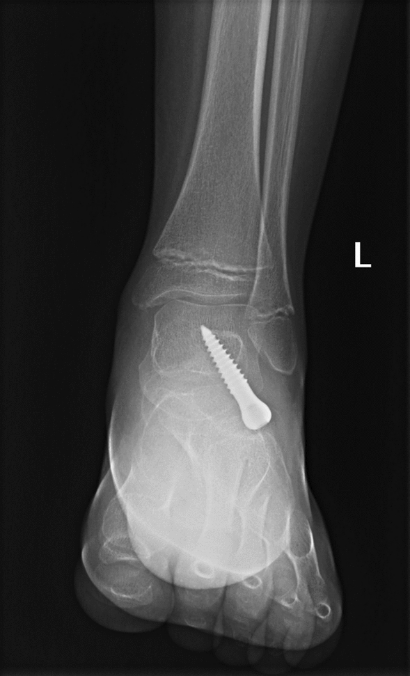
AP radiographic images, 8 months after pes planovalgus correction with the talus screw, male, 12 years old
All procedures were performed by two experienced orthopedic surgeons (co-authors of the article). The procedures were performed under general anesthesia and regional hemostasis, achieved with a thigh tourniquet. The patients were in a supine position. An oblique 1–2 cm incision, which corresponded to skin tension lines, was made on the lateral aspect of the foot, somewhat distal and anterior to the tip of the lateral malleolus, at the level of the sinus tarsi. After cutting through the subcutaneous tissue, the surgeons dissected away the soft tissues of the sinus tarsi, to expose the inferior surface of the talus. With the foot held at the right angle and maximally inverted, as ensured via fluoroscopy, a pilot hole for the screw was created in the inferior surface of the talus with a straight awl being directed obliquely, proximally and medially. Once the pilot hole was created, the right screw was selected, with its length and diameter dependent on the size of the patient’s talus, the length of the foot, and age. With the patient’s foot held steadily at a right angle and maximally inverted, the screw was inserted into the prepared pilot hole, starting from the inferior surface of the talus. The screw was advanced, under fluoroscopy, until the desired degree of correction was achieved, with the spherical bulge of the screw protruding toward the sinus tarsi and resting against its floor and walls. Achilles tendon lengthening (Z plasty) was performed in eight patients as a concomitant procedure. The indication for Achilles tendon lengthening was a dorsiflexion of less than 5°–10° in the neutral position of the foot [1, 2, 5, 14, 15, 18]. Following surgery, full3 weight-bearing was allowed on day one after surgery, provided no pain was felt. If there was pain on walking, the patient used elbow crutches for 5–14 days. In cases of concomitant Achilles tendon lengthening, the limb was immobilized in a short leg cast for 6 weeks. For 6 weeks following surgery, the patients were discouraged from strenuous exercise and sports [7, 11]. The uniform rehabilitation protocol for all patients was started on the first day after surgery, in patients without Achilles tendon lengthening, and 6 weeks after surgery, in the group of patients with concomitant Achilles tendon lengthening.
Patients underwent gait assessment with a G-Sensor prior to surgery and after a minimum of 14 months after surgery. The G-Sensor is a wireless system with an inertial sensor placed on the patient’s body, Fig. 2. The system is composed of a triaxial accelerometer, magnetic sensor, triaxial gyroscope, and G-studio software to analyze the data obtained from the sensor in real time. The G-Sensor is used for objective, accurate, and repeatable gait assessment [27–29]. The G-Sensor is suitable for assessing gait and physical activity, as shown by the coefficient of variation between instruments 2.5% and analyses of the inter-instrument correlation coefficient from 0.90 to 0.99 [32–34]. The obtained data, which illustrate all the phases of the gait cycle, help objectively assess the patient’s gait and detect possible gait deviations [27–29].
Fig. 2.
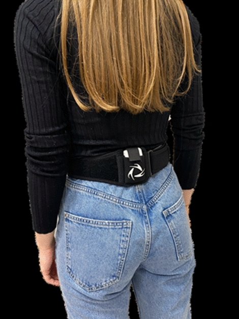
The G-Sensor placed on the patient’s body
In this study we analyzed the following gait parameters:
gait cycle duration (s),
step length (%),
support phase duration (%),
swing phase duration (%),
double support duration (%),
single support duration (%),
cadence (steps/min),
velocity (m/s),
step length (m).
The G-Sensor was placed at the level of the L4–L5 intervertebral disc. The patients walked barefoot at their normal speed along a distance of 5 m, turning back and forth. All patients were assessed in the same room, with the same device. The data collected from the device were processed with G-studio software (BTS Bioengineering, Quincy, MA, USA); exported to a Microsoft Excel spreadsheet, and analyzed. The pre-operative and post-operative data and the data from the operated and non-operated foot were compared.
Statistical Analysis
Data were statistically analyzed using the Statistica 13.1. The Shapiro–Wilk test was used to check for normality of distribution. The student's t-test was used to compare quantitative variables. The level of statistical significance was set at p < 0.05.
Results
The mean follow-up period was 15.5 months (ranging from 14 to 18 months).
A comparison of the pre-operative gait parameters for the operated and non-operated foot showed no differences in terms of gait cycle duration, step length, support phase duration, swing phase duration, double support duration, or single support duration; Table 1.
Table 1.
Spatio-temporal parameters of gait before treatment
| Analyzed variable | OL | NOL | p value* |
|---|---|---|---|
| Mean ± standard deviation | |||
| Gait cycle duration (s) | 1.54 ± 0.23 | 1.54 ± 0.27 | 0.957 |
| Step length (%) | 49.07 ± 4.91 | 51.58 ± 4.08 | 0.114 |
| support phase duration (%) | 58.45 ± 6.97 | 59.67 ± 5.38 | 0.57 |
| Swing phase duration (%) | 41.54 ± 6.97 | 40.31 ± 5.37 | 0.568 |
| Double support duration (%) | 9.47 ± 3.14 | 9.69 ± 2.62 | 0.868 |
| Single support duration (%) | 39.49 ± 5.57 | 41.41 ± 7.42 | 0.401 |
| Steps analyzed | 10.23 ± 1.21 | 10.11 ± 1.26 | 0.783 |
OL operated limb, NOL non-operated limb
*Student's t-test
The post-operative gait parameter assessment for the operated and non-operated foot showed a significant difference only in terms of step length (p = 0.047); Table 2; Fig. 3. There were no post-operative differences between the two feet in gait cycle duration, support phase duration, swing phase duration, double support duration, or single support duration; Table 2.
Table 2.
Spatio-temporal parameters of gait after treatment
| Analyzed variable | OL | NOL | p value* |
|---|---|---|---|
| Mean ± standard deviation | |||
| Gait cycle duration (s) | 1.24 ± 0.24 | 1.21 ± 0.21 | 0.749 |
| Step length (%) | 48.84 ± 3.74 | 51.15 ± 3.74 | 0.047 |
| Support phase duration (%) | 55.58 ± 11.66 | 59.08 ± 3.85 | 0.188 |
| Swing phase duration (%) | 42.11 ± 3.15 | 40.91 ± 3.85 | 0.27 |
| Double support duration (%) | 9.25 ± 2.81 | 8.25 ± 2.21 | 0.194 |
| Single support duration (%) | 40.61 ± 4.24 | 41.53 ± 2.69 | 0.388 |
| Steps analyzed | 10.45 ± 1.89 | 10.59 ± 1.84 | 0.810008740662566 |
OL operated limb, NOL non-operated limb
*Student's t-test
Fig. 3.

Post-operative step length for the operated and non-operated limb
Cadence increased from the pre-operative mean of 82.29 steps/min to a post-operative mean of 102.94 steps/min, p < 0.001); Table 3; Fig. 4. Gait velocity increased significantly from 0.81 m/s before to 0.96 m/s after surgery, (p = 0.046); Table 3; Fig. 5. Step length showed no change when the pre-operative and post-operative values were compared; Table 3.
Table 3.
Selected gait parameters before and after surgery
| Analyzed variable | Before surgery | After surgery | p value* |
|---|---|---|---|
| Mean ± standard deviation | |||
| Cadence (steps/min) | 82.29 ± 13.59 | 102.94 ± 14.09 | < 0.001 |
| Velocity (m/s) | 0.81 ± 0.21 | 0.96 ± 0.24 | 0.046 |
| Step length (m) | 0.63 ± 0.14 | 0.57 ± 0.11 | 0.185 |
| Analysis duration (s) | 48.72 ± 10.51 | 39.29 ± 9.51 | 0.005 |
*Student's t-test
Fig. 4.

Pre-operative and post-operative cadence
Fig. 5.

Gait velocity before after surgery
We observed a significant reduction in post-operative gait cycle duration in the operated limb (1.24 s) in comparison with the pre-operative value (1.54 s), (p < 0.001); Table 4. The pre-operative and post-operative gait cycle duration differed for the non-operated limb (p < 0.001); Table 4. The other evaluated gait parameters showed no statistically significant differences between the operated and non-operated limbs either before or after surgery; Table 4.
Table 4.
Detailed gait parameters before and after surgery
| Analyzed variable | Before surgery | After surgery | p value* |
|---|---|---|---|
| Mean ± standard deviation | |||
| Gait cycle duration OL (s) | 1.54 ± 0.23 | 1.24 ± 0.24 | < 0.001 |
| Step length OL (%) | 49.07 ± 4.91 | 48.84 ± 3.74 | 0.873 |
| Support phase duration OL (%) | 58.45 ± 6.97 | 55.58 ± 11.66 | 0.375 |
| Swing phase duration OL (%) | 41.54 ± 6.97 | 42.11 ± 3.15 | 0.741 |
| Double support duration OL (%) | 9.47 ± 4.14 | 9.25 ± 2.81 | 0.845 |
| Single support duration OL (%) | 39.49 ± 5.57 | 40.61 ± 4.24 | 0.486 |
| Steps analyzed OL | 10.23 ± 1.21 | 10.45 ± 1.89 | 0.679 |
| Gait cycle duration NOL (s) | 1.54 ± 0.27 | 1.21 ± 0.21 | < 0.001 |
| Step length NOL (%) | 59.67 ± 5.38 | 59.08 ± 3.85 | 0.689 |
| Support phase duration NOL (%) | 40.31 ± 5.37 | 40.91 ± 3.85 | 0.686 |
| Swing phase duration NOL (%) | 9.69 ± 3.62 | 8.25 ± 2.22 | 0.131 |
| Double support duration NOL (%) | 41.41 ± 7.42 | 41.53 ± 2.69 | 0.941 |
| Single support duration NOL (%) | 51.58 ± 4.08 | 51.15 ± 3.74 | 0.734 |
| Steps analyzed NOL | 10.11 ± 1.28 | 10.59 ± 1.84 | 0.371 |
OL operated limb, NOL non-operated limb
*Student's t-test
Discussion
In our study, we assessed gait parameters before and after surgical treatment of symptomatic pes planovalgus. We observed improved gait parameters following pes planovalgus treatment with the talus screw, which supports our research hypothesis. We observed post-operative increase in gait velocity and cadence values; a reduced gait cycle duration in the operated limb, in comparison with the pre-operative value.
Symptomatic pes planovalgus is a common reason for orthopedic consultations, with various techniques for surgical treatment for this deformity reported in the literature [1–24]. Treatment methods include expandable sinus tarsi implants (subtalar arthroereisis), calcaneal stop screws, bone grafts, tendon lengthening, tendon transfer, talar or calcaneal osteotomy, and arthrodesis (permanent stiffening of the joint) [1–24]. Osteotomies and arthrodesis are used in adolescents and adults;. they involve significant intervention and are irreversible. Removable implants inserted to block the sinus tarsi or implants inserted into the calcaneal bone (subtalar arthroereisis) are minimally invasive, but they may be displaced, which causes pain and the need for re-operation.
The lack of adequate tension of soft tissues and greater laxity of ligaments and muscles may result in an incorrect spatial configuration of the foot and the development of symptomatic pes planovalgus in children. The deformity referred to as pes planovalgus encompasses forefoot supination and abduction resulting from excessive subtalar joint eversion, hindfoot varus, a flattened longitudinal arch, and Achilles tendon and calf muscle contracture, with all of these pathologies potentially negatively affecting gait biomechanics [7, 12–15, 17, 18]. Anthropometric dynamic, isokinetic, and kinematic measurements help precisely differentiate between a normal foot and a flat foot [7]. Analysis of biomechanical and gait parameters is an important component of evaluating the outcomes of foot deformity correction [2, 7, 11, 12, 14, 18–20, 22, 27–31].
Unlike other implants designed for surgical correction of pes planovalgus, the Spherus screw is to be inserted into the talus. The advantage of this screw placement is the support for the screw base and its large area of contact with the floor and walls of the sinus tarsi. The Spherus screw stabilizes the talus by distributing the weight of the body onto a larger area, at the same time reducing the risk of screw migration into the talus and its penetration into the calcaneus. In light of the lack of available reports on the outcomes of inserting talar screws, such as the Spherus screw, from the direction of the sinus tarsi in the treatment of symptomatic pes planovalgus, we decided to address this subject with our study.
Martinelli et al. posited that since pes planovalgus is associated with abnormal kinetics and kinematics in comparison with those in normal feet, it is important to assess kinematic parameters after arthroereisis [13]. The gait of patients with pes planovalgus is characterized by the following abnormalities a plantar-flexed ankle during foot landing, increased foot dorsiflexion during terminal stance, foot eversion throughout the stance phase, early activation of the peroneus longus and the gastrosoleus complex during heel strike [7]. These pathologies in patients with pes planovalgus reduce push-off force and load absorption ability [7]. Dynamic analysis of biomechanical parameters showed that arthroereisis may change foot function within several months after6 operation [11, 12, 18]. Hagen et al. evaluated static and gait parameters in 7 patients 3–28 days after arthroereisis with calcaneal screw implantation [11]. During bipedal stance, the plantar force increased in lateral areas and decreased in medial areas of the foot [11]. Those authors also observed a lateral shift in the center of gravity and a lateral shift in the contact area [11]. Franz et al. reported a load shift from the medial to the lateral mid- and forefoot after arthroereisis [12]. Six months after arthroereisis, there was a reduced forefoot load and a hindfoot overload, in comparison with the healthy controls [12]. Vogt et al. assessed pedobarographic gait parameters in patients after arthroereisis with expandable sinus tarsi implants or calcaneus stop screws [19]. Those authors reported a post-operative reduction in the medial midfoot contact area, arch index, and medial forefoot peak pressure [19]. In a systematic review Smith et al. reported improved kinematic and pedobarographic parameters following arthroereisis procedures [20]. Vescio et al. in their systematic review stated that some authors observed an improved post-operative ground-reaction-force in the form of increased ground-force in the lateral areas of the foot, in comparison with pre-operative data. However, other authors noted no differences in the post-operative ground-reaction-force in comparison with the pre-operative data [22]. In comparison with osteotomy and arthrodesis, arthroereisis requires a shorter immobilization period and a shorter period of reduced weight-bearing, which may result in less range-of-motion limitation and better gait parameters [2]. Arthroereisis increases the foot range of motion, reduces ankle–foot pronation during gait, increases foot stability in single leg stance, and normalizes the load absorption capacity [18]. All of these aspects of post-operative lower limb biomechanics analysis after pes planovalgus correction were performed with various measuring devices. Therefore a strict comparison of the results is not possible.
Gieysztor et al. assessed the gain of healthy children with the use of a G-Sensor [27]. The support phase duration, swing phase duration, double support duration, single support duration, cadence, and velocity reported by Gieysztor [27] were comparable with the respective values measured in our study. This indicates normalization of gait parameters following the use of the Spherus screw in pes planovalgus treatment. Gait during normal child development should be symmetrical for normal skeletal biomechanics [27, 29–31]. In our study, most gait parameters, except for step length, were symmetrical post-operatively, which suggests good treatment outcomes.
Our study results show improvement in the assessed gait parameters, which may indicate normalized foot anatomy, increased range of motion, and reduced pain following pes planovalgus treatment with the talus screw. The group of patients evaluated in our study showed post-operative increase in gait velocity and cadence values, in comparison with the respective pre-operative values. We observed a reduced gait cycle duration in the operated limb, in comparison with the pre-operative value. These results indicate a positive effect of treatment on lower limb biomechanics.
Following treatment with the talus screw, post-operative full weight-bearing was allowed from the first post-operative day. However, some other surgical techniques are associated with 2–8 weeks of reduced weight-bearing and 2–8 weeks of immobilization in a cast [3, 5, 13–15]. We believe that the lack of cast immobilization and the possibility of walking with full weight-bearing from the first day after surgery with the talus screw also have a positive effect on gait parameters.
Some authors performed other procedures within the foot and ankle joint in addition to arthroereisis [1–3, 8, 10, 12, 14, 15, 18, 19]. The most common procedure, performed in 17%–100% of patients, was Achilles tendon and gastrocnemius lengthening [1–3, 5, 10, 12, 14, 15, 18, 19]. Achilles tendon and gastrocnemius muscle contracture often accompany flexible flatfoot. We believe that the presence of Achilles tendon contracture, requires tendon lengthening. In our study, 33% of patients underwent Achilles tendon lengthening due to tendon contracture. Literature reports indicate that the use of a concomitant surgical procedure has no effect on radiographic or clinical outcomes following subtalar arthroereisis [15, 16].
The purpose of pes panovalgus treatment is to improve gait as well as clinical and functional7 parameters. In our study, we observed post-operative improvement in gait parameters after talus screw placement, which indicates good treatment outcomes.
One limitation of our study was the relatively small sample size, which is due to the fact that we aimed to perform a prospective gait assessment. Arthroereisis is not a very common procedure and, therefore, the number of patients is limited. Other authors also evaluated similar-sized groups of patients [1, 5, 8, 10, 15, 17]. Another limitation of our study is the relatively short follow-up; however, as evidenced by other studies, most patients have normal foot function 4 weeks after arthroereisis [11]. Moreover, some studies had a follow-up period duration comparable to that in our study [11, 15, 17, 19]. We did not assess the effects of the body mass index (BMI) or sex on gait parameters; however, few authors analyze these variables, and the available reports indicate no effect of either sex or BMI on gait parameters [25]. The strengths of our study include its prospective nature, while some earlier studies had been retrospective [1, 2, 5, 8, 13, 15, 17, 19], only two experienced orthopedic surgeons being involved in surgeries, the use of an objective and accurate measuring device (G-Sensor), and a uniform rehabilitation protocol for all patients. In the future, we are planning a study on the use of the talus screw in a larger group of patients and with a longer follow-up period for post-operative gait parameters.
In our research, we provided new findings on the detailed analysis of gait parameters after arthroeresis with the use of the talus screw. Like other authors [11, 19], we also believe that gait assessment ensures an objective, radiation-free method for dynamic evaluation of the outcomes following pes planovalgus surgery. The use of a G-Sensor offers a graphic record of gait parameter improvement following pes planovalgus treatment, which makes it easier to present surgical treatment outcomes to the patients and their parents. Gait analysis is an important component of treatment outcomes for surgeons, rehabilitation specialists, and patients and their parents. The results of our study can be crucial in the decision-making process involved in qualifying patients with pes planovalgus for surgical treatment.
Conclusions
The use of a G-Sensor helps assess dynamic gait parameters and perform follow-up assessments following sinus tarsi implant insertion (subtalar arthroereisis).
Arthroereisis with the talus screw helps improve gait parameters following surgery.
Post-operatively, we observed increased gait velocity and cadence and decreased gait cycle duration in the operated limb.
Short-term biomechanical outcomes of pes planovalgus treatment with the talus screw are good.
Acknowledgements
Sources of funding: Internal project of the Institute of Medical Sciences of the University of Opole P-2021-003
Data availability
The data presented in this study are available on request from the corresponding author.
Declarations
Conflict of Interest
There is no conflict of interest.
Ethical standard statement
This article does not contain any studies with human or animal subjects performed by the any of the authors.
Informed consent
For this type of study informed consent is not required.
Footnotes
Publisher's Note
Springer Nature remains neutral with regard to jurisdictional claims in published maps and institutional affiliations.
References:
- 1.Jerosch J, Schunck J, Abdel-Aziz H. The stop screw technique–a simple and reliable method in treating flexible flatfoot in children. Foot and Ankle Surgery. 2009;15(4):174–178. doi: 10.1016/j.fas.2009.01.004. [DOI] [PubMed] [Google Scholar]
- 2.Kubo H, Lipp C, Hufeland M, Ruppert M, Westhoff B, Krauspe R, Pilge H. Outcome after subtalar screw arthroereisis in children with flexible flatfoot depends on time of treatment: Midterm results of 95 cases. Journal of Orthopaedic Science. 2020;25(3):497–502. doi: 10.1016/j.jos.2019.06.007. [DOI] [PubMed] [Google Scholar]
- 3.Leonchuk SS, Dyachkov K, Neretin AS, Blanchard AJ, Popkov D. Subtalar arthroereisis for treatment of children with flexible planovalgus foot deformity and analysis of CT data in long-term period. Journal of Orthopaedics. 2020;8(22):478–484. doi: 10.1016/j.jor.2020.10.005. [DOI] [PMC free article] [PubMed] [Google Scholar]
- 4.Arbab D, Frank D, Bouillon B, Lüring C, Wingenfeld C, Abbara-Czardybon M. Subtalare screw arthroereisis for the treatment of symptomatic. Flexible Pes Planovalgus. Z Orthop Unfall. 2018;156(1):93–99. doi: 10.1055/s-0043-120071. [DOI] [PubMed] [Google Scholar]
- 5.Robin de Bot TAL, Stevens J, Hermus JP, Staal HM, van Rhijn LW, Witlox AM. Clinical and radiological outcomes of subtalar Kalix II arthroereisis for a symptomatic pediatric flexible flatfoot. Foot & Ankle Specialist. 2021;14(1):9–18. doi: 10.1177/1938640019892062. [DOI] [PubMed] [Google Scholar]
- 6.Bernasconi A, Lintz F, Sadile F. The role of arthroereisis of the subtalar joint for flatfoot in children and adults. EFORT Open Reviews. 2017;2(11):438–446. doi: 10.1302/2058-5241.2.170009. [DOI] [PMC free article] [PubMed] [Google Scholar]
- 7.Faldini C, Mazzotti A, Panciera A, Perna F, Stefanini N, Giannini S. Bioabsorbable implants for subtalar arthroereisis in pediatric flatfoot. Musculoskeletal Surgery. 2018;102(1):11–19. doi: 10.1007/s12306-017-0491-y. [DOI] [PubMed] [Google Scholar]
- 8.Hong J, Dai G, Weng Q, Liu Y. Interference screw for the treatment of pediatric flexible flatfoot. The Journal of Foot and Ankle Surgery. 2020;59(6):1209–1214. doi: 10.1053/j.jfas.2020.04.016. [DOI] [PubMed] [Google Scholar]
- 9.Elmarghany M, El-Ghaffar TMA, Elgeushy A, Elzahed E, Hasanin Y, Knörr J. Is subtalar extra articular screw arthroereisis (SESA) reducing pain and restoring medial longitudinal arch in children with flexible flat foot? Journal of Orthopaedics. 2020;20:147–153. doi: 10.1016/j.jor.2020.01.038. [DOI] [PMC free article] [PubMed] [Google Scholar]
- 10.Elbarbary HM, Arafa AS, Said ABZ, Hegaz M, Reiad MW, Basha NY, Fahmy M. Clinical and radiological outcomes of subtalar arthroereisis for management of planovalgus foot in children with cerebral palsy: 3-year follow-up. Foot & Ankle Specialist. 2022;15(6):536–544. doi: 10.1177/1938640020980911. [DOI] [PubMed] [Google Scholar]
- 11.Hagen L, Pape JP, Kostakev M, Peterlein CD. Pedobarographic changes during first month after subtalar extra-articular screw arthroereisis (SESA) operation of juvenile flexible flatfoot. Archives of Orthopaedic and Trauma Surgery. 2020;140(3):313–320. doi: 10.1007/s00402-019-03230-7. [DOI] [PubMed] [Google Scholar]
- 12.Franz A, Herz D, Raabe J, Seeberger U, Bollmann C. Pedobarographic outcome after subtalar screw arthroereisis in flexible juvenile flatfoot. Foot and Ankle Surgery. 2021;27(4):389–394. doi: 10.1016/j.fas.2020.05.003. [DOI] [PubMed] [Google Scholar]
- 13.Martinelli N, Bianchi A, Martinkevich P, Sartorelli E, Romeo G, Bonifacini C, Malerba F. Return to sport activities after subtalar arthroereisis for correction of pediatric flexible flatfoot. Journal of Pediatric Orthopedics. Part B. 2018;27(1):82–87. doi: 10.1097/BPB.0000000000000449. [DOI] [PubMed] [Google Scholar]
- 14.Fernández de Retana P, Alvarez F, Viladot R. Subtalar arthroereisis in pediatric flatfoot reconstruction. Foot and Ankle Clinics. 2010;15(2):323–335. doi: 10.1016/j.fcl.2010.01.001. [DOI] [PubMed] [Google Scholar]
- 15.Wang S, Chen Li, Jian Yu, Zhang C, Jia-Zhang Huang Xu, Wang XM. Mid-term results of subtalar arthroereisis with talar-fit implant in pediatric flexible flatfoot and identifying the effects of adjunctive procedures and risk factors for sinus tarsi pain. Orthopaedic Surgery. 2021;13(1):175–184. doi: 10.1111/os.12864. [DOI] [PMC free article] [PubMed] [Google Scholar]
- 16.Tan JHI, Tan SHS, Lim AKS, Hui JH. The outcomes of subtalar arthroereisis in pes planus: a systemic review and meta-analysis. Archives of Orthopaedic and Trauma Surgery. 2021;141(5):761–773. doi: 10.1007/s00402-020-03458-8. [DOI] [PubMed] [Google Scholar]
- 17.Ruiz-Picazo D, Jiménez-Ortega P, Doñate-Pérez F, Gaspar-Aparicio N, García-Martín V, Ramírez-Villaescusa J, Losa-Palacios S. Radiographic and functional results following subtalar arthroereisis in pediatric flexible flatfoot. Advances in Orthopedics. 2019;1(2019):5061934. doi: 10.1155/2019/5061934. [DOI] [PMC free article] [PubMed] [Google Scholar]
- 18.Memeo A, Verdoni F, Rossi L, Ferrari E, Panuccio E, Pedretti L. Flexible juvenile flat foot surgical correction: A comparison between two techniques after ten years' experience. Journal of Foot and Ankle Surgery. 2019;58(2):203–207. doi: 10.1053/j.jfas.2018.07.013. [DOI] [PubMed] [Google Scholar]
- 19.Vogt B, Toporowski G, Gosheger G, Rölfing JD, Rosenbaum D, Schiedel F, Laufer A, Kleine-Koenig MT, Theil C, Roedl R, Frommer A. Subtalar arthroereisis for flexible flatfoot in children-clinical, radiographic and pedobarographic outcome comparing three different methods. Children (Basel). 2021;8(5):359. doi: 10.3390/children8050359. [DOI] [PMC free article] [PubMed] [Google Scholar]
- 20.Smith C, Zaidi R, Bhamra J, Bridgens A, Wek C, Kokkinakis M. Subtalar arthroereisis for the treatment of the symptomatic paediatric flexible pes planus: A systematic review. EFORT Open Rev. 2021;6(2):118–129. doi: 10.1302/2058-5241.6.200076. [DOI] [PMC free article] [PubMed] [Google Scholar]
- 21.De Pellegrin M. Subtalar screw-arthroereisis for correction of flat foot in children. Der Orthopäde. 2005;34(9):941–953. doi: 10.1007/s00132-005-0835-4. [DOI] [PubMed] [Google Scholar]
- 22.Vescio A, Testa G, Amico M, Lizzio C, Sapienza M, Pavone P, Pavone V. Arthroereisis in juvenile flexible flatfoot: Which device should we implant? A systematic review of literature published in the last 5 years. World Journal of Orthopedics. 2021;12(6):433–444. doi: 10.5312/wjo.v12.i6.433. [DOI] [PMC free article] [PubMed] [Google Scholar]
- 23.Mattesi L, Ancelin D, Severyns MP. Is subtalar arthroereisis a good procedure in adult-acquired flatfoot? A systematic review of the literature. Orthopaedics & Traumatology, Surgery & Research. 2021;107(6):103002. doi: 10.1016/j.otsr.2021.103002. [DOI] [PubMed] [Google Scholar]
- 24.Baryeh KW, Ismail H, Sobti A, Harb Z. Outcomes following the use of subtalar arthroereisis in the correction of adult acquired flatfoot: A systematic review. Foot & Ankle Specialist. 2022;15(4):384–393. doi: 10.1177/1938640020987775. [DOI] [PubMed] [Google Scholar]
- 25.Bobiński A, Tomczyk Ł, Reichert P, Morasiewicz P. Short-term and medium-term radiological and clinical assessment of patients with symptomatic flexible flatfoot following subtalar arthroereisis with Spherus screw. Journal of Clinical Medicine. 2023;12(15):5038. doi: 10.3390/jcm12155038. [DOI] [PMC free article] [PubMed] [Google Scholar]
- 26.Bobiński A, Tomczyk Ł, Pelc M, Chruścicki DA, Śnietka B, Morasiewicz P. Arthroereisis with a talar screw in symptomatic flexible flatfoot in children. Journal of Clinical Medicine. 2023;12(23):7475. doi: 10.3390/jcm12237475. [DOI] [PMC free article] [PubMed] [Google Scholar]
- 27.Gieysztor E, Kowal M, Paprocka-Borowicz M. Gait parameters in healthy preschool and school children assessed using wireless inertial sensor. Sensors (Basel). 2021;21(19):6423. doi: 10.3390/s21196423. [DOI] [PMC free article] [PubMed] [Google Scholar]
- 28.Gieysztor E, Kowal M, Paprocka-Borowicz M. Primitive reflex factors influence walking gait in young children: An observational study. International Journal of Environmental Research and Public Health. 2022;19(7):4070. doi: 10.3390/ijerph19074070. [DOI] [PMC free article] [PubMed] [Google Scholar]
- 29.Gieysztor E, Pecuch A, Kowal M, Borowicz W, Paprocka-Borowicz M. Pelvic symmetry is influenced by asymmetrical tonic neck reflex during young children's gait. International Journal of Environmental Research and Public Health. 2020;17(13):4759. doi: 10.3390/ijerph17134759. [DOI] [PMC free article] [PubMed] [Google Scholar]
- 30.Morasiewicz P, Dragan SF. Pedobarographic evaluation of body weight distribution on the lower limbs and balance after derotation corticotomies using the Ilizarov method. Acta of Bioengineering and Biomechanics. 2013;15(2):91–96. [PubMed] [Google Scholar]
- 31.Morasiewicz P, Konieczny G, Dejnek M, Morasiewicz L, Urbański W, Kulej M, Dragan SŁ, Dragan SF, Pawik Ł. Pedobarographic analysis of body weight distribution on the lower limbs and balance after ankle arthrodesis with Ilizarov fixation and internal fixation. Biomedical Engineering Online. 2018;17(1):174. doi: 10.1186/s12938-018-0608-z. [DOI] [PMC free article] [PubMed] [Google Scholar]
- 32.Blythe SG. INPP screening test for signs of neuromotor immaturity in adults. In: Blythe SG, editor. Neuromotor immaturity in children and adults. Wiley; 2014. pp. 83–110. [Google Scholar]
- 33.Del Din S, Godfrey A, Rochester L. Validation of an accelerometer to quantify a comprehensive battery of gait characteristics in healthy older adults and Parkinson’s disease: Toward clinical and at home use. IEEE Journal of Biomedical and Health Informatics. 2016;20:838–847. doi: 10.1109/JBHI.2015.2419317. [DOI] [PubMed] [Google Scholar]
- 34.Sankarpandi SK, Baldwin AJ, Ray J, Mazzà C. Reliability of inertial sensors in the10 assessment of patients with vestibular disorders: A feasibility study. BMC Ear, Nose Throat Disord. 2017;17:1. doi: 10.1186/s12901-017-0034-z. [DOI] [PMC free article] [PubMed] [Google Scholar]
Associated Data
This section collects any data citations, data availability statements, or supplementary materials included in this article.
Data Availability Statement
The data presented in this study are available on request from the corresponding author.