Abstract
Canine kobuvirus (CaKoV) is a pathogen associated with canine gastrointestinal disease (GID). This study examined 327 rectal swabs (RS), including 113 from Vietnam (46 healthy, 67 with GID) and 214 from Thailand (107 healthy and 107 with GID). CaKoV was detected in both countries, with prevalences of 28.3% (33/113) in Vietnam and 7.9% (17/214) in Thailand. Additionally, CaKoV was found in both dogs with diarrhea and healthy dogs. CaKoV was mainly found in puppies under six months of age (30.8%). Co-detection with other canine viruses were also observed. The complete coding sequence (CDS) of nine Vietnamese and four Thai CaKoV strains were characterized. Phylogenetic analysis revealed a close genetic relationship between Vietnamese and Thai CaKoV strains, which were related to the Chinese strains. CDS analysis indicated a distinct lineage for two Vietnamese CaKoV strains. Selective pressure analysis on the viral capsid (VP1) region showed negative selection, with potential positive selection sites on B-cell epitopes. This study, the first of its kind in Vietnam, provides insights into CaKoV prevalence in dogs of different ages and healthy statuses, updates CaKoV occurrence in Thailand, and sheds light on its molecular characteristics and immune evasion strategies.
Keywords: B-cell epitope predictions, Canine kobuvirus, Phylogenetic tree, Selective pressure analysis, Thailand, Vietnam
Subject terms: Infectious diseases, Viral infection, Viral genetics
Introduction
Canine kobuvirus (CaKoV) was initially discovered in diarrheal fecal samples from dogs in the United States in 2011, using next-generation sequencing (NGS) methods1,2. CaKoV is characterized as a small, nonenveloped, icosahedral virus with a single-stranded, positive-sense RNA genome, approximately 30 nm in diameter. Taxonomically, it belongs to the Kobuvirus genus within the Picornaviridae family3–5. Currently, the Kobuvirus genus comprises six species (Aichivirus A–F): Aichivirus A (human kobuvirus), Aichivirus B (bovine kobuvirus), Aichivirus C (porcine kobuvirus), Aichivirus D (kagovirus 1), Aichivirus E (rabbit picornavirus) and Aichivirus F (bat kobuvirus)6–9. CaKoV belongs to the Aichivirus A species, along with human kobuvirus10, murine kobuvirus11, kathmandu wastewater kobuvirus12, rolling kobuvirus13, and feline kobuvirus14.
Similar to other Aichi viruses, the genome of CaKoV ranges from 8.1 to 8.2 kb and follows a specific organization: (1) a 5′ untranslated region (UTR) with an internal ribosome entry site (IRES) facilitating direct translation of the polyprotein, (2) the viral genome protein (VPg—a small covalently linked polypeptide with the 5′UTR), (3) a single open reading frame (ORF) consisting of 7332–7341 nucleotides (nt), followed by (4) the 3′ UTR region, and (5) the poly (A) tail. This single ORF encodes a polyprotein of 2442–2475 amino acids (aa), subject to post-translational cleavage1,2. The polyprotein comprises a nonstructural leader protein (L); the P1 region (encoding three structural proteins VP0, VP3 and VP1); the P2 and the P3 regions (encoding seven nonstructural proteins 2A–2C and 3A–3D, respectively)3. CaKoV’s structural proteins construct the virion capsid, facilitating virus particles adhesion and entry into host cells. In contrast, the nonstructural proteins and mediators control RNA replication polyprotein cleavage, and virion assembly within infected cells3. In the genetic structure of CaKoV, the 3D segment acts as the viral RNA-dependent RNA polymerase, representing a conserved region essential for kobuvirus replication. On the other hand, the viral VP1 protein, the most variable structural region, plays a crucial role and is the most exposed part of the virus in terms of immunodominance7,15. The 3C protein, characterized by conserved motifs, functions as a cysteine protease, mediating polyprotein cleavages15. The nonstructural L protein, lacking self-catalytic activity and protease function, likely participates in viral RNA replication and encapsidation16. Additionally, the nonstructural 2A protein, featuring conserved motifs like H-box/NC and a transmembrane domain, associates with cellular proteins of the H-rev107 family and regulates cell proliferation17, as well as viral RNA replication18.
Following its initial detection in the USA, CaKoV has been identified in various countries, including Italy19, the United Kingdom20, China21–24, South Korea25,26, Japan27, Thailand28, Brazil29 and Germany30. The pathological role of CaKoV in canine gastrointestinal diseases (GID) remains a topic of debate2,21,27,31. Furthermore, CaKoV has been detected in extraintestinal organs such as the liver, lungs, brain, and tonsils of puppies32. It was also found in various organs including stomach, brain, lungs, bladder, trachea, and intestinal lymph nodes of infected foxes30.
Genetic recombination, a well-documented phenomenon in many picornavirus genera, plays a significant role in their diversification15,33. However, recombination events among CaKoVs were reported in only one recent study34. Therefore, information regarding the genetic characterization, recombination, and bioinformatics analyses of CaKoV is still very limited, especially in countries that have not been studied.
Therefore, the purpose of this study was to investigate the presence of CaKoV in rectal swab (RS) samples collected from dogs in Vietnam and Thailand. The study also analyzed risk factors, and explored the relationship between clinical manifestations related to CaKoV-positive dogs. Furthermore, the genetic and bioinformatic analyses of the CaKoV strains obtained in Vietnam and Thailand provided additional crucial information for understanding CaKoV evolution, as well as assessing the impact of selective pressure on the host's immune response.
Results
Prevalence of CaKoV detection in domestic dogs in Vietnam and Thailand
CaKoV was identified in RS samples collected from dogs in both Vietnam and Thailand, exhibiting a prevalence of 28.3% (33/113) and 7.9% (17/214), respectively. Furthermore, CaKoV was presented in RS samples from two categories of surveyed dogs: those without diarrhea (8.7% and 6.5%) and those with diarrhea (41.8% and 9.3%), in Vietnam and Thailand, respectively. The amalgamated data from both countries indicated that the group of dogs with diarrhea (21.8%) exhibited a higher CaKoV prevalence than the group without diarrhea (7.2%). Additionally, CaKoV was detected across all age groups, except for senior dogs (Table 1). The highest prevalence of CaKoV detection was observed in puppies (30.8%), with a significantly higher prevalence compared to juveniles, mature adults, and geriatric dogs, showing differences of (p = 0.0039; OR = 5.02; 95% CI: 11.6801–14.9694); (p = 0.0004; OR = 9.21; 95% CI: 2.7152–31.2595), and (p = 0.0284; OR = 9.8; 95% CI: 1.2737–75.5110), respectively.
Table 1.
Prevalence of canine kobuvirus (CaKoV) detection in Vietnam and Thailand by age of investigated dogs.
| Age groups (month) | Number of CaKoV-positive dogs/Total collected dogs (%) | ||||||||
|---|---|---|---|---|---|---|---|---|---|
| Vietnam | Thailand | Overall | |||||||
| Non-diarrhea | Diarrhea | Total | Non-diarrhea | Diarrhea | Total | Non-diarrhea | Diarrhea | Total | |
| Puppies (≤ 6) | 3/15 (20%) | 22/46 (47.8%) | 25/61 (41%) | 5/38 (13.2%) | 7/21 (33.3%) | 12/59 (20.3%) | 8/53 (15.1%) | 29/67 (43.3%) | 37/120 (30.8%) |
| Juveniles (> 6–12) | 1/15 (6.7%) | 2/9 (22.2%) | 3/24 (12.5%) | 0/8 (0%) | 1/17 (5.9%) | 1/25 (4%) | 1/23 (4.3%) | 3/26 (11.5%) | 4/49 (8.2%) |
| Young adults (> 12–24) | 0/2 (0%) | 4/4 (100%) | 4/6 (66.7%) | 0/9 (0%) | 1/11 (9.1%) | 1/20 (5%) | 0/12 (0%) | 4/14 (28.6%) | 5/26 (19.2%) |
| Mature adults (> 24–72) | 1/12 (8.3%) | 0/9 (0%) | 1/21 (4.7%) | 1/21 (4.8%) | 1/23 (4.3%) | 2/44 (4.5%) | 2/33 (6.1%) | 1/32 (3.1%) | 3/65 (4.6%) |
| Senior (> 72–132) | 0/1 (0%) | – | 0/1 (0%) | 0/18 (0%) | 0/25 (0%) | 0/43 (0%) | 0/19 (0%) | 0/25 (0%) | 0/44 (0%) |
| Geriatric (≥ 132) | – | – | – | 1/13 (7.7%) | 0/10 (0%) | 1/23 (4.3%) | 1/13 (7.7%) | 0/10 (0%) | 1/23 (4.3%) |
| Total | 4/46 (8.7%) | 28/67 (41.8%) | 33/113 (28.3%) | 7/107 (6.5%) | 10/107 (9.3%) | 17/214 (7.9%) | 11/153 (7.2%) | 38/174 (21.8%) | 49/327 (15%) |
–, no data available.
In addition to CaKoV, positive samples were concurrently identified with other common enteric viruses, including canine parvovirus (CPV), canine distemper virus (CDV), canine coronavirus (CCoV), and canine astrovirus (CaAstV). Details of these co-detection instances are outlined in Supplementary Table 2. The prevalence of single CaKoV detection was 21.2% in Vietnam and 52.9% in Thailand. Notably, co-detection of CaKoV-positive samples with three or four viruses mentioned above were observed exclusively in Vietnam and not in Thailand.
Genetic characterization of the CaKoV in Vietnam and Thailand
The CDS of CaKoV from Vietnam (9 sequences) and Thailand (4 sequences) were successfully characterized and deposited in GenBank database under accession numbers PP320358-PP320370. The CaKoV genome spanned a length of 7571 nt, featuring a single ORF that encoded 2444 aa. The ORF initiated with the methionine codon (Met) and ended with the alanine codon (Ala). Details outlining the structure of the CaKoV genome, as identified in this study, are presented in Table 2. Additionally, information about the RS samples from dogs that were used to assemble the complete CaKoV genome is provided (Supplementary Table 3).
Table 2.
Structure and length of the complete coding sequences (CDS) of canine kobuvirus (CaKoV) in Vietnam and Thailand.
| Genome structure CaKoV | Length (nt) | ||
|---|---|---|---|
| 5′UTRs | 113 | ||
| Leader protein (L) | 513 | ||
| Structural protein region | P1 region (2649 nt) | VP0 | 1146 |
| VP3 | 669 | ||
| VP1 | 834 | ||
| Non-structural protein region | P2 region (1833 nt) | 2A | 333 |
| 2B | 495 | ||
| 2C | 1005 | ||
| P3 region (2340 nt) | 3A | 282 | |
| 3B | 81 | ||
| 3C | 1170 | ||
| 3D | 807 | ||
| 3′UTRs | 123 |
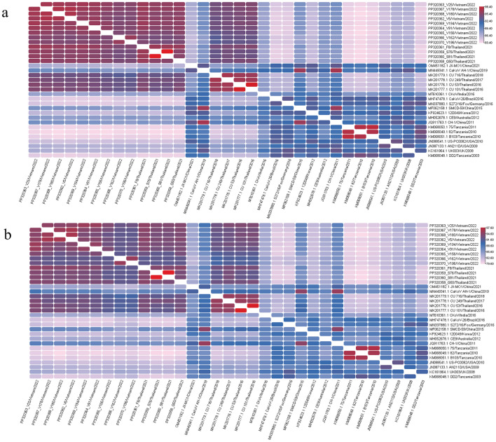
The nucleotide and amino acid similarities between the CaKoV sequences from Vietnam and those from Thailand in this study were compared with strains previously available in the GenBank database, originating from diverse countries such as Thailand, China, Germany, India, South Korea, the UK, Australia, Tanzania, Brazil, and the United States (Fig. 1, Supplementary Table 4, Supplementary Table 9). Within the CDS of Vietnamese CaKoV sequences, a similarity of 95–97.3% in nt and 92–96.4% in aa was observed. The four CDS of Thai CaKoV sequences exhibited a similarity of 96.3–99.9% in nt and 93.6–99.9% in aa. When comparing CDS of CaKoV obtained from Vietnam and Thailand, they displayed similarities of 94.6%-96.5% in nt and 91.9%-95% in aa. Remarkably, they showed the highest nt similarity with the Thai strains from the previous study (MK201176-79) and the Chinese strain (MN449341), while exhibiting the lowest nt similarity to Tanzanian strains (KM068048-51) (Fig. 1, Supplementary Table 4). As expected, the VP1 gene was the most variable region for CaKoV, with nt similarities ranging from 81.6 to 96.5% and aa similarities ranging from 81.1 to 98.5% (Supplementary Table 4). Notably, no recombination events were identified in the CaKoV strains obtained in this study.
Figure 1.
Heatmap of the nucleotide and amino acid similarity of complete coding sequences of canine kobuvirus (CaKoV) among isolates from Vietnam and Thailand, along with reference strains. (a) The similarity of nucleotide sequences; (b) the similarity of amino acid sequences.
Ten putative cleavage sites within the CaKoV genomic structure were predicted (Supplementary Table 5). In particular, the putative cleavage sites of the polyprotein mostly contained amino acid residues Q/G, except Q/H, Y/V and Q/A for the cleavage sites between VP0/VP3, VP1/2A, and 3A/3B, respectively. The putative cleavage site between VP3/VP1 was Q/A, conserved in almost all strains, except for wild carnivore strains (the spotted hyena and the side-striped jackals) in Tanzania (KM068049.1 and KM068051.1), where it was replaced by Q/T. A putative proline-rich region was identified at aa position 228-240 of the VP1 gene, with the motif 228PRAPPPLPPLPTP240 conserved in almost all strains, except for strains JN387133.1/USA (228PRAPP-LPPLPTP240), KM068049.1/Tanzania, KM068050.1/Tanzania, JN088541.1/USA (228CPVPPPLPPLPTP240), MH747478.1/Brazil (228HGAPPPLPPLPTP240), (Supplementary Table 6). Amino acid positions 60, 65, 67, 69, 150, 151, 153, 201, 204, 205, 210, 213 were conserved in almost Vietnamese and Thailand CaKoV strains (in this study), Thailand CaKoV strains (in a previous study), and Chinese strains. However, for two strains PP320366_V162/Vietnam/2022 and PP320370_V196/Vietnam/2022, the aa positions mentioned above were different and not conserved when compared to the remaining Vietnam and Thailand strains. Interestingly, these positions were conserved with Tanzanian and USA strains (Supplementary Table 6).
Phylogeny of the Vietnamese and Thai CaKoV
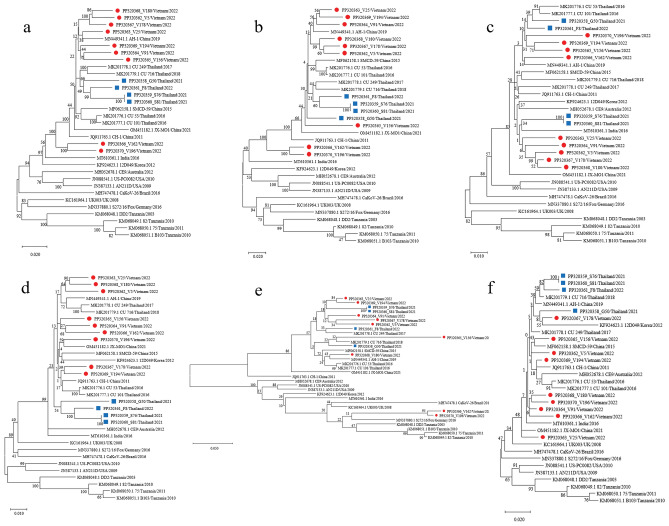
The phylogenetic analysis, based on the nucleotide sequences of the CDS, P1–P2–P3 region, VP, and 3D genes of nine CaKoV Vietnamese strains and four CaKoV Thai strains, is presented in Fig. 2. The tree based on the CDS indicated a close genetic relationship between Vietnam and Thailand strains, showing genetic affinities with Chinese strains. Interestingly, two Vietnamese strains (PP320366_V162/Vietnam/2022 and PP320370_V196/Vietnam/2022) formed a distinct subgroup, sharing genetic characteristics with both Chinese and Thai strains. The phylogenetic tree based on the P1 region exhibited similar characteristics to the CDS tree; however, the PP320365_V156/Vietnam/2022 strain formed a separate clade. Furthermore, the phylogenetic tree based on the VP1 gene, known for its high variability and belonging to the P1 region, suggested that the two CaKoV strains from Vietnam (PP320366_V162/Vietnam/2022 and PP320370_V196/Vietnam/2022) were closely related to strains MN337880 (isolated from foxes in Germany), and KM068049-068051 (isolated from wild carnivores in Tanzania), rather than strains isolated from domestic dogs in Asia and the Americas. Examining the P2 and P3 regions, the phylogenetic tree revealed that the Vietnamese and Thai strains in this study tended to cluster together, sharing genetic characteristics with strains from China, India, Australia, and previous Thai strains. They were separated from strains in Europe, the USA, Brazil, and Tanzania. For the 3D gene of CaKoV, belonging to the P2 region, the phylogenetic tree displayed less genetic variation, with the strains in this study forming a cohesive subgroup.
Figure 2.
Phylogenetic trees of canine kobuvirus (CaKoV) for various regions: (a) the complete coding sequences (CDSs), (b) P1 region, (c) P2 region, (d) P3 region, (e) VP1 gene, and (f) 3D gene. Trees of CDSs, P1, P2, and P3 region were constructed using MEGA v10.0 with the neighbor-joining algorithm employing the general time-reversible model. For VP1 and 3D genes, the trees were constructed with the Hasegawa–Kishono–Yano method, incorporating a gamma distribution and invariable sites (G + I) for complete coding sequences, P1, P2, P3 region, and the 3D gene. The VP1 gene tree employed a gamma distribution (G). All trees underwent 1,000 replications of bootstrap analysis. Vietnamese CaKoV isolates are marked with red circle, and Thai CaKoV isolates are noted by blue square.
B-cell epitope prediction for Vietnamese and Thai CaKoV
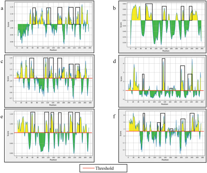
The VP1 capsid gene of CaKoV was employed for the prediction of both continuous and discontinuous B-cell epitopes. Multiple methods were utilized for predicting continuous B-cell epitopes, and the outcomes from each method were visualized through distinct charts and tables (refer to Table 3 and Fig. 3).
Table 3.
Analysis of B-cell epitopes of the VP1 gene of canine kobuvirus (CaKoV) sequences obtained in this study using the Kolaskar and Tongaonkar antigenicity method.
| No. | Start | End | Peptidea | Conserved amino acid/ total amino acid |
|---|---|---|---|---|
| 1 | 49 | 55 | FYRLLPL | 7/7 |
| 2 | 62 | 68 | SIPVPDG | 4/7 |
| 3 | 70 | 81 | VAQLPLDPLHWQ | 11/12 |
| 4 | 83 | 90 | SADVAGLT | 8/8 |
| 5 | 92 | 102 | MLSCFTYIAAD | 11/11 |
| 6 | 117 | 128 | TSLLIAYAPPGA | 10/12 |
| 7 | 156 | 173 | LISFSIPYTSPLSAIPTS | 18/18 |
| 8 | 195 | 210 | TLLFLPSYPQQDVQPQ | 10/16 |
| 9 | 228 | 238 | PRAPPPLPPLP | 11/11 |
| 10 | 247 | 254 | SVAVVKQG | 8/8 |
| 11 | 267 | 273 | RVYIVRA | 7/7 |
aThe diversity of amino acid residues within the predicted epitopes from the deduced amino acid sequences of PP320363_V25/Vietnam/2022 (VP1 gene) was analyzed Vietnamese and Thai CaKoV sequences.
Figure 3.
The physicochemical properties of canine kobuvirus (CaKoV) VP1 protein were evaluated using various methods: (a) Kolaskar & Tongaonkar antigenicity, (b) BepiPred linear epitope 2.0, (c) Chou & Fasman Beta-turn prediction, (d) Emini surface accessibility, (e) Karplus & Schulz flexibility prediction, and (f) Parker hydrophilicity. The Y-axis represents the corresponding score for each residue, while the X-axis corresponds to the residue positions in the sequence.
Residues with scores above the threshold (depicted by the red line on the chart) were considered indicative of having a higher likelihood of being potential epitopes. Additionally, the overlapping aa regions from all plots, based on the results of all prediction methods, served as the basis for suggesting potential continuous B-cell epitopes (Fig. 3).
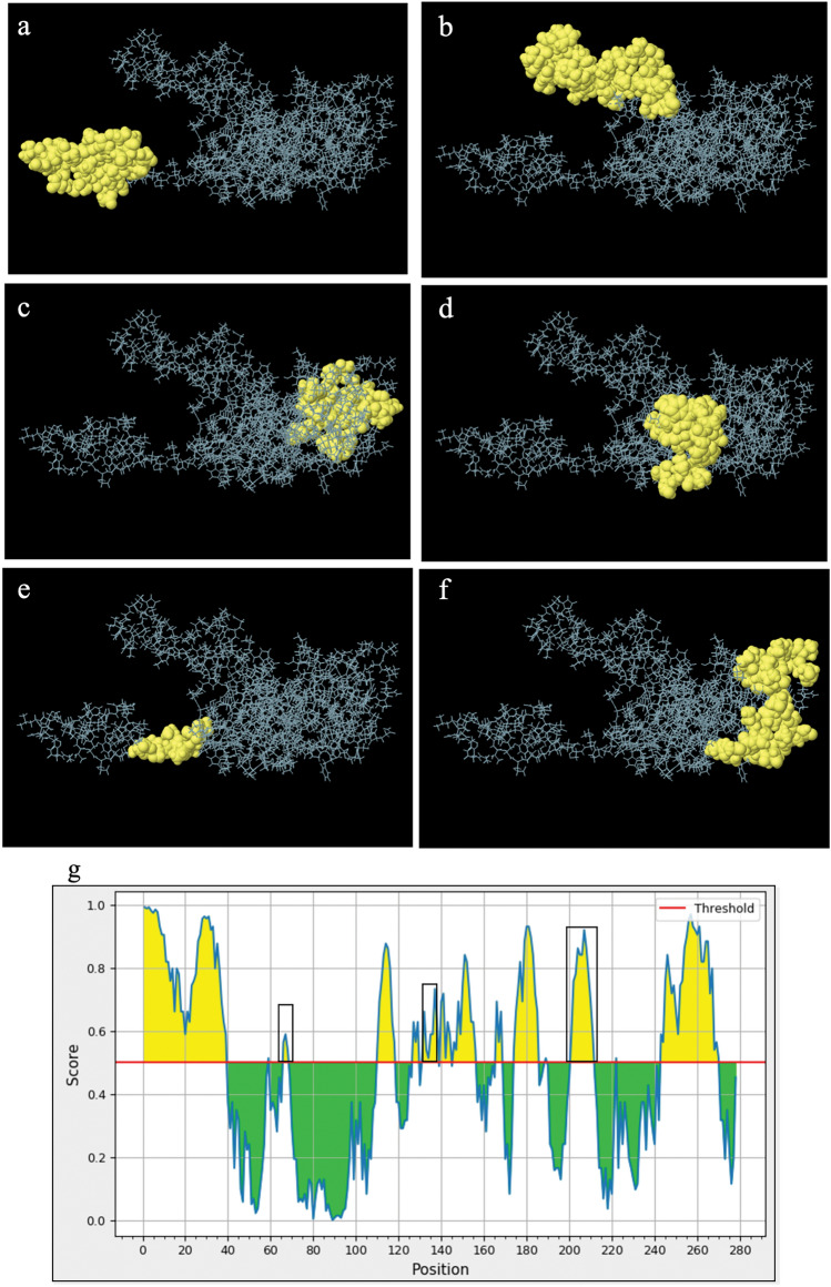
The ElliPro tool identified six discontinuous epitopes from the CaKoV VP1 gene capsid structure (Fig. 4). These epitopes varied in size from 8 to 36 residues, with scores ranging from 0.608 to 0.848. Information on the residues associated with discontinuous epitopes, including the quantity, positions, and scores, were detailed in Supplementary Table 8. Notably, the ElliPro predictions for discontinuous B-cell epitopes, based on the CaKoV VP1 region, overlapped with the linear B-cell epitopes. Furthermore, the Ellipro chart depicting continuous B-cell epitopes (Fig. 4g) revealed yellow areas, aligning with antigenic characteristics as indicated by the linear epitope predictions from the BepiPred linear epitope 2.0 method (Fig. 3b).
Figure 4.
The presentation of B-cell epitopes derived from the VP1 capsid of canine kobuvirus (CaKoV) employing ElliPro server: (a–f) Predicted discontinuous B-cell epitopes, and (g) Predicted B-cell linear structure.
Selection pressure for Vietnamese and Thai CaKoV
The analysis of the dN and dS ratios derived from the CaKoV VP1 gene alignment was conducted using the MEME and SLAC methods. Both analyses suggested that the VP1 gene of CaKoV from Vietnam and Thailand underwent negative selection pressure (with dN/dS < 1), indicating a tendency towards purifying or stabilizing selection. However, it is noteworthy that potential positive selection sites were also identified, implying regions where positive selection might be occurring (Table 4).
Table 4.
The selection sites of Vietnamese and Thai canine kobuvirus (CaKoV) based on VP1 region.
| CaKoV VP1 gene | Selection pressure | MEME† | SLAC | FEL | FUBAR‡ |
|---|---|---|---|---|---|
| Vietnamese strains | Positive selection | 2 | 0 | 0 | 1 |
| Negative selection | – | 28 | 108 | 107 | |
| Overall dN/dS | 0.0549 | 0.0707 | N/A | N/A | |
| Thai strains | Positive selection | 4 | 0 | 0 | 0 |
| Negative selection | – | 19 | 74 | 80 | |
| Overall dN/dS | 0.0488 | 0.0628 | N/A | N/A |
†MEME: positive selection for Vietnamese strains at sites 204th and 206th; for Thai strains at sites 69th, 82nd, 138th and 206th.
‡FUBAR: positive selection for Vietnamese strains at site 206th.
The analysis of selective pressure on the VP1 region of CaKoV revealed specific codon positions that were under positive selection. For Vietnamese CaKoV, the 204th and 206th codon positions were identified as positive selected, with the 206th codon being detected by both MEME and FUBAR methods, while the 204th codon was identified only by the FUBAR method. For Thai CaKoV, four codon positions (69th, 82nd, 138th, and 206th) were found to be positively selected, but they were only detected by the MEME method. The analysis also explored the relationship between the impact of selective pressure and B-cell epitopes. In both Vietnamese and Thai CaKoV, there was a positively selected position at the 206th codon, which corresponds to positions within predicted linear and conformational B-cell epitopes; the Thai CaKoV strain exhibited the additional presence of two positively selected positions, 69th and 138th codons. This suggests a potential interplay between selective pressure and the regions of the virus that are recognized by the immune system.
Discussion
This study revealed a prevalence of CaKoV in Vietnam and provided an update status of CaKoV in Thailand, showing a lower prevalence compared to a previous study28. Our investigation found that the highest detection rate of CaKoV was in dogs under 6 months old, aligning with similar patterns observed in previous reports, where the highest prevalence occurred in dogs under 4 months old in Japan (Soma et al.27) and 1 year old in Thailand28. Notably, human studies have also reported a higher incidence of Human Aichi Virus (AiV) infection in children35–37. In addition to affecting young dogs, CaKoV was found across various age groups in this study, consistent with findings in both humans38 and dogs28. However, a study in Japan reported that CaKoV was not detected in dogs over 5 months old27. These variations suggest that the prevalence of CaKoV may differ between countries, influenced by factors such as sample sizes and the specific age groups sampled.
The detection of CaKoV in both diarrhea and non-diarrhea dogs aligns with similar findings in studies from China2,23, Korea26, Japan27. The CaKoV prevalence in dogs with diarrhea tends to be higher than in dogs without diarrhea, but due to the small sample size in this study, further research is needed to clarify this point. Moreover, in dogs with diarrhea, CaKoV was identified either as a single detection or in co-detection with other common enteric viruses. This pattern is consistent with previous research where CaKoV was found either as a sole detection2,19,22,26,27 or in co- detection with viruses such as CPV2, rotavirus, CCoV, CDV, CaAstV, and Canine bocavirus2,19,20,22,25–27,39, as well as bacteria or parasites like Campylobacter upsaliensis, Toxocara canis, Toxocara leonina20 and giardiasis32. Similarly, studies on AiV have shown a high rate of single AiV infection, suggesting its potential role in gastrointestinal tract diseases in humans38. However, the true pathogenic role of CaKoV remains uncertain. It is unclear whether CaKoV can independently cause disease or if it collaborates with other enteric pathogens to induce gastroenteritis in dogs40. A global study suggests that CaKoV might play a sporadic pathogenic role in specific dog populations or interact with other enteric pathogens29. Therefore, additional observational studies or experimental infections are crucial to understanding the pathogenic mechanism of CaKoV in the gastrointestinal tract and its interactions with other pathogens4. However, the inability to cultivate CaKoV poses a significant challenge in the pathobiological research of this virus20,26.
In the comparison of CaKoV structure between this study and AiV, it was noted that the proline-rich region at aa position 50-63 of the VP0 gene of CaKoV was absent. Only the proline-rich region at aa position 228-240 (P228XPPPPXPPXPXP240), which belong to the VP1 gene, showed similarity between CaKoV and AiV41. This distinctive proline-rich region plays a crucial role in mediating host-virus interactions, precise localization of adhesion, infectivity, and viral pathogenesis42,43. In the phylogenetic analysis, the results of this study revealed a significant tendency for strains from the same country to cluster together, consistent with previous reports19,29,34,39. The phylogenetic tree of CDS indicated that the sequences from China, Vietnam and Thailand formed an independent group, separated from sequences found in Africa, the United States, and Europe28,39. However, two Vietnamese CaKoV strains (PP320366_V162/Vietnam/2022, PP320370_V196/Vietnam/2022) showed a tendency to form a new monophyletic subgroup. Moreover, the VP1 gene of strains PP320366_V162/Vietnam/2022 and PP320370_V196/Vietnam/2022 exhibited genetic heterogeneity compared to other Vietnamese CaKoV strains. Importantly, recombination events were not recorded for the Vietnamese and Thai CaKoV strains in this study. These findings suggest that strains PP320366_V162/Vietnam/2022 and PP320370_V196/Vietnam/2022 may have regional characteristics or may be ancestral strains. However, the availability of complete CaKoV genome sequences was relatively limited, and increasing the number of CaKoV CDSs in various countries would help clarify this issue.
B-cell epitopes, or antigenic determinants, are distinct clusters of amino acids on an antigen that are recognized by secreted antibodies or B-cell receptors, inducing cellular or humoral immune responses44,45. The analyis and prediction of B-cell epitopes were conducted using IEDB resources, focusing on the VP1 gene region of kobuvirus. This region is essential for receptor interaction, exhibits high immunogenicity among picornavirus capsid proteins, and experiences significant selective pressure7,15,46–48. The impact of selective pressure on the host immune response to CaKoV was explored. The analysis revealed that negative selection predominantly influenced the evolutionary dynamics of the VP1 gene region of CaKoV in Vietnam and Thailand. This suggests that external selection pressure may not be closely linked to the presence of mutations. Negative selection reflects an inclination to maintain the overall antigenic stability of CaKoV, indicating a low evolutionary rate and a delicate balance between virus-host interaction and survival ability49–53. However, potential positive selection positions in the VP1 gene were still identified. This suggests that selective pressure tends to alter the B-cell epitope of CaKoV, making it less conserved. Consequently, the host's immune response to CaKoV may undergo changes, potentially leading to immune evasion.
In summary, this study found that CaKoV was prevalent in both diarrheic and non-dirrheric dogs of all ages, with the highest prevalence observed in dogs under 6 months old. CaKoV was not only detected as a single infection but also in co-detection with other common gastrointestinal viral pathogens. The phylogenetic tree and genomic characteristics analyses revealed a close relationship between Vietnamese and Thai CaKoV strains, and they were also closely related to Chinese strains. Notably, Vietnamese strains formed a unique sub-branch. Selective pressure analysis showed that negative selection was the predominant force shaping the evolution of Vietnamese and Thai CaKoVs. However, potential positive selection sites were identified within the VP1 gene, particularly in regions associated with predicted B-cell epitopes. This suggests that while there is overall pressure to maintain stability, certain regions of the virus might be evolving under positive selection, possibly impacting host immune responses. The need for further research to elucidate the pathogenic mechanisms and immune responses associated with CaKoV infections in dogs.
Materials and methods
Sample collection and RNA extraction
A total of 327 RS samples were collected from pet dogs visiting veterinary clinics in Vietnam and Thailand between August 2021 and August 2022. This comprised 113 samples from Ho Chi Minh City, Vietnam (46 from healthy dogs and 67 from GID dogs) and 214 samples from Bangkok and Nakhon Si Thammarat Province, Thailand (107 from healthy dogs and 107 from GID dogs). The RS samples were randomly collected from both healthy dogs and those with diarrhea. However, dogs with clinical manifestations of gastrointestinal issues due to food poisoning, hepatobiliary disease, or those vaccinated within the last 4 weeks were excluded from the study. Information about the sampled dogs, including age, breed, sex, and vaccination status, was recorded using a questionnaire. To analyze age-related risk factors associated with CaKoV detection, all dogs were categorized into six age groups, adjusted based on established age classifications for dogs54.
The animal sampling and research protocols for this study were reviewed and approved by the Institutional Animal Care and Use Committee (IACUC) (No. 2231006) and the Institutional Biosafety Committee (IBC) (No. 2131019) of Chulalongkorn University (Bangkok, Thailand). Approval was also obtained from the Animal Ethics Committee (AEC) (No. NLU-220217) of Nong Lam University (Ho Chi Minh City, Vietnam). All procedures were performed in accordance with the relevant guidelines and regulations. The authors complied with the ARRIVE guidelines. Written informed consent was obtained from the owners for the participation of their animals in this study.
The RS sampling procedure involved inserting a sterile disposable cotton swab (Puritan, Guilford, USA) into the dog’s rectum, followed by immersion in a 1.5 mL Eppendorf tubes containing 0.7 mL of 1% (v/v) sterile phosphate-buffered salt (PBS) at pH 7.4. The samples were stored at − 80 °C until the extraction step. Viral RNA extraction was carried out using the Nucleic Acid Extraction Kit II (Geneaid, Ltd., Taipei, Taiwan) according to the manufacturer's protocol. Subsequently, the extracted RNA samples were quantified and assessed for quality using a Nanodrop® Lite spectrophotometer (Thermo Fisher Scientific Inc., Waltham, MA, USA) at the absorbance ratio A260/A280 and stored at − 80 °C until further use.
CaKoV detection
The extracted RNA samples underwent screening for the presence of CaKoV using the reverse transcription-polymerase chain reaction (RT-PCR) method with specific primers. These primers were self-designed to target the 3D gene region of CaKoV, referencing the available CaKoV sequence JQ911763 (Supplementary Table S1). The total volume was adjusted to 25 μl for the QIAGEN® One-Step RT-PCR Kit (Qiagen GmbH, Hilden, Germany), including 5 μl of QIAGEN 5 × buffer, 1 μl of 10 mM dNTP Mix, 2 μl of 0.6 μM forward and reverse primers, 1 µl of enzyme mixture, 3 µl of extracted RNA, and 11 µl of distilled water.
The thermal cycling conditions were conducted on a PCR thermocycler (SensoQuest GmbH, Göttingen, Germany), involving a complementary DNA (cDNA) synthesis step at 50 °C for 30 min, followed by inactivation of reverse transcription (RT) enzyme and initial PCR activation step at 95 °C for 15 min. Subsequently, 40 cycles of denaturation at 95 °C for 30 s, annealing at 57 °C for 30 s, and extension at 72 °C for 1 min were carried out, followed by a final extension step at 72 °C for 10 min. The positive control for CaKoV was synthesized using GeneArt™ Strings™ DNA fragments based on the 3D region of strain JQ911763 (Thermo Fisher Scientific GmbH, Darmstadt, Germany). A no-template control (NTC) was employed as a negative control. PCR products were visualized using the QIAxcel® DNA Screening Kit (Qiagen GmbH, Hilden, Germany) and the Qiaxcel® high-resolution capillary electrophoresis device (Qiagen GmbH, Hilden, Germany). Installation and analysis procedures were followed a previous report protocol55. The presence of the 438 bp amplicon product was considered as CaKoV positive. Subsequently, the PCR products were sent for genetic sequencing using a NGS-based method (Celemics, Inc., Seoul, Korea) to double-confirm the presence of CaKoV. The derived nucleotide sequences were analyzed and aligned with previously described CaKoV sequences depositing in the GenBank database using BLASTn analysis. CaKoV-positive samples were also tested for co-detection with other common canine enteric viruses by PCRs, including CPV56, CDV57, CCoV58, and CaAstV59.
CaKoV whole genome characterization
CaKoV-positive samples, initially identified by CaKoV-PCR screening, underwent further investigation for full-length genome analysis using multiple PCR assays. Seven primer pairs, self-designed based on nucleotide alignments from previously described CaKoV sequences available in the GenBank database (Supplementary Table 1), were employed to amplify the full-length CaKoV genome. The initial step involved the construction of cDNA from the extracted RNA samples, using the Omniscript® Reverse Transcription Kit (Qiagen GmbH, Hilden, Germany). For cDNA synthesis, the final volume was 20 μl, comprising 2 μl of 10 × RT buffer, 2 μl of 5 mM dNTP mixture, 1 μl of 10 μM random primer (Promega, Madison, Wisconsin, USA), 1 μl of RNase-free water, 0.75 μl of 1 × buffer, 0.25 μl of RNase inhibitor (10 units/μl), 1 μl of Omniscript reverse transcriptase, and 12 μl of extracted RNA sample.
Subsequently, the PCR reaction was performed with a final volume of 25 μl, including 3 μl of cDNA, 1 μl of 1 μM for each forward and reverse primers, 12.5 μl of GoTaq® Green Master Mix (Promega, Madison, Wisconsin, USA), and 7.5 μl of distilled water. The thermal cycling conditions included an initial denaturation at 95 °C for 5 min, followed by 40 cycles of 95 °C for 1 min, 55–58 °C for 1 min, and 72 °C for 1 min, and then a final extension at 72 °C for 10 min. PCR products were visualized using the QIAxcel® DNA Screening Kit, as mentioned above. Positive amplicons were then subjected to genomic sequencing according to the protocol described above. Subsequently, the obtained gene sequences were aligned and assembled using the BioEdit software package version 7.2 with the ClustalW function.
Phylogenetic and genetic analyses
Genetic analysis involved comparing the similarity of the CaKoV nucleotide sequence obtained from this study with the CaKoV sequences available in the database. Phylogenetic trees were constructed based on the complete coding sequence (CDS), P1–P2–P3 regions, and two genes, VP1 and 3D of CaKoV. The MEGA software package version 10.0 was used for this purpose. The software utilized the Find Best DNA/Protein Models (ML) option to select the substitution model for the constructed phylogram. The maximum likelihood (ML) method was applied, utilizing the General Time Reversible model (GTR) for CDS, P1, P2, P3 region, and the Hasegawa–Kishono–Yano (HKY) for VP1 and 3D genes. Gamma distribution and invariable sites (G + I) were considered for CDS, P1, P2, P3 region, and 3D gene, while a gamma distribution (G) was used for VP1 gene. The analysis included 1000 bootstrap replicates to assess the robustness of the phylogenetic relationships. To further evaluate the relationship between the CaKoV strains obtained in this study and other strains, deduced amino acid sequences of CaKoV were aligned and compared using the BioEdit software package, version 7.2. Additionally, TBtools was employed to create a heatmap60 via https://github.com/CJ-Chen/TBtools/releases.
Recombination analysis
All CaKoV strains obtained in Vietnam and Thailand underwent screening for potential genetic recombination events using the Recombination Detection Program version 4.0 (RDP4) software package. This comprehensive package incorporates seven different integrated recombination detection algorithms, namely RDP, GeneConv, Chimera, MaxChi, SiScan, 3Seq, and BootScan. To ensure reliability and consistency in results, potential recombinant sequences were considered only when positive results were obtained in at least 4 out of 7 methods with a p-value of ≤ 0.0161,62. This approach minimizes the risk of detecting false-positive signals. Subsequently, to visualize recombination breakpoints, potentially recombinant sequences were subjected to detailed analysis using SimPlot software version Beta 4.94. This involved employing two methods: similarity plot analysis and Bootscan analysis. The procedures for implementing this analytical method adhered to a previously published procedure55.
B-cell epitope prediction
The nucleotide sequences of VP1 capsid gene of CaKoV capsids were in-silico translated into amino acid sequences, and these sequences underwent analysis using the B-Cell Epitope Online Prediction Tool: “Analysis Resources and immune epitope database” (IEDB-AR) version 2.27 available at http://www.iedb.org/63. The tool was employed to identify both continuous B-cell epitopes (linear B-cell epitopes) and discontinuous B-cell epitopes (3D structure of the antigen) derived from the primary amino acid sequence.
For the prediction of continuous B-cell epitopes, the Kolaskar & Tongaonkar antigenicity method was used64. Additionally, to enhance the accuracy of predicting continuous B-cell epitopes, the correlation of all given single physicochemical properties was evaluated using five other propensity scales: (1) BepiPred 2.0 linear epitopes (for predicting specific regions in the protein that bind to the B-cell receptor)65, (2) Parker hydrophilicity (highlighting hydrophilic epitope regions)66, (3) Emini surface accessibility (indicating epitope surface accessibility)67, (4) Chou & Fasman Beta turn prediction (determining beta-turn regions as epitopes)68, and (5) Karplus & Schulz mobility prediction (assessing the mobility of protein segments)69. The setting parameters of all above methods, including the threshold, average score, minimum score, and maximum score of each method, are shown in Supplementary Table 7.
For predicting discontinuous B-cell epitopes, the online Ellipro tool of IEDB-AR server v2.27 (http://tools.iedb.org/ellipro/) was used70. In this approach, the minimum score and maximum distance (Angstrom) were set at 0.5 and 6, respectively.
Selection pressure analysis
The assessment of non-neutral nucleotide substitution involved calculating the ratio between synonymous substitutions (dS) and non-synonymous substitutions (dN). This evaluation was conducted through phylogenetic reconstruction using a ML model with general reversible nucleotide substitutions and performed using the Datamonkey web server (http://www.datamonkey. org). The Bayes factor was set at 50 to evaluate the dN and dS rates in each specific codon56,61. Positive selection (representing adaptive molecular evolution), neutral mutation, and negative selection (representing purifying selection) were defined as dN/dS > 1, dN/dS = 1, and dN/dS < 1, respectively. Sites under selection pressure were predicted using the HyPhy software package, available on the Datamonkey web server (http://www.datamonkey.org/), which includes four methods: mixed effects (MEME), Single Likelihood Ancestry Counting (SLAC), Fixed Effects Likelihood (FEL), and Fast Unconstrained Bayesian Approximation for Choice Inference (FUBAR). To confirm sites with positive selection pressure for each method, a p-value threshold < 0.1 for SLAC, FEL and MEME was established, while for FUBAR the posterior probability exceeded 0, 9 was used. Sites showing positive selection were identified when consistent results were obtained from a minimum of two of these methods. The CaKoV Thai strains from a previous study in Thailand (MK201776-79)28 were also included in the analysis of selective pressure, in conjunction with the CaKoV Thai strains from the current study.
Statistical analysis
Statistical analysis was performed using SAS® Studio software 3.81 (SAS® OnDemand for Academics) (https://www.sas.com/el_gr/software/on-demand-for-academics.html) (© 2012-2020 SAS Institute Inc., Cary, NC, USA). The correlation between the presence of CaKoV and age group was analyzed using Pearson's chi-square test or Fisher’s exact test (depending on the population size for each variable). A p-value < 0.05 was considered statistically significant to evaluate the relationship. Additionally, odds ratios (ORs) were calculated to quantify the strength of association between each factor and the presence of CaKoV.
Supplementary Information
Acknowledgements
TVN is supported by the Second Century Fund (C2F), Chulalongkorn University. CP is supported by the Ratchadapisek Somphot Fund for Postdoctoral Fellowship, Chulalongkorn University. ST is partly supported by National Research Council of Thailand (NRCT): R. Thanawongnuwech NRCT Senior scholar 2022 #N42A650553. This research is funded by Thailand Science research and Innovation Fund Chulalongkorn University (HEAF67310040) (to ST).
Author contributions
CP and ST designed the research. TVN conducted the experiments, analyzed the result, and wrote the first draft of manuscript. TK analyzed the result, CP and ST finalized the manuscript. All authors read and approved the final manuscript.
Data availability
The data used to support the findings of this study have been deposited in NCBI GenBank under accession numbers PP320358-PP320370.
Competing interests
The authors declare no competing interests.
Footnotes
Publisher's note
Springer Nature remains neutral with regard to jurisdictional claims in published maps and institutional affiliations.
Contributor Information
Chutchai Piewbang, Email: alkaline_eart@hotmail.com.
Somporn Techangamsuwan, Email: somporn62@hotmail.com.
Supplementary Information
The online version contains supplementary material available at 10.1038/s41598-024-62833-2.
References
- 1.Kapoor A, et al. Characterization of a canine homolog of human Aichivirus. J. Virol. 2011;85:11520–11525. doi: 10.1128/JVI.05317-11. [DOI] [PMC free article] [PubMed] [Google Scholar]
- 2.Li L, et al. Viruses in diarrhoeic dogs include novel kobuviruses and sapoviruses. J. Gen. Virol. 2011;92:2534–2541. doi: 10.1099/vir.0.034611-0. [DOI] [PMC free article] [PubMed] [Google Scholar]
- 3.Khamrin P, Maneekarn N, Okitsu S, Ushijima H. Epidemiology of human and animal kobuviruses. Virusdisease. 2014;25:195–200. doi: 10.1007/s13337-014-0200-5. [DOI] [PMC free article] [PubMed] [Google Scholar]
- 4.Caddy SL. New viruses associated with canine gastroenteritis. Vet. J. 2018;232:57–64. doi: 10.1016/j.tvjl.2017.12.009. [DOI] [PMC free article] [PubMed] [Google Scholar]
- 5.Decaro N. Enteric viruses of dogs. Adv. Small Anim. Care. 2020;1:143–160. doi: 10.1016/j.yasa.2020.07.012. [DOI] [Google Scholar]
- 6.Yamashita T, et al. Complete nucleotide sequence and genetic organization of Aichi virus, a distinct member of the Picornaviridae associated with acute gastroenteritis in humans. J. Virol. 1998;72:8408–8412. doi: 10.1128/JVI.72.10.8408-8412.1998. [DOI] [PMC free article] [PubMed] [Google Scholar]
- 7.Yamashita T, et al. Isolation and characterization of a new species of kobuvirus associated with cattle. J. Gen. Virol. 2003;84:3069–3077. doi: 10.1099/vir.0.19266-0. [DOI] [PubMed] [Google Scholar]
- 8.Reuter G, Boldizsar A, Pankovics P. Complete nucleotide and amino acid sequences and genetic organization of porcine kobuvirus, a member of a new species in the genus Kobuvirus, family Picornaviridae. Arch. Virol. 2009;154:101–108. doi: 10.1007/s00705-008-0288-2. [DOI] [PubMed] [Google Scholar]
- 9.Chang JT, Chen YS, Chen BC, Chao D, Chang TH. Complete genome sequence of the first aichi virus isolated in taiwan. Genome Announc. 2013;1:e00107–00112. doi: 10.1128/genomeA.00107-12. [DOI] [PMC free article] [PubMed] [Google Scholar]
- 10.Yamashita T, et al. Isolation of cytopathic small round viruses with BS-Cl cells from patients with gastroenteritis. J. Infect. Dis. 1991;164:954–957. doi: 10.1093/infdis/164.5.954. [DOI] [PubMed] [Google Scholar]
- 11.Phan TG, et al. The fecal viral flora of wild rodents. PLoS Pathog. 2011;7:e1002218. doi: 10.1371/journal.ppat.1002218. [DOI] [PMC free article] [PubMed] [Google Scholar]
- 12.Ng TF, et al. High variety of known and new RNA and DNA viruses of diverse origins in untreated sewage. J. Virol. 2012;86:12161–12175. doi: 10.1128/JVI.00869-12. [DOI] [PMC free article] [PubMed] [Google Scholar]
- 13.Pankovics P, Boros A, Kiss T, Reuter G. Identification and complete genome analysis of kobuvirus in faecal samples of European roller (Coracias garrulus): For the first time in a bird. Arch. Virol. 2015;160:345–351. doi: 10.1007/s00705-014-2228-7. [DOI] [PubMed] [Google Scholar]
- 14.Chung JY, et al. Detection and genetic characterization of feline kobuviruses. Virus Genes. 2013;47:559–562. doi: 10.1007/s11262-013-0953-8. [DOI] [PMC free article] [PubMed] [Google Scholar]
- 15.Reuter G, Boros Á, Pankovics P. Kobuviruses–a comprehensive review. Rev. Med. Virol. 2011;21:32–41. doi: 10.1002/rmv.677. [DOI] [PubMed] [Google Scholar]
- 16.Sasaki J, Nagashima S, Taniguchi K. Aichi virus leader protein is involved in viral RNA replication and encapsidation. J. Virol. 2003;77:10799–10807. doi: 10.1128/jvi.77.20.10799-10807.2003. [DOI] [PMC free article] [PubMed] [Google Scholar]
- 17.Hughes PJ, Stanway G. The 2A proteins of three diverse picornaviruses are related to each other and to the H-rev107 family of proteins involved in the control of cell proliferation. J. Gen. Virol. 2000;81:201–207. doi: 10.1099/0022-1317-81-1-201. [DOI] [PubMed] [Google Scholar]
- 18.Sasaki J, Taniguchi K. Aichi virus 2A protein is involved in viral RNA replication. J. Virol. 2008;82:9765–9769. doi: 10.1128/JVI.01051-08. [DOI] [PMC free article] [PubMed] [Google Scholar]
- 19.Di Martino B, Di Felice E, Ceci C, Di Profio F, Marsilio F. Canine kobuviruses in diarrhoeic dogs in Italy. Vet. Microbiol. 2013;166:246–249. doi: 10.1016/j.vetmic.2013.05.007. [DOI] [PMC free article] [PubMed] [Google Scholar]
- 20.Carmona-Vicente N, et al. Phylogeny and prevalence of kobuviruses in dogs and cats in the UK. Vet. Microbiol. 2013;164:246–252. doi: 10.1016/j.vetmic.2013.02.014. [DOI] [PMC free article] [PubMed] [Google Scholar]
- 21.Kong N, et al. Molecular characterization of new described kobuvirus in dogs with diarrhea in China. Springerplus. 2016;5:2047. doi: 10.1186/s40064-016-3738-4. [DOI] [PMC free article] [PubMed] [Google Scholar]
- 22.Li C, et al. Prevalence and phylogenetic analysis of canine kobuviruses in diarrhoetic dogs in northeast China. J. Vet. Med. Sci. 2016;78:7–11. doi: 10.1292/jvms.15-0414. [DOI] [PMC free article] [PubMed] [Google Scholar]
- 23.Liu D, et al. Emerging of canine kobuvirus in dogs in China, 2015. Pol. J. Vet. Sci. 2017;2017:20. doi: 10.1515/pjvs-2017-0088. [DOI] [PubMed] [Google Scholar]
- 24.Wang Y, et al. Identification and full-genome sequencing of canine kobuvirus in canine fecal samples collected from Anhui Province, eastern China. Arch. Virol. 2020;165:2495–2501. doi: 10.1007/s00705-020-04773-6. [DOI] [PMC free article] [PubMed] [Google Scholar]
- 25.Choi S, et al. Phylogenetic analysis of astrovirus and kobuvirus in Korean dogs. J. Vet. Med. Sci. 2014;76:1141–1145. doi: 10.1292/jvms.13-0585. [DOI] [PMC free article] [PubMed] [Google Scholar]
- 26.Oem JK, Choi JW, Lee MH, Lee KK, Choi KS. Canine kobuvirus infections in Korean dogs. Arch. Virol. 2014;159:2751–2755. doi: 10.1007/s00705-014-2136-x. [DOI] [PMC free article] [PubMed] [Google Scholar]
- 27.Soma T, Matsubayashi M, Sasai K. Detection of kobuvirus RNA in Japanese domestic dogs. J. Vet. Med. Sci. 2016;78:1731–1735. doi: 10.1292/jvms.16-0217. [DOI] [PMC free article] [PubMed] [Google Scholar]
- 28.Charoenkul K, et al. First detection and genetic characterization of canine Kobuvirus in domestic dogs in Thailand. BMC Vet. Res. 2019;15:254. doi: 10.1186/s12917-019-1994-6. [DOI] [PMC free article] [PubMed] [Google Scholar]
- 29.Miyabe FM, Ribeiro J, Alfieri AF, Alfieri AA. Detection of canine kobuvirus RNA in diarrheic fecal samples of dogs with parvoviruses. Braz. J. Microbiol. 2019;50:871–874. doi: 10.1007/s42770-019-00095-1. [DOI] [PMC free article] [PubMed] [Google Scholar]
- 30.Kaiser FK, et al. Detection of systemic canine kobuvirus infection in peripheral tissues and the central nervous system of a fox infected with canine distemper virus. Microorganisms. 2021;9:2521. doi: 10.3390/microorganisms9122521. [DOI] [PMC free article] [PubMed] [Google Scholar]
- 31.Caddy SL, Goodfellow I. Complete genome sequence of canine astrovirus with molecular and epidemiological characterisation of UK strains. Vet. Microbiol. 2015;177:206–213. doi: 10.1016/j.vetmic.2015.03.011. [DOI] [PMC free article] [PubMed] [Google Scholar]
- 32.Ribeiro J, et al. Extra-intestinal detection of canine kobuvirus in a puppy from Southern Brazil. Arch. Virol. 2017;162:867–872. doi: 10.1007/s00705-016-3164-5. [DOI] [PMC free article] [PubMed] [Google Scholar]
- 33.Lukashev AN, et al. Genetic variation and recombination in Aichi virus. J. Gen. Virol. 2012;93:1226–1235. doi: 10.1099/vir.0.040311-0. [DOI] [PubMed] [Google Scholar]
- 34.Deng B, et al. Detection and genetic characterization of canine kobuvirus from stray dogs in Shanghai, China. Arch. Virol. 2023;168:112. doi: 10.1007/s00705-023-05710-z. [DOI] [PMC free article] [PubMed] [Google Scholar]
- 35.Reuter G, Boldizsar A, Papp G, Pankovics P. Detection of Aichi virus shedding in a child with enteric and extraintestinal symptoms in Hungary. Arch. Virol. 2009;154:1529–1532. doi: 10.1007/s00705-009-0473-y. [DOI] [PubMed] [Google Scholar]
- 36.Sdiri-Loulizi K, et al. Detection and genomic characterization of Aichi viruses in stool samples from children in Monastir, Tunisia. J. Clin. Microbiol. 2009;47:2275–2278. doi: 10.1128/JCM.00913-09. [DOI] [PMC free article] [PubMed] [Google Scholar]
- 37.Kaikkonen S, Rasanen S, Ramet M, Vesikari T. Aichi virus infection in children with acute gastroenteritis in Finland. Epidemiol. Infect. 2010;138:1166–1171. doi: 10.1017/S0950268809991300. [DOI] [PubMed] [Google Scholar]
- 38.Drexler JF, et al. Aichi virus shedding in high concentrations in patients with acute diarrhea. Emerg. Infect. Dis. 2011;17:1544–1548. doi: 10.3201/eid1708.101556. [DOI] [PMC free article] [PubMed] [Google Scholar]
- 39.Wang Z, et al. Epidemiological and phylogenetic analysis of canine kobuviruses in Tangshan. China. Arch. Virol. 2020;165:2317–2322. doi: 10.1007/s00705-020-04727-y. [DOI] [PMC free article] [PubMed] [Google Scholar]
- 40.Li M, et al. Prevalence and genome characteristics of canine astrovirus in southwest China. J. Gen. Virol. 2018;99:880–889. doi: 10.1099/jgv.0.001077. [DOI] [PubMed] [Google Scholar]
- 41.Zhu L, et al. Structure of human Aichi virus and implications for receptor binding. Nat. Microbiol. 2016;1:16150. doi: 10.1038/nmicrobiol.2016.150. [DOI] [PubMed] [Google Scholar]
- 42.Adzhubei AA, Sternberg MJ, Makarov AA. Polyproline-II helix in proteins: Structure and function. J. Mol. Biol. 2013;425:2100–2132. doi: 10.1016/j.jmb.2013.03.018. [DOI] [PubMed] [Google Scholar]
- 43.Berisio R, Vitagliano L. Polyproline and triple helix motifs in host-pathogen recognition. Curr. Protein. Pept. Sci. 2012;13:855–865. doi: 10.2174/138920312804871157. [DOI] [PMC free article] [PubMed] [Google Scholar]
- 44.Getzoff ED, Tainer JA, Lerner RA, Geysen HM. The chemistry and mechanism of antibody binding to protein antigens. Adv. Immunol. 1988;43:1–98. doi: 10.1016/s0065-2776(08)60363-6. [DOI] [PubMed] [Google Scholar]
- 45.Jakhar R, Gakhar SK. An immunoinformatics study to predict epitopes in the envelope protein of SARS-CoV-2. Can. J. Infect. Dis. Med. Microbiol. 2020;2020:7079356. doi: 10.1155/2020/7079356. [DOI] [PMC free article] [PubMed] [Google Scholar]
- 46.Rossmann MG, et al. Structure of a human common cold virus and functional relationship to other picornaviruses. Nature. 1985;317:145–153. doi: 10.1038/317145a0. [DOI] [PubMed] [Google Scholar]
- 47.Oberste MS, Maher K, Kilpatrick DR, Pallansch MA. Molecular evolution of the human enteroviruses: Correlation of serotype with VP1 sequence and application to picornavirus classification. J. Virol. 1999;73:1941–1948. doi: 10.1128/JVI.73.3.1941-1948.1999. [DOI] [PMC free article] [PubMed] [Google Scholar]
- 48.Pham NT, et al. Sequence analysis of the capsid gene of Aichi viruses detected from Japan, Bangladesh, Thailand, and Vietnam. J. Med. Virol. 2008;80:1222–1227. doi: 10.1002/jmv.21193. [DOI] [PubMed] [Google Scholar]
- 49.Bush RM. Predicting adaptive evolution. Nat. Rev. Genet. 2001;2:387–392. doi: 10.1038/35072023. [DOI] [PubMed] [Google Scholar]
- 50.Yang Z, Nielsen R. Codon-substitution models for detecting molecular adaptation at individual sites along specific lineages. Mol. Biol. Evol. 2002;19:908–917. doi: 10.1093/oxfordjournals.molbev.a004148. [DOI] [PubMed] [Google Scholar]
- 51.Kosiol C, Bofkin L, Whelan S. Phylogenetics by likelihood: Evolutionary modeling as a tool for understanding the genome. J. Biomed. Inform. 2006;39:51–61. doi: 10.1016/j.jbi.2005.08.003. [DOI] [PubMed] [Google Scholar]
- 52.Sangula AK, et al. Evolutionary analysis of foot-and-mouth disease virus serotype SAT 1 isolates from east Africa suggests two independent introductions from southern Africa. BMC Evol. Biol. 2010;10:371. doi: 10.1186/1471-2148-10-371. [DOI] [PMC free article] [PubMed] [Google Scholar]
- 53.Zang Y, et al. Epidemiologic and genomic characterizations of porcine kobuviruses in diarrheic and healthy pigs. Anim. Basel. 2023;13:3129. doi: 10.3390/ani13193129. [DOI] [PMC free article] [PubMed] [Google Scholar]
- 54.Harvey ND. How old is my dog? Identification of rational age groupings in pet dogs based upon normative age-linked processes. Front. Vet. Sci. 2021;8:643085. doi: 10.3389/fvets.2021.643085. [DOI] [PMC free article] [PubMed] [Google Scholar]
- 55.Piewbang C, Techangamsuwan S. Phylogenetic evidence of a novel lineage of canine pneumovirus and a naturally recombinant strain isolated from dogs with respiratory illness in Thailand. BMC Vet. Res. 2019;15:300. doi: 10.1186/s12917-019-2035-1. [DOI] [PMC free article] [PubMed] [Google Scholar]
- 56.Nguyen-Manh T, Piewbang C, Rungsipipat A, Techangamsuwan S. Molecular and phylogenetic analysis of Vietnamese canine parvovirus 2C originated from dogs reveals a new Asia-IV clade. Transbound. Emerg. Dis. 2021;68:1445–1453. doi: 10.1111/tbed.13811. [DOI] [PubMed] [Google Scholar]
- 57.Piewbang C, Rungsipipat A, Poovorawan Y, Techangamsuwan S. Development and application of multiplex PCR assays for detection of virus-induced respiratory disease complex in dogs. J. Vet. Med. Sci. 2017;78:1847–1854. doi: 10.1292/jvms.16-0342. [DOI] [PMC free article] [PubMed] [Google Scholar]
- 58.Ksiazek TG, et al. A novel coronavirus associated with severe acute respiratory syndrome. N. Engl. J. Med. 2003;348:1953–1966. doi: 10.1056/NEJMoa030781. [DOI] [PubMed] [Google Scholar]
- 59.Van Nguyen T, Piewbang C, Techangamsuwan S. Genetic characterization of canine astrovirus in non-diarrhea dogs and diarrhea dogs in Vietnam and Thailand reveals the presence of a unique lineage. Front. Vet. Sci. 2023;10:145. doi: 10.3389/fvets.2023.1278417. [DOI] [PMC free article] [PubMed] [Google Scholar]
- 60.Chen C, et al. TBtools: An integrative toolkit developed for interactive analyses of big biological data. Mol. Plant. 2020;13:1194–1202. doi: 10.1016/j.molp.2020.06.009. [DOI] [PubMed] [Google Scholar]
- 61.Piewbang C, Radtanakatikanon A, Puenpa J, Poovorawan Y, Techangamsuwan S. Genetic and evolutionary analysis of a new Asia-4 lineage and naturally recombinant canine distemper virus strains from Thailand. Sci. Rep. 2019;9:3198. doi: 10.1038/s41598-019-39413-w. [DOI] [PMC free article] [PubMed] [Google Scholar]
- 62.Dankaona W, Mongkholdej E, Satthathum C, Piewbang C, Techangamsuwan S. Epidemiology, genetic diversity, and association of canine circovirus infection in dogs with respiratory disease. Sci. Rep. 2022;12:15445. doi: 10.1038/s41598-022-19815-z. [DOI] [PMC free article] [PubMed] [Google Scholar]
- 63.Vita R, et al. The Immune Epitope Database (IEDB): 2018 update. Nucleic Acids Res. 2019;47:D339–D343. doi: 10.1093/nar/gky1006. [DOI] [PMC free article] [PubMed] [Google Scholar]
- 64.Kolaskar AS, Tongaonkar PC. A semi-empirical method for prediction of antigenic determinants on protein antigens. FEBS Lett. 1990;276:172–174. doi: 10.1016/0014-5793(90)80535-q. [DOI] [PubMed] [Google Scholar]
- 65.Jespersen MC, Peters B, Nielsen M, Marcatili P. BepiPred-2.0: Improving sequence-based B-cell epitope prediction using conformational epitopes. Nucleic Acids Res. 2017;45:W24–W29. doi: 10.1093/nar/gkx346. [DOI] [PMC free article] [PubMed] [Google Scholar]
- 66.Parker JM, Guo D, Hodges RS. New hydrophilicity scale derived from high-performance liquid chromatography peptide retention data: Correlation of predicted surface residues with antigenicity and X-ray-derived accessible sites. Biochem. 1986;25:5425–5432. doi: 10.1021/bi00367a013. [DOI] [PubMed] [Google Scholar]
- 67.Emini EA, Hughes JV, Perlow DS, Boger J. Induction of hepatitis A virus-neutralizing antibody by a virus-specific synthetic peptide. J. Virol. 1985;55:836–839. doi: 10.1128/JVI.55.3.836-839.1985. [DOI] [PMC free article] [PubMed] [Google Scholar]
- 68.Chou P, Fasman GD. Amino acid sequence. Adv. Enzymol. Relat. Areas. Mol. Biol. 2009;47:45. doi: 10.1002/9780470122921.ch2. [DOI] [PubMed] [Google Scholar]
- 69.Karplus P, Schulz G. Prediction of chain flexibility in proteins: A tool for the selection of peptide antigens. Sci. Nat. 1985;72:212–213. doi: 10.1007/BF01195768. [DOI] [Google Scholar]
- 70.Ponomarenko J, et al. ElliPro: A new structure-based tool for the prediction of antibody epitopes. BMC Bioinform. 2008;9:514. doi: 10.1186/1471-2105-9-514. [DOI] [PMC free article] [PubMed] [Google Scholar]
Associated Data
This section collects any data citations, data availability statements, or supplementary materials included in this article.
Supplementary Materials
Data Availability Statement
The data used to support the findings of this study have been deposited in NCBI GenBank under accession numbers PP320358-PP320370.


