



Abstract
Background
The Coronal Plane Alignment of the Knee (CPAK) classification system has been developed as a comprehensive system that describes 9 coronal plane phenotypes based on constitutional limb alignment and joint line obliquity (JLO). Due to the characteristics of Asian populations, which show more varus and wider distribution in lower limb alignment than other populations, modification of the boundaries of the arithmetic hip-knee-ankle angle (aHKA) and JLO should be considered. The purpose of this study was to determine the knee phenotype in a Korean population based on the original CPAK and modified CPAK classification systems.
Methods
We reviewed prospectively collected data of 500 healthy and 500 osteoarthritic knees between 2021 and 2023 using radiographic analysis and divided them based on the modified CPAK classification system by widening the neutral boundaries of the aHKA to 0° ± 3° and using the actual JLO as a new variable. Using long-leg standing weight-bearing radiographs, 6 radiographic parameters were measured to evaluate the CPAK type: the mechanical HKA angle, medial proximal tibial angle (MPTA), lateral distal femoral angle (LDFA), aHKA, JLO, and actual JLO.
Results
From 2 cohorts of 1,000 knees, the frequency distribution representing all CPAK types was different between the healthy and arthritic groups. The most common categories were type II (38.2%) in the healthy group and type I (53.8%) in the arthritic group based on the original CPAK classification. The left and upward shift in the distribution of knee phenotypes in the original classification was corrected evenly after re-establishing the boundaries of a neutral aHKA and the actual JLO. According to the modified CPAK classification system, the most common categories were type II (35.2%) in the healthy group and type I (38.0%) in the arthritic group.
Conclusions
Although the modified CPAK classification corrected the uneven distribution seen when applying the original classification system in a Korean population, the most common category was type I in Korean patients with osteoarthritic knees in both classification systems. Furthermore, there were different frequencies of knee phenotypes among healthy and arthritic knees.
Keywords: Knee phenotypes, Coronal plane alignment of the knee, Constitutional varus, Osteoarthritis, Total knee arthroplasty
Restoration of a neutral mechanical axis has historically been considered the main aim to establish a successful total knee arthroplasty (TKA).1) Although the outcomes of TKA with mechanical alignment (MA) are acceptable, patient dissatisfaction rates of up to 20% after conventional TKA have been reported in the literature.2) Despite the redesigning of the implants and developments in the wear characteristics of the bearing surface over the last several decades, questions regarding this patient dissatisfaction remain unanswered.3,4) One such question is whether using a standardized neutral MA approach provides the best solution for all patients undergoing TKA, regardless of the wide individual variation in limb alignment.5,6)
Due to the desire to overcome the problems of patient dissatisfaction after TKA, a more individualized realignment strategy has been introduced as an alternative approach to MA, which focuses on the pre-arthritic knee alignment of each patient.6,7) Recent studies have demonstrated their alternative alignment options that consider coronal deformity and joint line obliquity (JLO) to be reliable and reproducible techniques that better represent the preoperative knee phenotype.8,9)
MacDessi et al.10) were the first to propose a standardized classification system, the Coronal Plane Alignment of the Knee (CPAK), to enhance the nomenclature of coronal deformity of the knee. This is a simple and comprehensive system for describing knee phenotypes that integrate the independent variables, constitutional coronal limb alignment (or arithmetic hip-knee-ankle angle [aHKA]) and JLO. However, although this system has been validated in many Australian patients, previous analyses of this comprehensive classification system have not considered the patients’ racial backgrounds. Furthermore, racial differences have been shown to impact alignment.11,12,13)
Recently, Hsu et al.13) have reported knee phenotypes classified according to a modified CPAK classification system based on a wider range of a neutral aHKA and actual JLO in the Asian population. To our knowledge, information about the distribution of CPAK types and characteristics of coronal alignment in the Korean population is lacking. The aims of this study were as follows: to validate the CPAK classification in a Korean population and to determine whether the distribution of CPAK types would be different among countries.
METHODS
This retrospective study received approval from the Chonnam National University Hwasun Hospital Institutional Review Board (IRB No. CNUH-2023-141) and informed consent was obtained from all patients.
Patients
We performed a cross-sectional radiological analysis of a healthy and an arthritic cohort of patients who were evaluated at a single institution from 2021 to 2023. The healthy cohort included 250 young adults (500 non-arthritic knees) aged between 20 and 30 years. Only participants without a prior history of orthopedic disease or trauma were included. The arthritic cohort included 500 consecutive patients who were scheduled to undergo TKA for the treatment of knee osteoarthritis. Patients were excluded if they had prior ipsilateral knee surgery, posttraumatic osteoarthritis, inflammatory arthritis, severe arthritic bone loss on the preoperative anteroposterior (AP) longleg standing weight-bearing radiographs, or flexion contracture greater than 20°, were non-Korean patients, or had incomplete data. Finally, a total of 1,000 subjects (500 healthy knees and 500 arthritic knees) were included and used for radiological analysis based on the CPAK classification.
Radiologic Evaluation
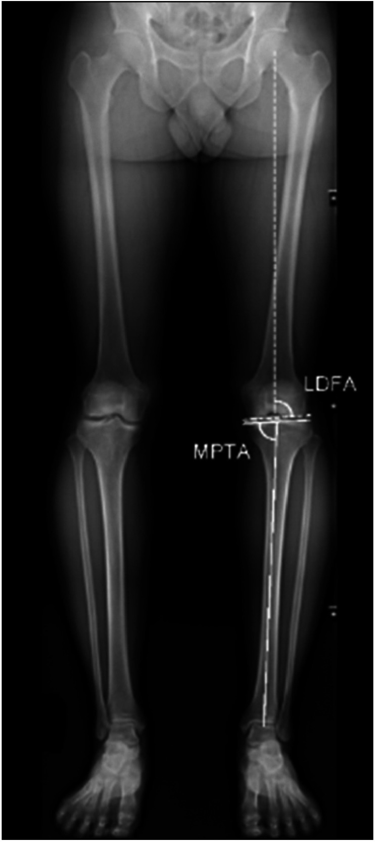
AP long-leg standing weight-bearing radiographs were taken following the method described by Paley and Pfeil.14) Radiographic evaluation included assessments of the coronal alignment of the lower limb (mechanical HKA [mHKA]), medial proximal tibial angle (MPTA), and lateral distal femoral angle (LDFA). The mHKA was measured as the angle between the mechanical axes of the femur and tibia. The MPTA was measured as the medial angle between the mechanical axis of the tibia and the proximal tibial joint line tangent, and the LDFA was defined as the lateral angle between the mechanical axis of the femur and the distal femoral joint line tangent (Fig. 1). All the radiographic parameters were measured by 2 independent orthopedic surgeons who were blinded to the purpose of this study (TWY and JYK). Each surgeon repeated the measurements twice with an interval of 4 weeks between the radiographic measurements to determine the intra- and interobserver reliability.
Fig. 1. Measurement of the medial proximal tibial angle (MPTA) and lateral distal femoral angle (LDFA) on an anteroposterior long-leg standing weight-bearing radiograph. The MPTA was defined as the medial angle between the mechanical axis of the tibia and the joint line of the proximal tibia (represented by the solid white line). The LDFA was defined as the lateral angle between the mechanical axis of the femur and the joint line of the distal femur (represented by the dashed white line).
CPAK Classification and CPAK Classification Matrix
The CPAK classification system was used for the categorization of constitutional knee phenotypes.10,15) The constitutional alignment was determined by the aHKA, which was calculated based on the following algorithm: aHKA = MPTA – LDFA. A negative aHKA indicates a varus alignment of the lower limb. Evaluation of JLO was calculated based on the following algorithm: JLO = MPTA + LDFA, and its obliquity was defined relative to the floor with the patient in double-leg stance.
Previous studies have shown that racial differences impact alignment, and due to the characterization of Asian populations, which show more varus and wider distribution in lower limb alignment, modification of the boundaries of the aHKA and JLO should be considered, as previously described by Hsu et al.11,12,13) Therefore, varus and valgus aHKAs were enlarged to less than −3° and greater than +3°, respectively, in this study series (standard deviation, 3.3°). Furthermore, the “actual JLO” was calculated according to the following algorithm: JLO = 90° − (MPTA + LDFA) / 2, and its obliquity reflected a certain degree of JLO parallel to the ground. The boundaries for a neutral actual JLO were 0° ± 3°, inclusive. With the 3 subgroups of the aHKA set against the 3 subgroups of JLO in a matrix, patients were matched to create 9 possible CPAK phenotypes (Fig. 2).
Fig. 2. Coronal Plane Alignment of the Knee classification system with 9 theoretical types of knees based on arithmetic hip-knee-ankle (aHKA) and joint line obliquity (JLO) measurements. MPTA: medial proximal tibial angle, LDFA: lateral distal femoral angle.

Statistical Analysis
Scatterplots were created to describe the proportions of healthy and arthritic knees classified based on the original and modified CPAK systems. Continuous variables are presented as means with standard deviations and were compared using the independent t-tests. The chi-square test or Fisher’s exact test was used to compare differences in categorical variables. The threshold for significance was p < 0.05.
RESULTS
The patient demographic characteristics and clinical data are summarized in Table 1. The geographic variation in knee phenotypes according to the CPAK classification system is shown in Table 2. All intra- and interobserver intraclass correlation coefficients of the radiographic measurements showed good agreement (> 0.75).
Table 1. Demographic Characteristics between the Groups.
| Variable | Healthy group (n = 500) | Arthritic group (n = 500) | p-value* |
|---|---|---|---|
| Age (yr) | 23.8 ± 8.2 | 75.0 ± 4.0 | < 0.001 |
| Female sex | 84 (16.8) | 419 (83.8) | < 0.001 |
| Body mass index (kg/m2) | 24.8 ± 8.9 | 26.1 ± 4.0 | < 0.001 |
| mHKA (°)† | 1.0 ± 2.9 | 10.3 ± 4.6 | < 0.001 |
| MPTA (°) | 87.1 ± 2.6 | 84.7 ± 3.0 | < 0.001 |
| LDFA (°) | 87.9 ± 2.3 | 89.3 ± 3.0 | < 0.001 |
| aHKA (°) | −0.8 ± 3.0 | −4.6 ± 3.8 | < 0.001 |
| JLO (°) | 175.0 ± 3.0 | 174.0 ± 3.5 | < 0.001 |
Values are presented as mean ± standard deviation or number (%).
mHKA: mechanical hip-knee-ankle angle, MPTA: medial proximal tibial angle, LDFA: lateral distal femoral angle, aHKA: arithmetic hip-knee-ankle, JLO: joint line obliquity.
*An independent t-test was used for analysis of differences in age, body mass index, and radiologic parameters. The chi-square test was used to analyze differences in sex. Statistically significant (p < 0.05). †A positive angle represents varus alignment, and a negative angle represents valgus alignment.
Table 2. CPAK Classification Distributions of Previously Published Literature of Other Ethnicities.
| Variable | MacDessi et al.10) | Toyooka et al.16) | Hsu et al.13) | Sappey-Marinier et al.17) | Steele et al.18) | |
|---|---|---|---|---|---|---|
| Healthy knees (n = 500) | Arthritic knees (n = 500) | Arthritic knees (n = 500) | Healthy knees (n = 214) | Arthritic knees (n = 1,078) | Arthritic knees (n = 1,946) | |
| Country | Belgium | Australia | Japan | Taiwan | France | USA |
| Type I | 132 (26.4) | 97 (19.4) | 269 (53.8) | 78 (36.4) | 360 (33.4) | 193 (19.8) |
| Type II | 196 (39.2) | 161 (32.2) | 127 (25.4) | 84 (39.3) | 210 (19.5) | 335 (34.5) |
| Type III | 49 (9.8) | 77 (15.4) | 41 (8.2) | 29 (13.6) | 115 (10.6) | 170 (17.5) |
| Type IV | 27 (5.4) | 49 (9.8) | 36 (7.2) | 12 (5.6) | 110 (10.2) | 62 (6.4) |
| Type V | 77 (15.4) | 73 (14.6) | 22 (4.4) | 10 (4.7) | 204 (18.9) | 122 (12.6) |
| Type VI | 17 (3.4) | 37 (7.4) | 5 (1.0) | 1 (0.5) | 68 (6.3) | 80 (8.2) |
| Type VII | 1 (0.2) | 3 (0.6) | 0 | 0 | 4 (0.4) | 4 (0.4) |
| Type VIII | 0 | 8 (1.6) | 0 | 0 | 6 (0.6) | 2 (0.2) |
| Type IX | 1 (0.2) | 2 (0.4) | 0 | 0 | 1 (0.1) | 5 (0.5) |
Values are presented as number (%).
CPAK: Coronal Plane Alignment of the Knee.
Original CPAK Classification
The knee phenotype distribution in both groups according to the CPAK classification area is shown in Tables 3 and 4. The frequencies of all CPAK types were different between the healthy and arthritic groups (Fig. 3). In the healthy group, the most common distribution was type II (38.2%, n = 191) with a neutral alignment and apex distal JLO orientation, followed by type I (28.6%, n = 143), type III (16.4%, n = 82), type V (7.6%, n = 38), type VI (4.2%, n = 21), and type IV (4.0%, n = 20). There were few patients (1.0%, n = 5) with an apex proximal JLO.
Table 3. Overall Absolute and Relative Preoperative Knee Phenotype Distribution of Healthy Knees with the Original CPAK Classification.
| MPTA – LDFA (aHKA) | Total | ||||
|---|---|---|---|---|---|
| < –2° | –2° to 2° | > 2° | |||
| MPTA + LDFA (JLO) | |||||
| < 177° | 143 (28.6) | 191 (38.2) | 82 (16.4) | 416 (83.2) | |
| 177° to 183° | 20 (4.0) | 38 (7.6) | 21 (4.2) | 79 (15.8) | |
| > 183° | 0 | 2 (0.4) | 3 (0.6) | 5 (1.0) | |
| Total | 163 (32.6) | 231 (46.2) | 106 (21.2) | 500 | |
Values are presented as number (%).
CPAK: Coronal Plane Alignment of the Knee, MPTA: medial proximal tibial angle, LDFA: lateral distal femoral angle, aHKA: arithmetic hip-knee-ankle, JLO: joint line obliquity.
Table 4. Overall Absolute and Relative Preoperative Knee Phenotype Distribution of Arthritic Knees with the Original CPAK Classification.
| MPTA – LDFA (aHKA) | Total | ||||
|---|---|---|---|---|---|
| < –2° | –2° to 2° | > 2° | |||
| MPTA + LDFA (JLO) | |||||
| < 177° | 269 (53.8) | 88 (17.6) | 8 (1.6) | 365 (73.0) | |
| 177° to 183° | 87 (17.4) | 36 (7.2) | 3 (0.6) | 126 (25.2) | |
| > 183° | 8 (1.6) | 1 (0.2) | 0 | 10 (2.0) | |
| Total | 364 (72.8) | 125 (25.0) | 11 (2.2) | 500 | |
Values are presented as number (%).
CPAK: Coronal Plane Alignment of the Knee, MPTA: medial proximal tibial angle, LDFA: lateral distal femoral angle, aHKA: arithmetic hip-knee-ankle, JLO: joint line obliquity.
Fig. 3. Scatter plots representing the overall original Coronal Plane Alignment of the Knee classification phenotype distribution in healthy knees (A) and arthritic knees (B). aHKA: arithmetic hip-knee-ankle angle, MPTA: medial proximal tibial angle, LDFA: lateral distal femoral angle, JLO: joint line obliquity.

In the arthritic group, the most common distribution was type I (53.8%, n = 269) with a constitutional varus alignment and apex distal JLO orientation, followed by type II (17.6%, n = 88), type IV (17.4%, n = 87), type V (7.2%, n = 36), type III (1.6%, n = 8), and type VI (0.6%, n = 3). There were also a few patients (2.0%, n = 10) with an apex proximal JLO.
Modified CPAK Classification
The knee phenotype distribution in both groups according to the modified CPAK classification area is shown in Tables 5 and 6. The left and upward shift in the distribution of knee phenotypes based on the original CPAK classification was corrected evenly after re-establishing the boundaries of a neutral aHKA and actual JLO (Fig. 4).
Table 5. Overall Absolute and Relative Preoperative Knee Phenotype Distributions of Healthy Knees with the Modified CPAK Classification System.
| MPTA – LDFA (aHKA) | Total | ||||
|---|---|---|---|---|---|
| < –3° | –3° to 3° | > 3° | |||
| Actual JLO* | |||||
| > 3° | 107 (21.4) | 176 (35.2) | 57 (11.4) | 340 (68.0) | |
| –3° to 3° | 30 (6.0) | 100 (20.0) | 27 (5.4) | 157 (31.4) | |
| < –3° | 0 | 1 (0.2) | 2 (0.4) | 3 (0.6) | |
| Total | 137 (27.4) | 277 (55.4) | 86 (17.2) | 500 | |
Values are presented as number (%).
CPAK: Coronal Plane Alignment of the Knee, MPTA: medial proximal tibial angle, LDFA: lateral distal femoral angle, aHKA: arithmetic hip-knee-ankle, JLO: joint line obliquity.
*The equation for actual JLO is described as 90 – (MPTA + LDFA) / 2.
Table 6. Overall Absolute and Relative Preoperative Knee Phenotype Distributions of Arthritic Knees with the Modified CPAK Classification System.
| MPTA – LDFA (aHKA) | Total | ||||
|---|---|---|---|---|---|
| < –3° | –3° to 3° | > 3° | |||
| Actual JLO* | |||||
| > 3° | 190 (38.0) | 68 (13.6) | 3 (0.6) | 261 (52.2) | |
| –3° to 3° | 135 (27.0) | 99 (19.8) | 4 (0.8) | 238 (47.6) | |
| < –3° | 1 (0.2) | 0 | 0 | 1 (0.2) | |
| Total | 326 (65.2) | 167 (33.4) | 7 (1.4) | 500 | |
Values are presented as number (%).
CPAK: Coronal Plane Alignment of the Knee, MPTA: medial proximal tibial angle, LDFA: lateral distal femoral angle, aHKA: arithmetic hip-knee-ankle, JLO: joint line obliquity.
*The equation for actual JLO is described as 90 – (MPTA + LDFA) / 2.
Fig. 4. Scatter plots representing the overall modified Coronal Plane Alignment of the Knee classification phenotype distribution in healthy knees (A) and arthritic knees (B). aHKA: arithmetic hip-knee-ankle angle, MPTA: medial proximal tibial angle, LDFA: lateral distal femoral angle, JLO: joint line obliquity.

In the healthy group, the most common distribution was type II (35.2%, n = 176) with a neutral alignment and apex distal JLO orientation, followed by type I (21.4%, n = 107), type V (20.0%, n = 100), type III (11.4%, n = 57), type IV (6.0%, n = 30), and type VI (5.4%, n = 27). There were few patients (0.6%, n = 3) with an apex proximal JLO.
In the arthritic group, the most common distribution was type I (38.0%, n = 190) with a constitutional varus alignment and apex distal JLO orientation, followed by type IV (27.0%, n = 135), type V (19.8%, n = 99), type II (13.6%, n = 68), type VI (0.8%, n = 4), and type III (0.6%, n = 3). There was also a patient (0.2%, n = 1) with an apex proximal JLO.
DISCUSSION
The principal findings of this study were as follows: (1) there were different frequencies of knee phenotypes between healthy and arthritic knees with the modified CPAK and original CPAK classification systems, (2) a significant discrepancy in the CPAK distributions existed between countries, and (3) although the modified CPAK classification corrected the uneven distribution when applying the original classification system in a Korean population, the most common categories were type I in the Korean patients with osteoarthritic knees in both classification systems. Our findings are clinically relevant and can facilitate the suggestion of more patient-specific recommendations when considering alignment strategies during the preoperative assessment of patients undergoing TKA.
To our knowledge, the present study is the first to elucidate the knee phenotype of healthy and arthritic Korean subjects according to the CPAK classification system. Song et al.12) evaluated Korean and Caucasian populations and showed that the MA was not neutral in the Korean population and that the majority of the Korean population had constitutional varus alignment. Furthermore, Asian populations show more varus and wider distribution in lower limb alignment.11,12,19) Therefore, we have modified the CPAK classification system by widening the neutral boundaries of the aHKA and JLO to resolve the left and upward shift in the distribution in our population. Compared with the original classification system, the modified classification system led to an increase in the distribution of knee phenotypes with a neutral aHKA and actual JLO. Differences in knee phenotypes between populations are nascent (Table 2);10,13,16,18) therefore, a comprehensive analysis of wide geographic variation in the prevalence of CPAK types among healthy and arthritic patients is essential, and different approaches should be used to provide a more individualized alignment approach for TKA.
Although systematically restoring a neutral MA is a classic goal for TKA, conflicting opinions have emerged, questioning the relationship between postoperative coronal misalignment and implant survivorship.20) Since Howell et al.21) have introduced the concept of KA, individualized or patient-specific techniques have been receiving increased interest to restore the pre-arthritic or native limb and joint line alignment of each patient.22) In this concept, KA provides a close match with the native patient anatomy and soft-tissue envelope and may enhance ligament balancing, reduce bone resection, and minimize soft-tissue releases.23,24) In fact, several clinical studies have demonstrated their modified alignment options and philosophies to be reliable techniques that better represent the preoperative knee phenotype.8,9,25) Hirschmann et al.6,7) have demonstrated the most compelling support for the use of a patient-specific alignment philosophy as an alternative to the MA using theoretical functional phenotypes of the native limb and knee joint line.
MacDessi et al.,10) established a standardized classification system to categorize knee phenotypes. They have demonstrated similar frequencies of knee phenotypes among healthy and arthritic populations and this system can be used in entire populations regardless of the presence of arthritic deformity. In their study, the most common categories were type ii (39.2% healthy vs. 32.2% arthritis) and type I (26.4% healthy vs. 19.4% arthritis). In contrast, we have demonstrated significant differences in knee phenotypes between patients with healthy and arthritic knees, and the majority of the patients (38.0%, n = 190) in the arthritic group were classified as type I. Measurements of the aHKA may change over time with advancing age, and Korean individuals with knee osteoarthritis have more varus alignment. Toyooka et al.16) assessed the CPAK distribution in 500 arthritic knees in a Japanese population, and CPAK type I (53.8%) was the most common phenotype, which is in line with our findings. Hsu et al.13) analyzed a healthy cohort including 214 healthy knees using the modified CPAK classification system, and the most common type was type II (39.3%), which is similar to our results. Although the CPAK classification system has pragmatic and universal characteristics, considerable racial variability is present, and discrepancies between healthy and arthritic knees have also been reported in Asian populations.13,16,26,27)
Some authors have demonstrated the importance of the restoration of JLO.17,28,29) Matsumoto et al.29) have reported that KA TKA resulted in parallel joint line orientation to the floor and neutral weight-bearing in the assessment of the true mechanical axis. They have concluded that these conditions may lead to better clinical outcomes in KA TKA. Sappey-Marinier et al.17) demonstrated the clinical relevance of considering JLO to better restore knee phenotypes to improve the TKA outcomes. They concluded that the KA technique appears to be a solution to improve functional outcomes after TKA. However, if the KA strategy can restore the patient’s knee phenotype among Korean patients with arthritic knees, the different frequencies of CPAK types between healthy and arthritic populations should be considered.
With the advancement of surgical precision through robotics, navigation, and balancing technologies, we are encouraged to assess patients’ knee phenotypes preoperatively and more closely approximate the native or constitutional alignment and soft-tissue balance.30) However, further studies are necessary to determine the effect of individualized alignment on patient satisfaction and longevity after TKA. Moreover, racial differences in the characteristics of the knee should be considered for surgical planning due to their impact on constitutional alignment traits.
This study has some limitations. First, the demographic characteristics of the patients in the present study might have been biased due to the disproportionate female sex predominance in the arthritic group, which is typical in Korean patients undergoing TKA. Second, the 2-dimensional radiological evaluation using long-leg standing weight-bearing radiographs is another limitation. Third, this was a retrospective cohort study based on the database of a single institute. Fourth, larger-scale multicenter studies are necessary to validate the distribution percentage and radiographic parameters; in particular, there were few patients with an apex proximal JLO in both cohorts. Despite these limitations, this study provided valuable information regarding the knee phenotype in an Asian population by re-establishing the boundaries of the aHKA to 0° ± 3° and applying the actual JLO as a new variable to correct the uneven distribution of the original CPAK classification system. Our findings may thus facilitate the establishment of a more individualized alignment approach for patients undergoing TKA.
Although the modified CPAK classification corrected the uneven distribution seen when applying the original classification system in a Korean population, the most common category was type I in the Korean patients with osteoarthritic knees in both classification systems. Furthermore, there were different frequencies of knee phenotypes among healthy and arthritic knees. Our findings suggest that a comprehensive analysis of the knee phenotype would help to optimize alignment strategies to restore constitutional alignment and joint line orientation during TKA.
Footnotes
CONFLICT OF INTEREST: No potential conflict of interest relevant to this article was reported.
References
- 1.Longstaff LM, Sloan K, Stamp N, Scaddan M, Beaver R. Good alignment after total knee arthroplasty leads to faster rehabilitation and better function. J Arthroplasty. 2009;24(4):570–578. doi: 10.1016/j.arth.2008.03.002. [DOI] [PubMed] [Google Scholar]
- 2.Gunaratne R, Pratt DN, Banda J, Fick DP, Khan RJ, Robertson BW. Patient dissatisfaction following total knee arthroplasty: a systematic review of the literature. J Arthroplasty. 2017;32(12):3854–3860. doi: 10.1016/j.arth.2017.07.021. [DOI] [PubMed] [Google Scholar]
- 3.Evans JT, Walker RW, Evans JP, Blom AW, Sayers A, Whitehouse MR. How long does a knee replacement last?: a systematic review and meta-analysis of case series and national registry reports with more than 15 years of follow-up. Lancet. 2019;393(10172):655–663. doi: 10.1016/S0140-6736(18)32531-5. [DOI] [PMC free article] [PubMed] [Google Scholar]
- 4.Jin QH, Lee WG, Song EK, Kim WJ, Jin C, Seon JK. No difference in the anteroposterior stability between the GRADIUS and multi-radius designs in total knee arthroplasty. Knee. 2020;27(4):1197–1204. doi: 10.1016/j.knee.2020.05.019. [DOI] [PubMed] [Google Scholar]
- 5.Howell SM, Howell SJ, Kuznik KT, Cohen J, Hull ML. Does a kinematically aligned total knee arthroplasty restore function without failure regardless of alignment category? Clin Orthop Relat Res. 2013;471(3):1000–1007. doi: 10.1007/s11999-012-2613-z. [DOI] [PMC free article] [PubMed] [Google Scholar]
- 6.Hirschmann MT, Moser LB, Amsler F, Behrend H, Leclercq V, Hess S. Phenotyping the knee in young non-osteoarthritic knees shows a wide distribution of femoral and tibial coronal alignment. Knee Surg Sports Traumatol Arthrosc. 2019;27(5):1385–1393. doi: 10.1007/s00167-019-05508-0. [DOI] [PubMed] [Google Scholar]
- 7.Hirschmann MT, Hess S, Behrend H, Amsler F, Leclercq V, Moser LB. Phenotyping of hip-knee-ankle angle in young non-osteoarthritic knees provides better understanding of native alignment variability. Knee Surg Sports Traumatol Arthrosc. 2019;27(5):1378–1384. doi: 10.1007/s00167-019-05507-1. [DOI] [PubMed] [Google Scholar]
- 8.Clark GW, Steer RA, Khan RN, Collopy DM, Wood D. Maintaining joint line obliquity optimizes outcomes of functional alignment in total knee arthroplasty in patients with constitutionally varus knees. J Arthroplasty. 2023;38(7 Suppl 2):S239–S244. doi: 10.1016/j.arth.2023.04.004. [DOI] [PubMed] [Google Scholar]
- 9.Zambianchi F, Bazzan G, Marcovigi A, et al. Joint line is restored in robotic-arm-assisted total knee arthroplasty performed with a tibia-based functional alignment. Arch Orthop Trauma Surg. 2021;141(12):2175–2184. doi: 10.1007/s00402-021-04039-z. [DOI] [PubMed] [Google Scholar]
- 10.MacDessi SJ, Griffiths-Jones W, Harris IA, Bellemans J, Chen DB. Coronal plane alignment of the knee (CPAK) classification. Bone Joint J. 2021;103(2):329–337. doi: 10.1302/0301-620X.103B2.BJJ-2020-1050.R1. [DOI] [PMC free article] [PubMed] [Google Scholar]
- 11.Lin YH, Chang FS, Chen KH, Huang KC, Su KC. Mismatch between femur and tibia coronal alignment in the knee joint: classification of five lower limb types according to femoral and tibial mechanical alignment. BMC Musculoskelet Disord. 2018;19(1):411. doi: 10.1186/s12891-018-2335-9. [DOI] [PMC free article] [PubMed] [Google Scholar]
- 12.Song MH, Yoo SH, Kang SW, Kim YJ, Park GT, Pyeun YS. Coronal alignment of the lower limb and the incidence of constitutional varus knee in Korean females. Knee Surg Relat Res. 2015;27(1):49–55. doi: 10.5792/ksrr.2015.27.1.49. [DOI] [PMC free article] [PubMed] [Google Scholar]
- 13.Hsu CE, Chen CP, Wang SP, Huang JT, Tong KM, Huang KC. Validation and modification of the coronal plane alignment of the knee classification in the Asian population. Bone Jt Open. 2022;3(3):211–217. doi: 10.1302/2633-1462.33.BJO-2022-0001.R1. [DOI] [PMC free article] [PubMed] [Google Scholar]
- 14.Paley D, Pfeil J. Principles of deformity correction around the knee. Orthopade. 2000;29(1):18–38. doi: 10.1007/s001320050004. [DOI] [PubMed] [Google Scholar]
- 15.MacDessi SJ, Griffiths-Jones W, Harris IA, Bellemans J, Chen DB. The arithmetic HKA (aHKA) predicts the constitutional alignment of the arthritic knee compared to the normal contralateral knee: a matched-pairs radiographic study. Bone Jt Open. 2020;1(7):339–345. doi: 10.1302/2633-1462.17.BJO-2020-0037.R1. [DOI] [PMC free article] [PubMed] [Google Scholar]
- 16.Toyooka S, Osaki Y, Masuda H, et al. Distribution of coronal plane alignment of the knee classification in patients with knee osteoarthritis in Japan. J Knee Surg. 2023;36(7):738–743. doi: 10.1055/s-0042-1742645. [DOI] [PubMed] [Google Scholar]
- 17.Sappey-Marinier E, Batailler C, Swan J, et al. Mechanical alignment for primary TKA may change both knee phenotype and joint line obliquity without influencing clinical outcomes: a study comparing restored and unrestored joint line obliquity. Knee Surg Sports Traumatol Arthrosc. 2022;30(8):2806–2814. doi: 10.1007/s00167-021-06674-w. [DOI] [PubMed] [Google Scholar]
- 18.Steele JR, Jang SJ, Brilliant ZR, et al. Deep learning phenotype automation and cohort analyses of 1,946 knees using the coronal plane alignment of the knee classification. J Arthroplasty. 2023;38(6S):S215–S221. doi: 10.1016/j.arth.2023.02.055. [DOI] [PubMed] [Google Scholar]
- 19.Hovinga KR, Lerner AL. Anatomic variations between Japanese and Caucasian populations in the healthy young adult knee joint. J Orthop Res. 2009;27(9):1191–1196. doi: 10.1002/jor.20858. [DOI] [PubMed] [Google Scholar]
- 20.Parratte S, Pagnano MW, Trousdale RT, Berry DJ. Effect of postoperative mechanical axis alignment on the fifteen-year survival of modern, cemented total knee replacements. J Bone Joint Surg Am. 2010;92(12):2143–2149. doi: 10.2106/JBJS.I.01398. [DOI] [PubMed] [Google Scholar]
- 21.Howell SM, Kuznik K, Hull ML, Siston RA. Results of an initial experience with custom-fit positioning total knee arthroplasty in a series of 48 patients. Orthopedics. 2008;31(9):857–863. doi: 10.3928/01477447-20080901-15. [DOI] [PubMed] [Google Scholar]
- 22.Bellemans J, Colyn W, Vandenneucker H, Victor J. The Chitranjan Ranawat award: is neutral mechanical alignment normal for all patients?: the concept of constitutional varus. Clin Orthop Relat Res. 2012;470(1):45–53. doi: 10.1007/s11999-011-1936-5. [DOI] [PMC free article] [PubMed] [Google Scholar]
- 23.Young SW, Sullivan NP, Walker ML, Holland S, Bayan A, Farrington B. No difference in 5-year clinical or radiographic outcomes between kinematic and mechanical alignment in TKA: a randomized controlled trial. Clin Orthop Relat Res. 2020;478(6):1271–1279. doi: 10.1097/CORR.0000000000001150. [DOI] [PMC free article] [PubMed] [Google Scholar]
- 24.McEwen PJ, Dlaska CE, Jovanovic IA, Doma K, Brandon BJ. Computer-assisted kinematic and mechanical axis total knee arthroplasty: a prospective randomized controlled trial of bilateral simultaneous surgery. J Arthroplasty. 2020;35(2):443–450. doi: 10.1016/j.arth.2019.08.064. [DOI] [PubMed] [Google Scholar]
- 25.Riviere C, Iranpour F, Auvinet E, et al. Alignment options for total knee arthroplasty: a systematic review. Orthop Traumatol Surg Res. 2017;103(7):1047–1056. doi: 10.1016/j.otsr.2017.07.010. [DOI] [PubMed] [Google Scholar]
- 26.Pagan CA, Karasavvidis T, Lebrun DG, Jang SJ, MacDessi SJ, Vigdorchik JM. Geographic variation in knee phenotypes based on the coronal plane alignment of the knee classification: a systematic review. J Arthroplasty. 2023;38(9):1892–1899. doi: 10.1016/j.arth.2023.03.047. [DOI] [PubMed] [Google Scholar]
- 27.Mulpur P, Desai KB, Mahajan A, Masilamani AB, Hippalgaonkar K, Reddy AV. Radiological evaluation of the phenotype of Indian osteoarthritic knees based on the coronal plane alignment of the knee classification (CPAK) Indian J Orthop. 2022;56(12):2066–2076. doi: 10.1007/s43465-022-00756-8. [DOI] [PMC free article] [PubMed] [Google Scholar]
- 28.Hutt J, Masse V, Lavigne M, Vendittoli PA. Functional joint line obliquity after kinematic total knee arthroplasty. Int Orthop. 2016;40(1):29–34. doi: 10.1007/s00264-015-2733-7. [DOI] [PubMed] [Google Scholar]
- 29.Matsumoto T, Takayama K, Ishida K, Hayashi S, Hashimoto S, Kuroda R. Radiological and clinical comparison of kinematically versus mechanically aligned total knee arthroplasty. Bone Joint J. 2017;99(5):640–646. doi: 10.1302/0301-620X.99B5.BJJ-2016-0688.R2. [DOI] [PubMed] [Google Scholar]
- 30.MacDessi SJ, Oussedik S, Abdel MP, Victor J, Pagnano MW, Haddad FS. The language of knee alignment : updated definitions and considerations for reporting outcomes in total knee arthroplasty. Bone Joint J. 2023;105(2):102–108. doi: 10.1302/0301-620X.105B2.BJJ-2022-1345. [DOI] [PubMed] [Google Scholar]