Abstract

Practical relevance Neurological diagnosis in veterinary practice can be very challenging, especially as many animals with neurological signs present as emergencies. Nevertheless, even in the absence of specialist facilities for definitively diagnosing neurological disorders, a great deal of information can be gained with some basic knowledge and a logical stepwise approach.
Clinical challenges A lack of initial consideration as to where exactly the problem might be localised within the nervous system, and what kind of disease processes may be in operation there, is the most common cause of failure in the diagnosis of neurological conditions in cats. Too often, this presents a hurdle that pushes the clinician into neglecting the neurological evaluation in favour of making the best guess at which diagnostic tests may achieve a diagnosis.
Audience This article is aimed at all first opinion practitioners who see cats as, undoubtedly, whatever the presentation, the approach to a suspected neurological case can be daunting for even the calmest and most patient clinician. It will provide the necessary tools to perform and make the most of the neurological examination of the feline patient.
MULTIMEDIA
Fifteen video recordings showing cats displaying a range of neurological signs, as well as a variety of neurological tests being performed, are included in the online version of this article
doi:10.1016/j.jfms.2009.03.002
What are you trying to achieve?
The aims of a neurological evaluation of any companion animal are to answer the following questions:
Do the clinical signs observed refer to a nervous system lesion?
What is the location of this lesion within the nervous system?
What are the main types of disease process that can explain the clinical signs?
How severe is the problem?
The first two questions are answered by performing a general physical examination and neurological examination with a view to defining the neuroanatomical diagnosis (ie, location and distribution of the lesion within the nervous system). The third question is addressed by correlating information on patient signalment and the history of the problem with the neuroanatomical diagnosis to determine the differential diagnosis. Disease severity helps to determine the prognosis for the animal with respect to the differential diagnoses.
Diagnostic tests are then carried out to investigate the differential diagnosis. These tests must be selected and interpreted in the light of a clear knowledge of the neuroanatomical diagnosis and the expected disease processes.
Do the clinical signs refer to a nervous system lesion?
A number of non-neurological conditions may mimic a nervous system lesion. Orthopaedic problems, cardiorespiratory diseases and metabolic disturbances, to name a few, can readily present with signs common to neurological cases, such as a gait abnormality, neuromuscular weakness or collapse. Furthermore, some inflammatory, infectious or neoplastic diseases of the nervous system may also affect other body systems. A detailed clinical examination should therefore be performed before embarking on the neurological examination. More specifically, orthopaedic examination and assessment of femoral pulses are important when evaluating a cat showing gait abnormalities; and ophthalmic (and especially retinal) examination can be particularly useful when evaluating a suspected case of feline infectious peritonitis (neurological form), lymphoma, systemic arterial hypertension or toxoplasmosis.
A number of non-neurological conditions may mimic a nervous system lesion. A detailed clinical examination should therefore be performed before embarking on the neurological examination.
Why attempt to localise the problem?
Having established that the purpose of the neurological examination is to determine the neurological abnormalities and, based on these, the location of the lesion(s) responsible (ie, the neuroanatomical diagnosis), why is this important?
Narrowing down the focus to the part(s) of the nervous system that may be affected can undeniably present a number of advantages. From a diagnostic point of view, the differential diagnosis is entirely dependent on the neuroanatomical diagnosis. As well as determining which part of the nervous system is affected, localising the lesion also involves determining if the problem is focal, multifocal (ie, affecting multiple parts of the nervous system) or diffuse (ie, affecting globally and symmetrically one or more parts of the nervous system). Such information can then be used to narrow down even further the differential list (see later section on how to establish a differential diagnosis list).
Furthermore, as alluded to earlier, a number of disease processes may only be diagnosed by exclusion of other causes that have a similar clinical history and presentation. Only by evaluating the correct part of the nervous system can these mimics be confidently ruled out. Failure to localise the lesion makes the task of interpreting any diagnostic test results very challenging for the clinician; particularly in the face of negative findings (as seen with some vascular or degenerative diseases of the central nervous system) or findings that do not match the clinical history.
Finally, running a limited number of investigations aimed at narrowing down the differential list to a specific part of the nervous system will of course mean less cost for the owner and less time spent reaching a diagnosis for the clinician (and cat).
What are the principles of lesion localisation?
Before rushing headlong into the specifics of the neurological examination, attention should be focused on what you are aiming to find out:
Are any neurological abnormalities detectable?
Which part(s) of the nervous system may be involved to explain these abnormalities?
Is the lesion localisation focal, multifocal or diffuse?
The first question does not require any detailed knowledge of neuroanatomy or neuroanatomical pathways. By simple observation and testing of a number of reflexes and responses (see later sections on hands-on and hands-off examination), the clinician should be able to determine whether or not the cat is neurologically sound.
The second and third questions are addressed in the neurological examination by testing the integrity of the various components of the nervous system and detecting any functional deficit(s) present. Normal findings are as important as abnormal ones in localising a lesion. Neurological abnormalities detected on examination should be added to the list of abnormal findings collected from the history. Attempts should then be made to explain all the abnormal findings by a single lesion within one of the following specific regions of the nervous system:
focal forebrain, brainstem, cerebellum;
C1-C5 spinal cord segments;
C6-T2 spinal cord segments;
T3-L3 spinal cord segments;
L4-L6 spinal cord segments;
L7-S3 spinal cord segments;
peripheral nerve, neuromuscular junction, muscle.
Lesions within these regions of the nervous system result in predictable and specific neurological signs. Note that in localising a lesion, it is not necessary that all the clinical signs referable to one location or syndrome are present. If a single lesion cannot explain all the abnormal findings identified, the lesion localisation is considered as being multifocal or diffuse.
Normal findings on neurological examination are as important as abnormal ones in localising a lesion.
How practically can the problem be localised within the nervous system?
The neurological examination can be divided into two main parts — a hands-off examination and a hands-on examination, which evaluate and assess the following:
Hands-off examination
State of consciousness
Awareness and behaviour
Posture and body position at rest
Gait
Hands-on examination
Postural reactions
Muscle tone and size
Spinal reflexes
Nociception
Cranial nerves
It is often tempting in the face of a neurological presentation to rush into testing an animal's spinal reflexes, postural reactions or cranial nerves and neglect the hands-off element of the examination. However, given the all-too-common fractious nature of our feline patients, spending more time simply observing the cat clearly makes sense. Not only can it prove valuable in its own right, but by simplifying the hands-on element, the examination becomes less time consuming, safer for the clinician and less upsetting for the animal.
Hands-off examination
This initial part of the examination can be performed while collecting historical information from the owner. The cat is best set free on the floor of the consultation room (ideally a non-slippery surface), allowing it to adapt to this new environment. This gives the clinician the opportunity to observe its awareness and behaviour, posture and gait.
State of consciousness, awareness and behaviour
The first step in the neurological examination should focus on evaluating the animal's state of consciousness, awareness to its environment and attitude to being handled.
Disturbances in the state of consciousness are classified in order of severity as depression, lethargy, obtundation, stupor (semicoma) and coma. As a rule, altered states of consciousness relate either to a diffuse lesion or widespread multifocal lesions of both cerebral hemispheres, or a focal lesion affecting the ascending reticular activating system (ARAS) of the brainstem. The ARAS functions to arouse the cerebral cortex and maintain the state of wakefulness.
Common changes in the patient's level of awareness and behaviour include disorientation, aggression, vocalising, circling, compulsive walking or head pressing. Signs such as these reflect disturbances in the ARAS and limbic system components of the cerebrum or rostral brainstem. Circling can be caused by lesions in the vestibular system as well as an asymmetrical or focal lesion in the forebrain (see video 1, doi:10.1016/j.jfms.2009.03.002).
The owner is usually the best judge of any subtle changes in the cat's behaviour in its normal environment and should be questioned about this.
Posture and body position at rest
Observation of the cat's posture and body position at rest helps the clinician to appreciate the general symmetry of the animal and its balance at the stance. It can also reveal abnormalities such as:
head tilt (associated with a vestibular disorder; see video 2, doi:10.1016/j.jfms. 2009.03.002);
head turn (associated with an ipsilateral forebrain lesion);
ventroflexion of the neck (associated with a neuromuscular disorder or severe cervical spinal cord grey matter lesion);
spinal curvature (ie, kyphosis or lordosis). Attention should also be paid to the position of the joints, limb stance, tail posture and the presence of voluntary tail movement. Cats often carry their tail elevated straight dorsally when they are suffering a significant loss of balance.
Gait evaluation
A normal gait requires intact function of the brainstem, cerebellum, spinal cord, and sensory and motor peripheral nerves, neuromuscular junction and muscles. The cerebrum's contribution to the gait is less important in cats compared with primates.
Ataxia
Ataxia is defined as an uncoordinated gait and can arise from a peripheral nerve or spinal cord lesion (general proprioceptive ataxia; see video 3, doi:10.1016/j.jfms. 2009.03.002), a vestibular lesion (vestibular ataxia; see videos 4 and 5, doi:10.1016/j.jfms.2009.03.002) or a cerebellar lesion (cerebellar ataxia).
Evaluation of the cat's gait should be performed with the aim of determining if the patient is ataxic (uncoordinated), paretic (weak) or lame (from either neuromuscular disease or an orthopaedic disorder), and identifying which limb(s) are involved.
Paresis
Paresis is defined as a loss of ability to support weight (lower motor neuron disease) or inability to generate a gait (upper motor neuron disease). The term paresis implies that some voluntary movement is still present, as compared with paralysis, which refers to a more severe paresis with complete (-plegia) loss of voluntary movement.
Depending on which limbs are affected, paresis/paralysis can be further defined as tetraparesis/plegia (all four limbs affected), para-paresis/plegia (pelvic limbs affected), monoparesis/plegia (only one limb affected) and hemiparesis/plegia (limbs on one side affected).
Two qualities of paresis can be distinguished: upper motor neuron (UMN) and lower motor neuron (LMN) paresis.
UMN paresis causes a delay in the onset of protraction, which is the swing phase of the gait (see video 6, doi:10.1016/j.jfms. 2009.03.002). Lesions at many different levels of the central nervous system can produce the same set of UMN clinical signs. Due to their close anatomical relationship within the caudal brainstem and spinal cord, most gait abnormalities involving the UMN pathways necessary for gait generation also cause some degree of general proprioceptive ataxia. From the point of view of lesion localisation, UMN paresis and general proprioceptive ataxia visible in the gait can occur as a consequence of a lesion affecting the brainstem or spinal cord. By contrast, lesions affecting the forebrain (aside from those caused by acute disease processes such as infarct, haemorrhage and head trauma) produce contralateral paresis that is so mild that it is usually not apparent in the gait.
LMN paresis manifests as degrees of difficulty in supporting weight, producing gait abnormalities varying from a short stride to collapse of the limb whenever weight is placed on it (see video 7, doi:10.1016/j.jfms.2009.03.002). Lesions may be localised in the peripheral nerves, neuromuscular junction and/or muscles. Motor deficits observed are ipsilateral to the lesion. Care must be taken in interpretation as many cats adopt an apparent plantigrade stance (crouched posture) in a hostile environment such as a consultation room. Contrary to UMN paresis, disorders of the LMN do not cause ataxia, only paresis.
Gait generation
Gait generation requires interaction between two motor systems: the upper motor neuron (UMN) and lower motor neuron (LMN) systems.
UMN system The UMN system is confined to the central nervous system. It is responsible for the initiation and maintenance of normal movements and for the maintenance of tone in the extensor muscles to support the body against gravity. The cell body of an UMN lies within the cerebral cortex, basal nuclei, brainstem or spinal cord. Nerve impulses travel through the brain and/or spinal cord white matter and synapse indirectly (via an interneuron) with a LMN to modulate its activity (essentially inhibitory).
LMN system The LMN system connects the central nervous system with the muscles to be innervated. The cell body of a LMN lies within the ventral horn of the spinal cord grey matter or within the cranial nerve nucleus of the brainstem. Its axon leaves the central nervous system by the ventral nerve roots to join successively a spinal nerve and a peripheral nerve before it synapses with an effector muscle. The LMN is the last neuron in the chain of neurons that produces muscular contraction necessary to maintain posture, support weight and provide the gait (final common pathway to the effector).
The UMN pathways are responsible for stimulating the appropriate LMNs that induce the postural and protraction phases of locomotion.
Hands-on examination
Postural reactions
The primary aim of testing postural reactions is to detect any subtle deficits that were not obvious on gait evaluation. These reactions reveal the cat's awareness of the precise position and movements of parts of its body (especially the limbs), as well as the cat's ability to generate movement in the part tested. The reactions commonly tested are the paw replacement (or ‘knuckling’) response, hopping response, and visual or tactile placing response. These responses are complex in their pathways but generally involve an afferent arm and an efferent arm (see box below). A lesion affecting any of these components could potentially result in abnormal postural reactions.
Paw position testing can be very difficult to assess in cats. Hopping (Fig 1; also see video 8, doi:10.1016/j.jfms.2009.03.002), wheelbarrowing (Fig 2; also see video 9, doi:10.1016/j.jfms.2009. 03.002) and tactile placing (Fig 3) are preferred postural reactions tests in feline patients. If the cat is reluctant to hop, it should be held with three limbs restrained and lowered suddenly to the ground surface with the limb to be tested extended. As soon as the paw strikes the ground the cat should be moved laterally to force it to hop on that limb.
Fig 1.
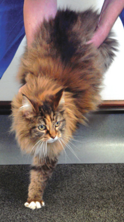
Hopping response. The normal cat responds to hopping by quickly replacing the limb under the body as it is moved laterally. The hopping movement should be smooth and fairly rapid, and not irregular or excessive. The thoracic limbs should be carefully compared
Fig 2.
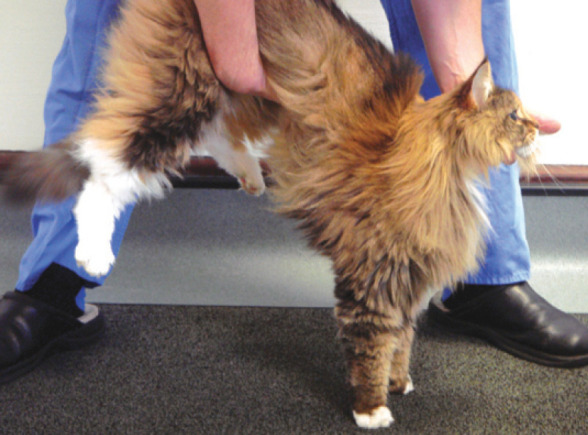
Wheelbarrowing is performed with the neck extended and the pelvic limbs elevated
Fig 3.
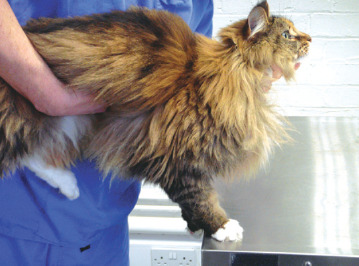
Tactile placing. When the carpus makes contact with the edge of the table, the cat should immediately place its foot on the surface
Postural reactions pathways
-
Afferent arm components
Joint proprioceptor
Peripheral sensory nerve
Spinal cord and brainstem ascending pathways
Contralateral forebrain
-
Efferent arm components
Contralateral forebrain
Descending motor pathways within the brainstem and spinal cord
Peripheral motor nerve and skeletal muscle
Spinal reflexes, muscle tone and size
Spinal reflex evaluation should be considered as a continuation of the gait and postural reactions assessments, and not as a sole entity. Following gait and postural reactions testing, the clinician should be in a position to narrow down the lesion localisation to being cranial to the T3 spinal cord segments, caudal to the T3 spinal cord segments, or within the peripheral nervous system (peripheral nerve, neuromuscular junction or muscles). Spinal reflex evaluation helps to narrow down further the lesion localisation by testing the integrity of the C6-T2 and L4-S3 intumescences, as well as the respective segmental sensory and motor nerves that form the peripheral nerve, and the muscles innervated (Table 1).
Table 1.
Neuroanatomical diagnosis based on combined evaluation of gait, postural reactions and segmental spinal reflexes testing
| Limb(s) presenting with abnormal gait and/or abnormal postural reactions | Segmental spinal reflexes in affected limbs | Likely neuroanatomical diagnosis |
|---|---|---|
| All four limbs | Normal to increased in all 4 limbs | Brainstem or C1-C5 spinal cord segments |
| Decreased to absent in all 4 limbs | Generalised polyneuropathy/junctionopathy/myopathy | |
| Decreased to absent in thoracic limbs; normal to increased in pelvic limbs | C6-T2 spinal cord segments | |
| Bilateral pelvic limbs | Normal to increased | T3-L3 spinal cord segments |
| Decreased to absent | L4-S3 spinal cord segments, peripheral nerve roots/nerves of the pelvic limbs | |
| Thoracic and pelvic limbs on the same side of the body | Normal to increased in thoracic and pelvic limbs | Ipsilateral brainstem or C1-C5 spinal cord segments |
| Decreased to absent in thoracic limbs; normal to increased in pelvic limbs | Ipsilateral C6-T2 spinal cord segments | |
| Unilateral thoracic limb | Normal to increased | Ipsilateral brainstem or C1-C5 spinal cord segments |
| Decreased to absent | Ipsilateral C6-T2 spinal cord segments, or the nerve roots, brachial plexus or peripheral nerves affecting that limb | |
| Unilateral pelvic limb | Normal to increased | Ipsilateral T3-L3 spinal cord segments |
| Decreased to absent | Ipsilateral L4-S3 spinal cord segments, or the nerve roots or peripheral nerves affecting that limb |
Spinal reflexes do not require consciousness and are segmental. Testing only evaluates the spinal segment(s) within the intumescences corresponding to the stimulated nerve. Lesions at the level of these intumescences result in loss of segmental spinal reflexes as well as reduced muscle tone and size.
Spinal reflex evaluation in cats is best performed with the animal positioned in dorsal recumbency between the thighs of the examiner (see video 10, doi:10.1016/j.jfms.2009.03.002). Although many spinal reflexes are described, the most reliable ones in cats are the withdrawal reflex and the patellar reflex (see boxes, page 345). Other spinal reflexes (triceps, biceps, extensor carpal radialis and gastrocnemius) are more difficult to perform and to interpret.
Spinal reflex evaluation should be considered as a continuation of the gait and postural reactions assessments, and not as a sole entity.
Withdrawal (flexor) reflex.
In the thoracic limb, the withdrawal reflex evaluates the integrity of the C6-T2 spinal cord segments (and associated nerve roots), brachial plexus, peripheral nerves (radial, axillary, musculocutaneous, median and ulnar) and the muscles innervated. In the pelvic limb, this reflex evaluates the integrity of the L4-S1 spinal cord segments (and associated nerve roots), the femoral and sciatic nerves, and the muscles innervated.
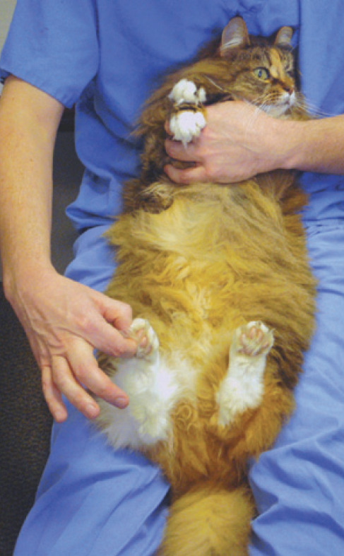
This test is performed with the cat in dorsal recumbency between the thighs of the examiner. A noxious stimulus is applied to the tested limb by pinching the nail bed or digit with the fingers or haemostat forceps. The stimulus causes a reflex contraction of the flexor muscles and withdrawal of the tested limb. If this withdrawal reflex is absent, individual toes can be tested to detect if specific nerve deficits are present.
It should be remembered that the withdrawal reflex in the thoracic or pelvic limbs does not depend on the animal's conscious perception of noxious stimuli (nociceptive function). It is a segmental spinal cord reflex that only depends on the function of the local spinal cord segments.
Nociception testing
Apart from conscious proprioception and evaluation of facial sensation (discussed later), evaluation of the sensory system in cats largely relies on tests for pain perception (nociception). The purpose of such testing is to detect and map out any areas of sensory loss. Assessment of pain sensation requires a noxious stimulus and evaluation of the animal's response.
Nociception is tested by pinching the digits with the fingers or with haemostats. Only a behavioural response to this noxious stimulus (ie, turning of the head and/or attempting to bite) indicates conscious pain perception. If no response is elicited when using fingers, the test should be repeated with haemostats to ensure that the response is absent. Withdrawal of the limb is only the flexor reflex, and should not be interpreted as evidence of pain perception.
Cranial nerve evaluation (Table 2)
Table 2.
Cranial nerve tests commonly used in cats
| Cranial nerve test | Afferent cranial nerve | Brain region | Efferent cranial nerve | Principal effect noted |
| Menace response | CN I (optic) | Forebrain Cerebellum Brainstem | CN VII (facial) | Blink elicited by a menacing gesture |
| Visual placing response | CN I (optic) | Forebrain | None | Reaching out for support on approaching the table |
| Pupillary light reflex | CN I (optic) | Brainstem | CN III (oculomotor) | Pupillary constriction elicited by shining a light in the eye |
| Dark adaptation test | CN I (optic) | Hypothalamus Brainstem | Sympathetic supply to the eye | Pupillary dilation in darkness |
| Palpebral reflex | CN V (trigeminal; ophthalmic or maxillary) | Brainstem | CN VII (facial) | Blink elicited by touching the medial or lateral canthus of the eye |
| Response to nasal stimulation | CN V (trigeminal; maxillary) | Forebrain Brainstem | None | Withdrawal of the head elicited by touching the nostril |
| Oculo-vestibular reflex | CN VIII (vestibular) | Brainstem | CN III (oculomotor) | Nystagmus induced by moving the head |
| CN IV (trochlear) | ||||
| CN VI (abducens) |
Menace response
The menace response is elicited by making a threatening gesture towards the eye of the cat with one hand (Fig 4). The afferent arm of this response involves the retina, optic nerve (cranial nerve [CN] II), contralateral optic tract and contralateral forebrain. The efferent pathway involves the contralateral forebrain, ipsilateral cerebellum and facial nerve (CN VII). The expected response is a blink. The contralateral eye must be covered with the other hand to assess each eye separately (see video 12, doi:10.1016/j.jfms.2009.03.002). Care must be taken not to touch the cat's eyelashes or to create air currents that might stimulate sensation of the face (trigeminal nerve, CN V) and elicit a palpebral or corneal reflex.
Fig 4.
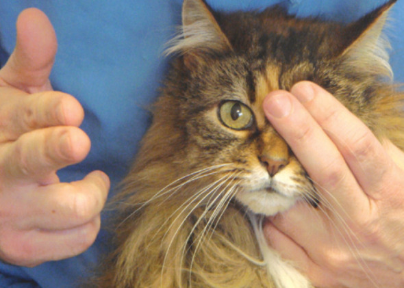
Menace response
Patellar reflex
The patellar reflex is elicited by tapping the patellar ligament and observing a reflex contraction of the quadriceps muscle and extension of the stifle joint. It is likewise performed with the cat in dorsal recumbency between the thighs of the examiner. This position allows the stifle to be slightly flexed, and the two sides of the cat to be compared.
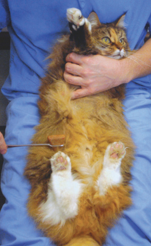
The patellar reflex evaluates the integrity of the L4-L6 spinal cord segments (and associated nerve roots), as well as the femoral nerve. A weak or absent reflex indicates a lesion within the L4-L6 spinal cord segments or the femoral nerve. A similarly weak or absent reflex can on occasion be seen in cats with stifle disease.
Evaluation of the extensor tone on the pelvic limb can be used as a control in cats with ambiguous patellar reflexes as it involves the same neuroanatomical components (femoral nerve and quadriceps muscle) (see video 11, doi:10.1016/j.jfms.2009. 03.002).
A lesion cranial to the L4 spinal cord segment can cause a normal or exaggerated reflex. In the absence of other neurological deficits, an exaggerated patellar reflex means little and can be observed in an excited or nervous cat. The patellar reflex can also appear hyper-reflexic in a cat with a sciatic nerve or L6-S1 spinal cord segment lesion. This pseudo-hyperreflexia is a result of decreased tone in the muscles that flex the stifle and normally counteract stifle extension during the patellar reflex.
In the absence of other neurological deficits, an exaggerated patellar reflex means little and can be observed in an excited or nervous cat.
The menace response can be particularly difficult to elicit in a normal cat and its absence should be interpreted with caution. It may be helpful to tap the cat lightly a few times to get its attention before evaluating this response.
Visual placing response
Carrying a cat towards a table top tests the visual placing response (see video 13, doi:10.1016/j.jfms.2009.03.002). On approaching the surface the cat will reach out to support itself on the table before the paw touches the table. This response requires intact visual and motor pathways and can be useful in assessing visual function in a cat where the menace response is ambiguous.
Pupil size and symmetry, pupillary light reflex and dark adaptation test
The size of the pupils represents a balance between the parasympathetic system, which is responsive to the amount of light entering the eye, and the sympathetic system, which is responsive to the emotional state of the cat. Through the parasympathetic nerve pathways that innervate the iris, the pupil regulates the amount of light that reaches the retina. The parasympathetic component of the oculomotor nerve (CN III) is involved in the control of pupillary constriction while the somatic efferent component of the oculomotor nerve is responsible for the motor innervation of the levator palpebrae superioris (elevation of the upper eyelid), ipsilateral dorsal, ventral and medial rectus extraocular muscles as well as the ventral oblique muscle (movement of the eyeball). The tone of the iris dilator muscle is maintained by the sympathetic system, which keeps the pupil partially dilated under normal conditions and dilates it during periods of stress and fear, and in response to painful stimuli. The ocular sympathetic nervous system also innervates and provides tone to the smooth muscle of the periorbita and eyelids. This tone keeps the eyeball protruded, the palpebral fissure widened and the third eyelid retracted.
Assessment of pupillary size and equality should be determined in ambient light as well as in darkness. Normally, the pupils of the eyes should be symmetrically shaped and equal to each other in size. In cats with anisocoria or dyscoria, determining which pupil is abnormal is achieved by checking the pupillary light reflex (PLR) and assessing if the asymmetry in pupil size increases in bright light or in complete darkness (dark adaptation test).
Cats with pupils of unequal size (anisocoria) or shape (dyscoria) must be found to be free of primary or secondary anatomical or mechanical abnormalities (eg, iris atrophy, uveitis or glaucoma) before a neurological dysfunction is assumed.
The PLR involves an afferent arm and an efferent arm. The afferent arm shares some common pathways (ipsilateral retina, optic nerve, optic chiasm and contralateral optic tract) with part of the afferent arm of the menace response and visual placing response. These tests use different integration centres within the brain and different efferent pathways. The PLR does not test the animal's vision and the cerebrum is not involved in the PLR pathway. The efferent arm of the PLR reflex is mediated by the parasympathetic portion of CN III. Combining the results of the menace response, visual placing response and PLR testing helps to determine whether or not the lesion is localised within this common pathway. The dark adaptation test allows the eyes to dark-adapt in complete darkness for a couple of minutes to allow complete relaxation of the pupillary sphincter muscle.
Trigeminal and facial nerve function
The trigeminal nerve (CN V) provides sensory innervation to the face (skin, cornea, and mucosae of the nasal septum and oral cavity) and motor innervation to the masticatory muscles (temporalis, masseter, medial and lateral pterygoid and rostral part of the digastric muscles). The motor function of CN V is assessed by evaluating the size and symmetry of the masticatory muscles and testing the resistance of the jaw to opening the mouth. The sensory function can be assessed by individually testing the corneal reflex (ophthalmic branch), the palpebral reflex (ophthalmic or maxillary branch when touching the medial or lateral canthus of the eye, respectively), the response to nasal stimulation (ophthalmic branch) (see below), and by pinching the skin of the face with haemostat forceps and observing an ipsilateral blink or facial twitch.
The facial nerve (CN VII) provides motor innervation to the muscles of facial expression. This motor function is primarily assessed by observing for movement of the ears, eyelids, lips and nostrils, and for symmetry of the lips. The facial nerve is also involved in the motor response (efferent part) of the following tests: palpebral reflex (CN V and VII; see video 14, doi:10.1016/j.jfms.2009.03.002); corneal reflex (CN V and VII); menace response (CN II and VII); and pinching of the face (CN V and VII). The Schirmer tear test can evaluate the parasympathetic supply to the lacrimal gland associated with CN VII.
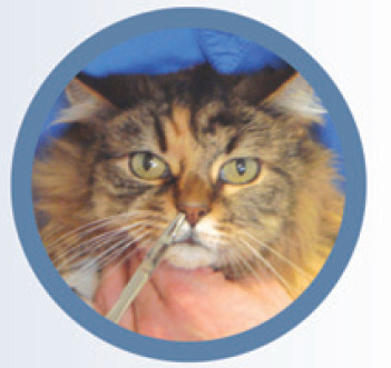
Response to nasal stimulation
As with the menace response, the response to nasal stimulation requires an intact contralateral forebrain. One of the two nostrils is stimulated using a pair of forceps or a pen while the cat's eyes are masked, or while making sure the animal is not able to see the stimulus, to prevent any visual input (Fig 5). The afferent arm involves the sensory component of the trigeminal nerve, which conducts the information to the brainstem, from where it continues to the contralateral forebrain. The expected response is a withdrawal movement of the head and neck. Like the menace response, this response might be abnormal in a cat with a structural contralateral forebrain lesion.
Fig 5.
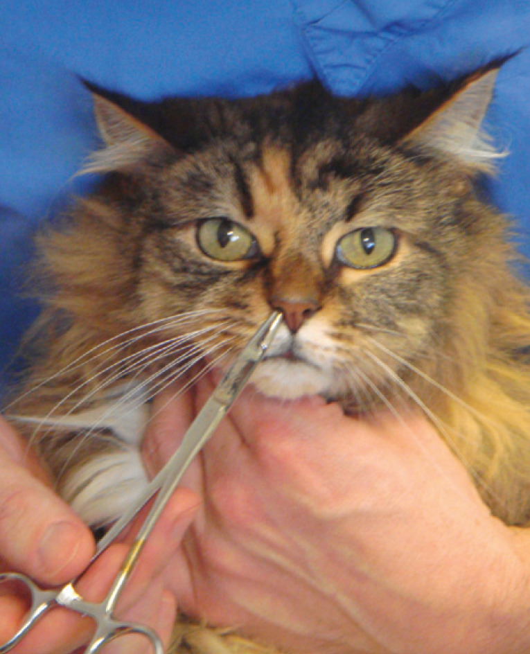
Assessing the response to nasal stimulation
Oculovestibular reflex and nystagmus
Observation of the animal's body and head posture at rest, and evaluation of its gait, can provide a lot of information about the vestibular function of CN VIII. This function can also be more specifically assessed by testing the oculovestibular reflex and looking for pathological nystagmus. Nystagmus is an involuntary rhythmic movement of the eyeballs. Physiological (or vestibular) nystagmus is a nystagmus that occurs in normal animals (to stabilise images on the retina during head movement), while pathological nystagmus reflects an underlying vestibular disorder. In both types of nystagmus there is a slow and fast phase (ie, jerk nystagmus).
A physiological nystagmus can be induced in normal individuals by rotating the head from side to side (oculovestibular reflex). With a feline patient, this is best done by holding the animal at arm's length and rotating it from side to side. The nystagmus is always observed in the plane of rotation of the head and consists of a slow phase away from the direction of head rotation, and a fast phase in the same direction as the head rotation. In the absence of any head movement, nystagmus should never be present in a normal animal.
Two types of pathological nystagmus can be observed in cats with vestibular disorders: spontaneous, which occurs when the head is in a normal position at rest; and/or positional, which is seen when the head is held in different positions (eg, to either side, dorsally or by placing the animal upside down on its back — Fig 6). Nystagmus is usually classified on the basis of its direction (the fast movement) and may be horizontal, vertical or rotatory. In disorders of the peripheral components of the vestibular system in the inner ear, the direction of the nystagmus is always opposite to the side of the lesion and is usually horizontal or rotatory. Lesions of the central components of the vestibular system can cause pathological nystagmus in any direction, and occasionally it changes direction with different positions of the head (see video 15, doi:10.1016/j.jfms.2009.03.002). A vertical nystagmus is most commonly due to a central lesion.
Fig 6.
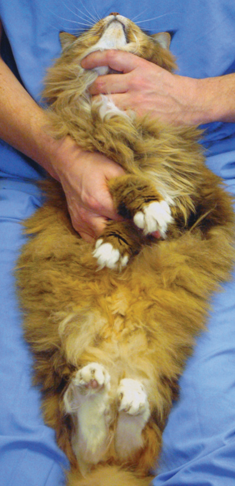
Testing for positional nystagmus. Extending the head and neck while the cat is in dorsal recumbency can help in detecting a positional nystagmus by challenging the vestibular system
Diagnostic tests should be performed solely to confirm or exclude the differentials in the list, and not in place of the clinical evaluation.
How to establish a differential diagnostic list
Determination of a list of differential diagnoses should be directed entirely by the neuroanatomical diagnosis, and is essential in choosing and interpreting any diagnostic tests, however sophisticated they may be. Diagnostic tests should be performed solely to confirm or exclude the differentials in the list, and not in place of the clinical evaluation.
The differential diagnosis list can be developed by taking into account:
Patient signalment
Historical data Questioning of the owner should aim to define the mode of onset (peracute, acute, subacute, chronic or episodic) and pattern of development of the condition. Historical data can also provide clues as to how widespread or focal the disease process is in the nervous system, whether there is evidence of asymmetry, and how severe the signs have been.
Neurological findings As has been discussed in this article, the aim of the neurological evaluation is to define the lesion localisation (forebrain, brainstem, cerebellum, spinal cord segments, peripheral nerve, neuromuscular junction and muscle) and distribution of the disease (focal, multifocal, diffuse) within the nervous system.
Disease processes that can affect the nervous system are traditionally classified according to the ‘VITAMIN D’ mnenomic (Vascular, Inflammatory/Infectious, Toxic/Traumatic, Anomalous, Metabolic, Idiopathic, Neoplastic/Nutritional, Degenerative). Each category has a typical signalment, onset and progression, and distribution within the nervous system (Table 3).
Table 3.
Disease processes that can affect the nervous system: mode of onset, pattern of development and expected distribution within the nervous system
| Pathological process | Mode of onset | Pattern of development | Distribution |
|---|---|---|---|
| Vascular | Peracute or acute (haemorrhage may cause subacute onset) | Non-progressive or regressive (haemorrhage may cause progression over a very short period) | Focal and often asymmetrical |
| Inflammatory/infectious | Acute, subacute or insidious | Progressive (waxes and wanes in some early cases) | Focal or multifocal Asymmetrical or symmetrical |
| Toxic | Acute | Progressive | Diffuse and bilaterally symmetrical |
| Traumatic | Peracute or acute | Static or improves over time | Often focal |
| Asymmetrical or symmetrical | |||
| Anomalous | Chronic (occasionally acute) | Non-progressive or slowly progressive early in life | Variable |
| Metabolic | Variable (often acute) | Waxes and wanes or progressive | Diffuse and bilaterally symmetrical |
| Idiopathic | Acute | Non-progressive or regressive | Specific to each syndrome |
| Neoplastic | Chronic (occasionally acute) | Progressive | Often focal Asymmetrical or symmetrical |
| Nutritional | Variable (acute or insidious) | Progressive | Diffuse and bilaterally symmetrical |
| Degenerative | Chronic | Progressive | Often diffuse and symmetrical |
What next?
With, by this stage, a clear knowledge of the region of the nervous system involved, and a differential list reduced to no more than three or four disease processes, consideration should be given only to those diagnostic tests that will help to narrow down the list further, and that can be afforded by the client. These tests should ideally be run in succession from least invasive through to more invasive.
KEY POINTS.
The neurological examination aims to determine if a clinical problem is neurological in origin and which part(s) of the nervous system may be involved to explain the abnormalities.
Adherence to a systematic stepwise approach to the neurological examination is essential to confidently make an accurate neuroanatomical diagnosis.
Precise localisation of the causative disorder within the nervous system (neuroanatomical diagnosis) and an understanding of the suspected disease processes (differential diagnosis) are the keys to an accurate neurological diagnosis.
Determination of a differential diagnosis list is essential in choosing and interpreting any diagnostic tests — however sophisticated they may be.
Acknowledgements
The author is grateful to Dr Alexander de Lahunta for reviewing this article and offering his comments.
Further reading
- 1.de Lahunta A. Glass E. Veterinary neuroanatomy and clinical neurology. 3rd edn. St. Louis, Missouri: WB Saunders Co, 2008. [Google Scholar]
- 2.Dewey CW. A practical guide to canine and feline neurology. Ames, Iowa: State Press, 2003. [Google Scholar]
- 3.Garosi LS. The neurological examination. In: Platt SR. Olby NJ. eds. BSAVA manual of canine and feline neurology. Quedgeley, UK, BSAVA, 2004: 1–23. [Google Scholar]
- 4.Garosi LS. Lesion localization and differential diagnosis. In: Platt SR. and Olby NJ, eds. BSAVA manual of canine and feline neurology. Quedgeley, UK, BSAVA, 2004: 24–34. [Google Scholar]
- 5.King AS. Physiological and clinical anatomy of domestic mammals. Volume 1. Central nervous system. Oxford: Oxford University Press, 1987. [Google Scholar]


