Abstract
A 6-month-old male domestic shorthair cat was presented for a 3-month history of dysphagia and upper respiratory signs. The cat was diagnosed with a generalized megaesophagus secondary to a large nasopharyngeal polyp that extended into the cervical esophagus. The polyp was removed by traction and a left ventral bulla osteotomy was performed to remove the polyp base. The cat's clinical signs resolved and follow-up radiographs 14 days after surgery revealed resolution of the megaesophagus. To the authors' knowledge, this is the first report of resolution of megaesophagus after removal of a nasopharyngeal polyp in a cat.
A 6-month-old male intact domestic shorthair cat was presented to the emergency service for a 3-month history of bilateral otitis, left head tilt, difficulty swallowing, stertor, and open-mouth breathing. The clinical signs were present since adoption by the owner at 12 weeks of age and their duration prior to that time was unknown. The referring veterinarian had cultured Pastuerella species from the external ear canals and had treated the cat with topical neomycin 0.25%, dexamethasone 0.1%, and thiabendazole 4%, and oral amoxicillin/clavulanic acid, clindamycin, and L-lysine with a partial response. The owner reported that the clinical signs waxed and waned during treatment. The cat had a normal appetite and water consumption, as well as a normal level of activity.
On physical examination the cat had a normal temperature, pulse and respiration and weighed 1.68 kg with a normal body condition score. The cat was assessed to be mildly dehydrated and had bilateral auricular discharge with the left worse than the right. The cat was open-mouth breathing with increased abdominal effort, had significant upper respiratory noise, and there was no air movement detected through either of the nares. The cat exhibited anisocoria with left-sided miosis, a left head tilt, and occasionally fell to the left when trying to ambulate. The remainder of the physical and neurologic examination was normal.
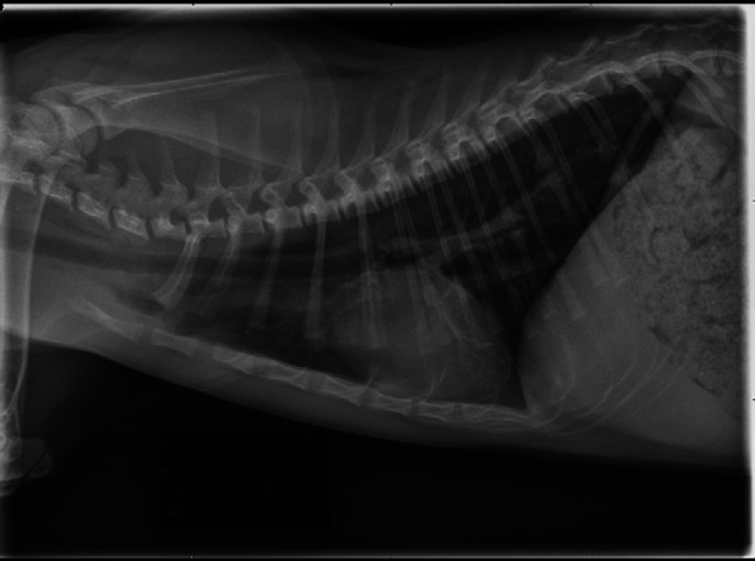
Complete blood count, serum chemistry, and urinalysis showed no significant abnormalities. Conscious thoracic radiography revealed a severe generalized megaesophagus and evidence of aerophagia (Fig. 1). Additional questioning of the owner did not reveal any history of regurgitation or vomiting.
Fig 1.
Left lateral thoracic radiograph on presentation demonstrating severe generalized megaesophagus. Note the significant aerophagia present.
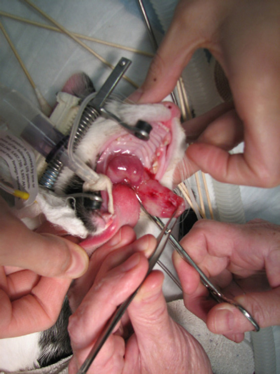
The patient underwent general anesthesia and laryngeal examination. A large soft tissue mass was noted in the oropharynx. Computer tomography (CT) was performed and demonstrated soft tissue attenuating material in the left tympanic bulla and a large rim-enhancing mass extending throughout the nasopharynx and into the cervical esophagus. Following the CT scan, a 7 cm mass extending from the nasopharynx was removed by gentle traction (Fig. 2). On histopathology, the mass was found to be a nasopharyngeal polyp with mixed inflammation.
Fig 2.

Gentle traction was used to remove the 7 cm nasopharyngeal polyp that was extending into the cervical esophagus.
The following day a fluoroscopic contrast esophogram was performed to assess esophageal motility. Esophageal dilation was similar to that noted on the previous radiographs. Motility was decreased, but productive peristaltic waves were noted with partial transport of ingesta to the stomach.
A left ventral bulla osteotomy was performed and a large amount of coagulated blood and a soft tissue mass was removed from within the tympanic bulla and submitted for histopathology. A bulla swab was submitted for aerobic culture. The cat was then neutered. Postoperatively the cat had a significant amount of epistaxis and developed a regenerative anemia which gradually resolved over 4 days. The cat also developed additional signs of Horner's syndrome including left-sided enophthalmos, elevation of the third eyelid and ptosis of the upper eyelid. Horner's syndrome is a common sequela to ventral bulla osteotomy and is due to inflammation or damage to the sympathetic postganglionic nerves, which travel through the tympanic bulla. The cat was released from the hospital 4 days after surgery.
No growth of aerobic bacteria was noted from the culture of the left bulla. Histopathology of the soft tissue removed from the bulla was similar to the findings in the polyp.
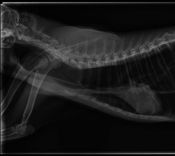
A recheck examination was performed 14 days postoperatively and the patient was reported to be doing well at home. The cat had an improved energy level and was eating well with no evidence of dysphagia or regurgitation. Physical examination revealed an improved, but persistent left head tilt, and resolved ptosis and anisocoria. No upper respiratory noise was noted and the cat was eupneic. Thoracic radiographs revealed a small amount of gas in the esophagus, but resolution of the previous dilation (Fig. 3). On follow-up contact with the owner 3 months postoperatively, the cat was doing well. No return of clinical signs was noted and the head tilt and Horner's syndrome were resolved. Three month follow-up radiographs at the referring veterinarian showed complete resolution of the megaesophagus.
Fig 3.

Left lateral thoracic radiograph demonstrating resolution of the megaesophagus 16 days after presentation and 15 days after removal of the nasopharyngeal polyp.
Nasopharyngeal polyps have traditionally been thought to affect primarily young cats; however, published median ages have ranged from 2 to 5.1 years and cats as old as 18 years have been reported to have nasopharyngeal polyps. 1–3 The most common clinical signs are respiratory stertor, dyspnea, and otic discharge; however, additional signs such as head tilt, ataxia, and decreased appetite are frequently reported. 1 There are rare reports of secondary complications including pulmonary hypertension. 4 The majority of these polyps originate in the middle ear or eustachian tube and they may extend into the oropharynx or through the tympanum into the external ear canal. Diagnostic evaluation for a suspected polyp may include an oropharyngeal examination under sedation and computed tomography imaging of the head to assess the tympanic bullae. Current therapeutic options include traction avulsion of the polyp which may result in a 33%–41% recurrence rate. 1,5 If bulla disease is present a ventral bulla osteotomy provides a significant reduction in recurrence rate to 0%–8%. 6–8
Only one other report of megaesophagus secondary to nasopharyngeal polyp in a cat has been published. 4 The cat had evidence of pulmonary hypertension and right-sided heart failure secondary to chronic upper airway obstruction. The cat did not survive beyond the immediate postoperative period and no information was gained as to the potential for resolution of the megaesophagus. In the report presented here, the cat did not have signs of megaesophagus such as regurgitation; however, it is possible that the difficulty swallowing reported by the owner may have been due in part to the esophageal dilation as well as the presence of the polyp in the pharynx.
Megaesophagus can have a variety of causes including myasthenia gravis, dysautonomia, esophagitis, and congenital and idiopathic processes. In the cat presented here, no additional signs of dysautonomia or myasthenia gravis were noted, although an acetylcholine receptor antibody titer was not performed. The spontaneous resolution of the megaesophagus in this cat after polyp removal implies that the megaesophagus was secondary to the obstructive nature of the polyp. It is suspected that in this cat the megaesophagus resulted from chronic aerophagia that developed due to the size of the polyp and its obstruction of the airway. Cats with megaesophagus should be evaluated for upper airway obstruction as well as other causes. Prognosis for resolution of megaesophagus in cats with upper airway obstruction may be good if the obstruction is treated and resolved, in contrast to the guarded prognosis with other causes of megaesophagus in cats, however, the prognostic impact of additional factors such as duration and presence of regurgitation is unknown at this time.
References
- 1.Anderson D.M., Robinson R.K., White R.A.S. Management of inflammatory polyps in 37 cats, Vet Rec 147, 2000, 684–687. [PubMed] [Google Scholar]
- 2.Veir J.K., Lappin M.R., Foley J.E., Getzy D.M. Feline inflammatory polyps: historical, clinical, and PCR findings for feline calici virus and feline herpes virus-1 in 28 cases, J Feline Med Surg 4, 2002, 195–199. [DOI] [PMC free article] [PubMed] [Google Scholar]
- 3.Kudnig S.T. Nasopharyngeal polyps in cats, Clin Tech Small Anim Pract 17, 2002, 174–177. [DOI] [PubMed] [Google Scholar]
- 4.MacPhail C.M., Innocenti C.M., Kudnig S.T., Veir J.K., Lappin M.R. Atypical manifestations of feline inflammatory polyps in three cats, J Feline Med Surg 9, 2007, 219–225. [DOI] [PMC free article] [PubMed] [Google Scholar]
- 5.Harvey C.E., Goldschmidt C.E. Inflammatory polypoid growths in the ear canals of cats, J Small Anim Pract 19, 1978, 669–677. [DOI] [PubMed] [Google Scholar]
- 6.Faulkner J.E., Budsberg S.C. Results of ventral bulla osteotomy for treatment of middle ear polyps in cats, J Am Anim Hosp Assoc 5, 1990, 496–499. [Google Scholar]
- 7.Kapatkin A.S., Matthiesen D.T., Noone K.E., et al. Results of surgery and long-term follow-up in 31 cats with nasopharyngeal polyps, J Am Anim Hosp Assoc 26, 1990, 387–392. [Google Scholar]
- 8.Trevor P.B., Martin R.A. Tympanic bulla osteotomy for treatment of middle ear disease in cats: 19 cases (1984–1991), J Am Vet Med Assoc 202, 1993, 123–128. [PubMed] [Google Scholar]


