Case Presentation
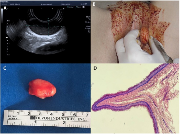
A 43-year-old female (G-1, A-1) presented to the clinic with a 2-year history of painless swelling in the left labia majora. Perineal examination was normal except for a 2 cm mass in the left labia majora, movable with soft rubbery consistency. The skin over the labia was normal, with no vascular changes. There was no inguinal lymphadenopathy. Her family history was unremarkable. Perineal ultrasound demonstrated a unicameral well-demarcated oval-shaped cyst measuring 29.5 x 13.0 mm in size, with homogeneous, hyperechogenic internal echotexture and no Doppler flow (Fig. 1A). The mass was enucleated with an intact capsule under anesthesia (Fig. 1B, C). The cyst was filled with thick, greasy material. The pathological findings of this mass were consistent with a steatocystoma (Fig. 1D). There was no recurrence during the 7-month follow-up.
Figure 1.

(A) Perineal ultrasound demonstrated a unicameral well-demarcated oval-shaped cyst. (B) Intraoperative view of mass in the left labia majora. (C) The cyst was excised intact. (D) Histopathology: the cystic wall is serpiginous, lined by thin squamous epithelium with an outer corrugated cuticle and minimal granular layer (H&E, x20).
Teaching Point
Steatocystoma is a lesion that results from a hamartomatous malformation of the pilosebaceous duct, leading to an epithelium-lined cystic lesion containing sebaceous lobules. Steatocystomas can be classified into steatocystoma multiplex (SCM) and steatocystoma simplex (SCS), according to the multiplicity. SCS was first reported in 1982 by Brownstein, who described 30 cases [1]. In order of decreasing frequency, these lesions are mostly located on the scalp, face, neck, axillae, chest, upper limbs, back, or lower limbs [1]. In contrast, SCM and SCS in the vulvo-perineal region is extremely rare, with only a few cases reported in the literature [2]. SCS is a rarely benign lesion occurring extremely rare on the vulva and should be considered in the differential diagnosis of painful/painless superficial vulvar mass.
Footnotes
Funding: None.
Competing Interests: None.
Authorship: All authors have contributed significantly to this publication.
References
- 1.Brownstein MH. Steatocystoma simplex. A solitary steatocystoma. Arch Dermatol. 1982;118(6):409–11. [PubMed] [Google Scholar]
- 2.Cunningham SC, Kao GF, Moore GW, Napolitano LM. Steatocystoma simplex. Surgery. 2004;136(1):95–7. doi: 10.1016/s0039-6060(03)00341-6. [DOI] [PubMed] [Google Scholar]


