The primary aims of plastic surgery are the restoration of appearance and function. This includes reconstruction after traumatic defects, congenital anomalies, and ablative surgery for malignancy, as well as surgery of the hand, breast surgery, and the field of aesthetic surgery. Considerable advances are being made as we begin to understand the biological basis for tissue regeneration, malignant neoplasia, and wound healing. The tools of the plastic surgeon now encompass the means of cellular manipulation of these biological processes.
Methods
Topics for this article were selected by reviewing the conference proceedings of the European Association of Plastic Surgeons and the American Society for Plastic and Reconstructive Surgery over the past four years. After a search of Medline, key articles relating to these topics were included.
Reconstructive flap surgery
Tissue used in reconstruction requires oxygen and nutrition to ensure its survival. A split thickness skin graft with low metabolic demands adheres to the wound bed and is initially nourished by diffusion, after which ingrowth of capillaries establishes a permanent blood supply. In wounds that are not able to support a graft by diffusion and many others that require tissues of better aesthetic and mechanical properties, reconstruction with tissue supported by its own blood supply is required. Such tissues transferred from one place to another are referred to as flaps. In the 1970s and 1980s anatomical research identified tissue units with an arterial and venous axis that allows them to be isolated or transferred. Advanced optical systems and microinstrumentation have permitted the transfer of these isolated tissue units with anastomosis of the axial blood supply at the recipient site for reconstruction.
Microsurgical free tissue transfer has become commonplace, and advances have been made in improving the benefit-risk ratio for free tissue transfer. The golden rule remains to achieve the maximum gain with the minimum risk and least morbidity at the donor site. Some of the commonest flaps used in free tissue transfer are based on an axial pedicle which supplies a muscle which in turn provides perforating vessels to supply the overlying skin (for example, rectus abdominis and latissimus dorsi myocutaneous flaps). Thus, to reconstruct a defect requiring skin it is often necessary to sacrifice the underlying muscle.
Recent advances
Improvements in the transfer of vascularised tissue from one part of the body to another can now restore both appearance and function by providing a dynamic component to the reconstruction
Reconstruction of certain tissues cannot easily be achieved by transfer from one part of the body to another, but better understanding of the cellular biology of these tissues has allowed regeneration to produce bone, cartilage, and epidermis
Selective photothermolysis with lasers allows removal of certain pigmented lesions and capillary vascular malformations with minimal non-specific skin damage, and the pulsed carbon dioxide laser allows precise dermabrasion for facial rejuvenation
An intermittent vacuum applied to a closed wound system has enhanced treatment of chronic wounds
Perforator flaps
The free vascularised transverse rectus abdominis myocutaneous (TRAM) flap has become a standard method for breast reconstruction after mastectomy. The transfer of the transversely oriented skin paddle along with the underlying rectus muscle allows anastomosis of the axial vessels which supply the muscle (and thus also the skin) to recipient vessels, usually in the axilla. The loss of the rectus muscle decreases the strength of the abdominal wall.1 The rectus muscle is transferred because it acts as a vehicle for the perforating branches of the inferior epigastric vascular pedicle. Thus, reducing morbidity at the donor site depends on raising the flap and dissecting out a major perforating branch to the skin and subcutaneous fat but leaving the muscle behind. Using the perforator in free tissue transfer has provided a further refinement to flap reconstruction. Transfer of the lower abdominal skin based on the perforator vessels is an effective flap for breast reconstruction2 and reduces the abdominal weakness produced by the previous method.3 With this principle, the skin overlying the latissimus dorsi muscle can be transferred without sacrifice of the muscle itself.4
Endoscopic flap harvest
Some defects of the lower limb and trunk wall which occur after trauma or ablative surgery require a muscle flap for reconstruction. Two of the most common muscles used for microsurgical transfer are the latissimus dorsi and the rectus abdominis. The large incisions needed to harvest these muscles can cause considerable morbidity. Instruments are now available for endoscopic harvest of such muscular flaps, and these techniques have reduced postoperative pain for both these sites.5 This procedure is currently limited by the ability to create an optical cavity, and this reduces its efficacy in the harvest of limb flaps where tension is often too great.
Functional reconstruction
One of the most challenging aspects of plastic surgery is to perform a dynamic reconstruction. Since the early development of free tissue transfer the techniques have evolved to restore function to the reconstructed part. Facial palsy causes considerable psychological morbidity owing to the facial asymmetry and inability to respond appropriately to emotion. In cases of congenital palsy or damage to the facial nerve by intracranial lesions such as acoustic neuroma, free vascularised functional muscle transfer has a role. Reanimation of the smile was achieved in 80% of cases in a recent large series with transfer of the pectoralis minor muscle to the weak side of the face after previous nerve grafting from the opposite intact facial nerve (fig 1).6 This success rate is even higher in children: function returns to near normal in over 90%.
Figure 1.
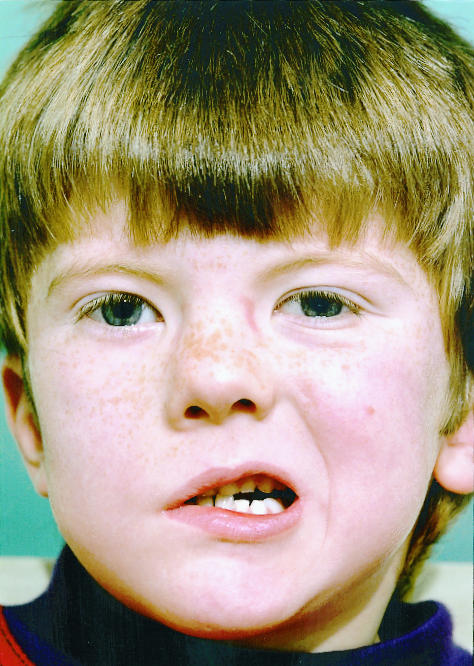
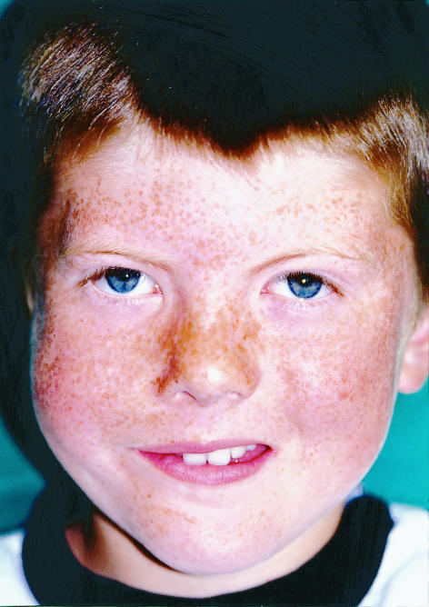
Reanimation of the smile after microvascular transfer of the pectoralis minor muscle to the face: (left) unilateral facial palsy before operation; (right) return of voluntary function as well as response to emotion after operation
Tissue regeneration
Cartilage
Research into cellular engineering has generated new tissues for reconstructive transfer. Cartilage is essential for reconstructing the ear after trauma or in congenital abnormalities such as microtia. Established techniques for total ear reconstruction exist using autologous costal cartilage. This is harvested and carved with reference to a mould of the opposite ear. This permits a good aesthetic reconstruction (fig 2) with long lasting results,7 but the donor site of the cartilage is not always without morbidity. More recently, human chondrocytes have been seeded onto a Vicryl scaffold to generate neocartilage with matrix.8 A Vicryl scaffold in the shape of auricular cartilage has been seeded and transplanted into a nude mouse (which is not immunocompetant) to subsequently generate a complete human auricular cartilage.9 Clinical studies applying this technology are eagerly awaited.
Figure 2.
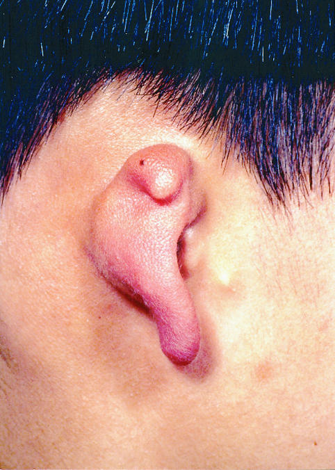
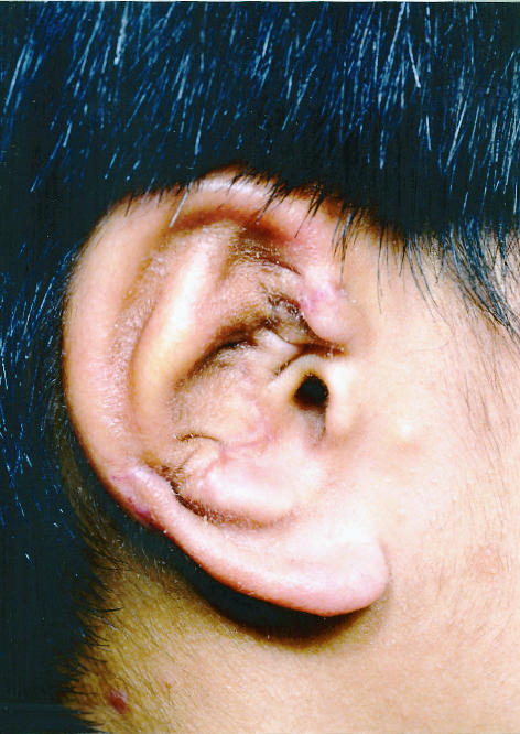
Reconstruction of the ear for congenital microtia using an autologous costal cartilage framework: (left) microtic ear remnants; (right) postoperative appearance after insertion of carved cartilage framework
Distraction osteogenesis
Requirement for bone generation and its remodelling have stimulated significant advance in the field of craniofacial plastic surgery. Bone lengthening by distraction has been applied to craniofacial surgery by McCarthy.10 A defect in cortical bone is produced (corticotomy) and percutaneous pins are inserted on either side. After a lag of five to seven days these pins are distracted at a rate of 1 mm a day until lengthening of up to 30 mm has been produced in a bone such as the mandible. During the first year of life periosteal bone regeneration may allow lengthening without corticotomy. Bony collapse, a problem in bone grafting, is infrequent with this method.11 The main limitations of distraction osteogenesis of the craniofacial skeleton have been technical, with bulky and cumbersome external appliances, but a system of miniature distraction devices has been successfully applied in mandibular, maxillary, and orbital expansion.12
Keratinocyte culture
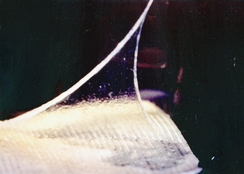
Each year in the United Kingdom there are an estimated 600 deaths and over 10 000 hospital admissions from burn injuries. Although advances in intensive care and early excision of burns have led to a reduction in mortality, there is often insufficient undamaged skin to provide autologous split skin grafts for patients with large burns. As a result these patients may die from uncontrolled sepsis and multiple organ failure. Human keratinocyte culture (fig 3), which can vastly expand an autologous graft,13 only partly solved the clinical problem as it takes three weeks to produce a large confluent sheet of cultured keratinocytes, and without dermal support the keratinocyte graft is relatively unstable.
Figure 3.
Sheet of cultured keratinocytes ready for transfer and clinical use.
A method to overcome these problems14 has been shown to be of value in clinical trials.15 The early burn wound is covered with allogeneic skin (dermis and epidermis). The epidermis, which is highly antigenic, is removed by dermabrasion before it can be rejected. The reduced antigenicity of the allogeneic dermis allows it to become incorporated in the wound bed. After two to three weeks the epidermal layer of the original allogeneic graft is replaced with cultured autologous keratinocytes. The burn wound can be covered at an early stage and is constituted by cultured epidermis with allogeneic dermal support.
Strategies currently under investigation to provide a cultured skin substitute include the use of collagen matrices seeded with cultured fibroblasts and covered with autologous keratinocytes. Such work is likely to lead to the ultimate goal of the “off the shelf skin replacement.”
Laser technology
Selective photothermolysis
Recent advances in computerised delivery systems have led to a wider field of application of lasers in surgery and more predictable results. The carbon dioxide laser was one of the first to be used, but the difficulty in limiting adjacent non-specific tissue damage, with the subsequent risk of scarring, limited its application in plastic surgery. The breakthrough in the development of medical lasers came with the principle of selective photothermolysis.16 If the laser’s wavelength corresponds to the absorption spectrum of a particular pigment in the skin (a chromophore—for example, haemoglobin, melanin, or tattoo pigments), the light energy is converted to heat energy, with consequent tissue destruction. Undesirable non-specific damage can be limited by using a pulse that allows the target chromophore to convert light into heat but not to conduct to adjacent tissue.
Capillary malformations
The advent of pulsed dye lasers with an output of yellow light (585 nm wavelength), which is preferentially absorbed by haemoglobin, has allowed clearing in excess of 82% of capillary vascular malformations (“port wine stains”).17 These results are long lasting, with little if any recurrence once clearing has been achieved. In adults these malformations may develop a rugose surface, and this limits the efficacy of treatment as the laser penetrates only a millimetre. Spider naevi and erythema associated with hypertrophic scarring also can benefit from laser treatment, but there is debate about the ability to improve the contour of such scars as published studies do not take into account the tendency of these lesions to resolve.
Pigmented lesions
Many superficial melanotic lesions can be treated with lasers. The absorption spectrum of melanin peaks at shorter wavelengths than that of haemoglobin, providing a range that limits damage to surrounding tissue. The lasers used penetrate only to the epidermis and more superficial layers of the dermis. Lesions with epidermal melanin pigment respond well to laser treatment: solar lentigines, cafe-au-lait macules, freckles, and dermal melanin pigmentation, including naevus of Ota and melasma.18 Punch biopsy of any pigmented lesion that is even remotely suspicious should be performed before laser treatment. This biopsy sample provides material that can be useful in predicting the outcome of laser treatment and the risk of complications.19
Facial wrinkles
The pulsed carbon dioxide laser is useful as an adjunct to facial rejuvenation. The wavelength of 10 600 nm, which is absorbed by the water in cell cytoplasm, combined with a pulse of less than a millisecond, causes little non-specific injury. This allows precise removal of the superficial layers of the skin, producing a 92% reduction in the depth of facial wrinkles with few adverse effects.20
Wound healing
Cellular biology
The highly visible effects of scarring have focused attention on wound healing in plastic surgery. Few tissues heal by regeneration after injury; most form a scar. Scar tissue interferes with limb function and, in children, with growth after burn injury as well as mid-facial growth after surgery for cleft lip and palate. In people of all ages, the deformity of scarring causes considerable psychological morbidity.
A sequence of growth factors and cytokines released after injury controls wound healing. In fetuses, wounds heal with a reduced inflammatory and angiogenic response and without scarring.21 By manipulating the concentrations of transforming growth factor β (TGFβ) in wounds in adult human skin, scar formation can be reduced.22 This has not yet been applied in regular clinical practice, but research in this area points the way towards a solution to this difficult problem.
Vacuum assisted wound closure
Chronic wounds provide one of the most difficult problems faced by surgeons: many chronic wounds occur in debilitated patients who are not good candidates for major reconstructive flap surgery. Intermittent negative pressure has recently been described as a method for treating chronic wounds,23 and a device for administering vacuum assisted closure is now available for routine clinical practice. It consists of an open cell foam which is roughly contoured to fit into the wound cavity. The open wound is converted to a closed wound by covering the foam and adjacent skin with a polyethylene adhesive film. A non-collapsible tube under the film connects the open cell foam to a vacuum pump and transmits negative pressure (an intermittent vacuum force of 75-125 mm Hg) uniformly to all areas of the wound. The foam needs to be replaced every 48-72 hours.
With this technique the rate of wound healing is speeded up, and many patients do not require surgery. Applying the vacuum removes interstitial fluid from the tissue adjacent to the wound, reducing induration. Removal of this fluid enhances the micro-oxygenation and nutrition of cells at the edge of the wound and promotes formation of granulation tissue. The uniform application of tension across the wound not only recruits adjacent skin and soft tissue to help close the defect but also stimulates mitosis in dermal fibroblasts. Wound healing is increased by 103% in comparison with controls treated without vacuum closure.23 Many wounds that would have otherwise required flap closure can be treated with a simple skin graft as a result of enhanced production of granulation tissue in the base of the wound.
Conclusions
Important advances in plastic surgery over the past four years include refinements of flap reconstruction and the applications of new technology such as the laser. This follows the evolutionary pattern of the specialty over the past 20 years, where the technical advance of the operating microscope, coupled with the anatomical identification of axial blood supply, led to free tissue transfer. Further advances in plastic surgery are likely to depend on a better understanding of cell biology and its use to manipulate the processes of wound healing, tissue regeneration, and neoplasia.
Acknowledgments
I thank Mr Douglas Harrison for supplying the photographs for figure 1 and Mr David Gault for supplying the photographs for figure 2.
Footnotes
Funding: None.
Conflict of interest: None.
References
- 1.Blondeel PN, Boeckx WD, Vanderstraeten GG, Lysens R, Van Landyut K, Tonnard P, et al. The fate the oblique muscles after free TRAM flap surgery. Br J Plastic Surg. 1997;50:315–321. doi: 10.1016/s0007-1226(97)90539-7. [DOI] [PubMed] [Google Scholar]
- 2.Allen RJ, Treece P. Deep inferior epigastric perforator flap for breast reconstruction. Annal Plastic Surg. 1994;32:32–38. doi: 10.1097/00000637-199401000-00007. [DOI] [PubMed] [Google Scholar]
- 3.Blondeel PN, Vanderstraeten GG, Monstrey SS, Van Landyut K, Tonnard P, Lysens R, et al. The donor site morbidity of free DIEP flaps and free TRAM flaps for breast reconstruction. Br J Plastic Surg. 1997;50:322–330. doi: 10.1016/s0007-1226(97)90540-3. [DOI] [PubMed] [Google Scholar]
- 4.Allen RJ, Tucker C. Superior gluteal artery perforator free flap for breast reconstruction. Plastic Reconstr Surg. 1995;95:1207–1212. doi: 10.1097/00006534-199506000-00010. [DOI] [PubMed] [Google Scholar]
- 5.Fine NA, Orgill DP, Pribaz JJ. Early clinical experience in endoscopic assisted muscle flap harvest. Annal Plastic Surg. 1994;33:465–469. doi: 10.1097/00000637-199411000-00001. [DOI] [PubMed] [Google Scholar]
- 6.Harrison DH. Transactions of the 11th congress of the International Confederation of Plastic, Reconstructive and Aesthetic Surgery. New York: Kugler; 1995. The treatment of unilateral facial palsy using free pectoralis minor transfer. ; pp. 664–665. [Google Scholar]
- 7.Gault DT. Transactions of the 2nd international congress on ear reconstrucion. New York: Kugler; 1997. Total ear reconstruction using autologous costal cartilage. ; pp. 34–36. [Google Scholar]
- 8.Casanova R, Sims CD, Randolph MA. Neocartilage formation usingaged human chondrocytes and polyglycholic acid polymer. 40th Annual Meeting of the Plastic Surgery Research Council, New York, 1995.
- 9.Cao Y, Vacanti JP, Paige KT, Upton J, Vacanti CA. Transplantation of chondrocytes utilizing a polymer-cell construct to produce tissue engineered cartilage in the shape of a human ear. Plastic Reconstr Surg. 1997;100:297–302. doi: 10.1097/00006534-199708000-00001. [DOI] [PubMed] [Google Scholar]
- 10.McCarthy JG, Shreiber J, Karp N. Lengthening of the human mandible by gradual distraction. Plastic Reconstr Surg. 1992;89:1–10. [PubMed] [Google Scholar]
- 11.Habal M. New bone formation by biological rhythmic distraction. J Craniofacial Surg. 1994;5:344–347. doi: 10.1097/00001665-199411000-00017. [DOI] [PubMed] [Google Scholar]
- 12.Cohen S, Bernstein F. Craniofacial distraction of the facial skeleton. J Craniofacial Surg. 1995;6:368–374. doi: 10.1097/00001665-199509000-00007. [DOI] [PubMed] [Google Scholar]
- 13.Rheinwald JG, Green H. Serial cultivation of strains of human epidermal keratinocytes: the formation of keratinizing colonies from single cells. Cell. 1975;6:331–334. doi: 10.1016/s0092-8674(75)80001-8. [DOI] [PubMed] [Google Scholar]
- 14.Cuono CB, Langdon R, Mcguire J. Use of cultured epidermal autografts and dermal allografts as skin replacement after burn injury. Lancet. 1986;i:1123–1124. doi: 10.1016/s0140-6736(86)91838-6. [DOI] [PubMed] [Google Scholar]
- 15.Compton CC, Hickerson W, Nadire K, Press W. Acceleration of skin regeneration from cultured epithelial autografts by transplantation to homograft dermis. J Burn Care Rehabil. 1994;14:653–662. doi: 10.1097/00004630-199311000-00010. [DOI] [PubMed] [Google Scholar]
- 16.Anderson RR, Parrish JA. Selective photothermolysis: precise microsurgery by selective absorption of pulsed radiation. Science. 1983;220:524–527. doi: 10.1126/science.6836297. [DOI] [PubMed] [Google Scholar]
- 17.Achauer BM, Vanderkam VM, Padilla JF. Clinical experience with the tunable pulsed dye laser (585nm) in the treatment of capillary vascular malformations. Plastic Reconstr Surg. 1993;92:1233–1238. [PubMed] [Google Scholar]
- 18.Grover R, Richman PI, Gault DT. Transactions of the 11th congress of the International Confederation of Plastic, Reconstructive and Aesthetic Surgery. New York: Kugler; 1995. Treatment of pigmented lesions using the lamda plus dye laser. ; pp. 43–44. [Google Scholar]
- 19.Grover R, Richman PI, Gault DT. Predicting the outcome of laser treatment for pigmented lesions. Br J Plastic Surg. 1998;51:8–13. doi: 10.1054/bjps.1997.0055. .. [DOI] [PubMed] [Google Scholar]
- 20.Grover R, Grobbelaar AO, Morgan BDG, Gault DT. A quantitative method for the assessment of facial rejuvenation: a prospective study investigating the carbon dioxide laser. Br J Plastic Surg. 1998;51:51–56. doi: 10.1054/bjps.1997.0090. [DOI] [PubMed] [Google Scholar]
- 21.Lorenz HP, Longaker MT, Perocha LA. Scarless wound repair: a human fetal skin model. Development. 1992;114:253–259. doi: 10.1242/dev.114.1.253. [DOI] [PubMed] [Google Scholar]
- 22.Shah M, Foreman DM, Ferguson MWJ. Neutralisation of TGFβ1 and TGFβ2 or exogenous addition of TGFβ3 to cutaneous wounds reduces scarring. J Cell Sci. 1995;108:983–1002. doi: 10.1242/jcs.108.3.985. [DOI] [PubMed] [Google Scholar]
- 23.Mullner T, Mrkonjic L, Kwasny O, Vecsei V. The use of negative pressure to promote the healing of tissue defects: a clinical trial using the vacuum sealing technique. Br J Plastic Surg. 1997;50:194–199. doi: 10.1016/s0007-1226(97)91369-2. [DOI] [PubMed] [Google Scholar]


