Abstract
Under ideal conditions, Escherichia coli cells divide after adding a fixed cell size, a strategy known as the adder. This concept applies to various microbes and is often explained as the division that occurs after a certain number of stages, associated with the accumulation of precursor proteins at a rate proportional to cell size. However, under poor media conditions, E. coli cells exhibit a different size regulation. They are smaller and follow a sizer-like division strategy where the added size is inversely proportional to the size at birth. We explore three potential causes for this deviation: degradation of the precursor protein and two models where the propensity for accumulation depends on the cell size: a nonlinear accumulation rate, and accumulation starting at a threshold size termed the commitment size. These models fit the mean trends but predict different distributions given the birth size. To quantify the precision of the models to explain the data, we used the Akaike information criterion and compared them to open datasets of slow-growing E. coli cells in different media. We found that none of the models alone can consistently explain the data. However, the degradation model better explains the division strategy when cells are larger, whereas size-related models (power-law and commitment size) account for smaller cells. Our methodology proposes a data-based method in which different mechanisms can be tested systematically.
Subject terms: Stochastic modelling, Cell biology, Cellular noise
Introduction
To maintain tight distributions on size, bacteria must control the division time based on their size1,2. Recent measurements indicate that bacteria, such as Escherichia coli and Bacillus subtilis, regularly divide using the adder division strategy3,4, where the size (Δ) added during a division cycle (birth to division) is not correlated with the size at birth (sb)5,6. The molecular mechanisms behind adder division in bacteria are complex including the coordination of several processes, such as septal ring formation, DNA replication, and cell wall synthesis4,7–11. A recent hypothesis suggests that a single factor, the accumulation of the FtsZ protein, is the main contributor to determining the timing of division under optimal growth conditions4. FtsZ forms a ring at the future division site and recruits other proteins to build a division apparatus12. Mathematical models describe the accumulation of FtsZ as a stochastic counting process with rates that depend on the size of the cell13–15 opening new frontiers in the modeling of cell dynamics16–18. However, the exact dynamics of the FtsZ accumulation and the conditions under which it is the main contributor to the division remain unclear.
The adder mechanism can be broken under slow growth conditions4,19. Then, Δ is negatively correlated with sb. This deviation from the adder is known as sizer-like strategy4,9,19,20 because it is an intermediate strategy between the adder and the sizer. In this last strategy, cells divide once they reach, on average, a specified size. This transition from adder to sizer-like by changing growth conditions could reveal more information on the division process and its regulation by different factors.
The study of possible mechanisms of division, especially under slow growth conditions, has recently received increasing attention. The discovery that FtsZ is one important factor for division in E. coli4, has led to several studies suggesting the origins of the sizer-like strategy. here is evidence supporting, mainly, the degradation of FtsZ by the clpX enzyme4,17 and the limitation of the initiation of division by the initiation of chromosome replication21–23. The importance of understanding the sizer-like has prompted multiple groups to propose data analysis methods to distinguish between different models of division7,24,25, opening a debate with no clear conclusions yet. Here, we consider a general model that unifies three competing models as particular cases: (a) degradation of FtsZ4,26, which assumes that these division regulator molecules have a life span shorter than the doubling time; (b) non-linear size dependence of the FtsZ accumulation rate13,19, and (c) additional size control mechanism9,23,27, which posits that FtsZ accumulation, and thus division, only starts when the cell reaches a minimal size. This commitment between the cell cycle stages and a certain size is common in the analysis of the cell cycle regulation for different microorganisms28,29.
We observe that the three proposed models can fit the profile of Δ versus sb by themselves4,19. However, it is difficult to experimentally test which mechanism better determines the origin of the sizer-like in each particular condition, as it requires experimental methods based on molecular biology that are not always easily accessible4,21,23, especially for organisms different from E. coli. This research will present a data-based method in which, by comparing the predictions statistically with the data and not needing the measurement of other variables such as the amount of division regulatory molecules or the instant of the initiation of chromosome replication, it is possible to estimate which model has higher probability of explain the observations.
The paper is organized as follows: we first introduce the three models of bacterial division as special cases of a general stochastic counting process. We obtain the distributions of size at division by numerically solving the corresponding forward Kolmogorov equation for each model. We evaluate the models against the existing data sets4,19, using not only their mean trends but also their full distributions. We apply the Akaike Information Criterion (AIC), a likelihood-based method, to measure the fit of the models penalizing their complexity. We find that none of the models can explain the sizer-like strategy consistently, but some models perform better than others depending on how negative the correlation Δ vs. sb. Finally, we discuss the implications of our findings and suggest further experiments to investigate more aspects of cell division.
Methods
Modeling division in rod-shaped bacteria
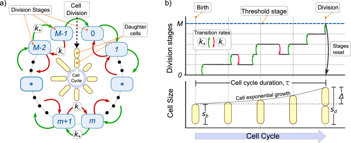
We consider the division process as the completion of a certain number of stages. During cell growth, as Fig. 1 shows, division occurs exactly when cell crosses a fixed number of stages M14,30. From a biological perspective, a possible interpretation of the division stages is related to the accumulation of a precursor protein, usually the FtsZ mentioned above4,31. However, other molecules could also be the main contributor to bacterial division32–35. Therefore, we use division stages instead of number of precursor proteins to explicitly keep our approach as general as possible.
Fig. 1. A general multistep model for triggering cell division.
a Diagram of the cell cycle explaining how division occurs at crossing M stages. b While bacteria grow exponentially (lower panel), the division stages accumulate at rate k+ and might revert at rate k− (upper panel). Once a number of steps M is reached, the cell divides, the steps are reset to zero, and the size is halved. The main variables of the bacterial division cycle are also shown: size at birth sb, size at division sd, and added size Δ = sd − sb.
Recent mathematical models have proposed cell-size based division rates36, multi-stages13,19 and back transitions4 to explain the division strategy. However, none of them can account for all the observed properties of cell division24. In this article, we present a model that encompasses each of these models as a special case and propose a likelihood-based method to test the predictions with data. We aim to provide a tool for hypothesis testing in future experiments.
A cell cycle is defined as the set of processes occurring during two consecutive divisions (Fig. 1). During the cell cycle, the cell size s grows exponentially over time t with growth rate μ. This means that the cell size follows:
| 1 |
In the adder strategy, the transition between stages occurs at a rate proportional to the current cell size14,19. To explain the sizer-like strategy, we will generalize the accumulation rates. The rate of stage increase k+ is considered nonhomogeneous and, depending on the model, stages can revert (by protein degradation) with a rate k−. At cell birth, that is, at the beginning of the cell cycle, the cell starts from stage m = 0 and size sb (which can differ from cell to cell). While cells grow exponentially, stages accumulate. When reaching the m = M stages, the cell divides. Exactly before division, the cell has a size sd such that the added size Δ is defined as the difference Δ = sd − sb. Finally, during cell splitting, cell size is halved and the stages are reset to m = 0.
To describe stage accumulation in the cell cycle, let Pm(t) be the probability that m ≤ M stages will be completed at time t with t = 0 being the beginning of the cycle. Given the rates k+ and k−, the dynamics of these probabilities are described by the master equation37:
| 2 |
PM is the probability of reaching the target step M or, equivalently, the probability of the division event to occur. Since after division the cell stars at stage m = 0, the initial condition (t = 0) is considered as Pm(t = 0) = δm,0 with δi,j being the Kronecker delta function.
As shown in Fig. 2a, we are interested in the estimate of the time to division τ. PM is related to τ as:
| 3 |
This is, PM(t) is also the probability that τ occurs in the interval (0, t). Hence, the probability density function PDF (also known as the distribution) of the division time ρτ is related to PM following:
| 4 |
Fig. 2. Process to predict the distribution of size at division and its comparison with data.
a The distribution of division times τ given the size at birth sb is estimated by solving numerically the master equation (2). b The size distribution at division sd is obtained from the distribution of division times and considering the exponential growth using (7). c The comparison with the data is made using methods based on likelihood. The distribution with higher likelihood fits the data better than a distribution with lower likelihood.
After estimating PM by solving (2), ρτ(t) can be calculated as follows38:
| 5 |
As shown in Fig. 2b, the PDF of the cell size at division sd can be estimated from ρτ, considering that the cells grow exponentially. This is, the cell size s is related to the size at birth sb through the time from birth t:
| 6 |
A transformation of variables allows us to obtain the PDF of sizes at division as:
| 7 |
where when exponential growth (1) is assumed. Observe how, since the cell size depends on sb, these distributions also depend on sb.
As explained in Fig. 2b, a comparison between experiment and theory requires the calibration of the model parameters. This is done by estimating the moments of sd using the estimated in (7). If 〈. 〉 defines the averaging operator, the α-moment of the distribution of sd, written as , is defined as follows,
| 8 |
where the mean size at division 〈sd〉, the moment with α = 1, can be used to calculate the mean added size per division cycle 〈Δ〉 = 〈sd〉 − sb as a function of the size at birth sb. The particular parameters of the model (as will be explained later) are adjusted, such as the predicted 〈sd〉 is constrained to the observed average 〈sd〉 assuming that the mean size at birth 〈sb〉 = 1.
After imposing this constraint, the best model parameters are adjusted to the data by maximizing the likelihood function. As shown in Fig. 2c, the likelihood function measures the precision of the PDF with the histogram associated with the data. With a higher likelihood, the predicted distribution fits the experiment better.
The moments of sd can also be used to quantify the noise in added size , the ratio between var(Δ) (the variance of Δ) and 〈Δ〉2, which is a measure of the stochastic variability of Δ. We can obtain as a function of sb from and its moments and 〈sd〉 using the formula19:
| 9 |
Observe how since depends on sb, different models may predict different trends on . For us, while the trend 〈Δ〉 vs sb is known as the division strategy, vs sb is the noise signature of the model. Next, we will explain these models as particular cases of k+ and k−.
The Adder strategy
The implementation of the adder, where 〈Δ〉 is independent on sb corresponds to the particular case of (2) where k+ and k− are given by19:
| 10 |
with k a constant and s = sbeμt is the cell size. Assuming exponentially growing cells and a division process defined by both (2) and (10), the mean added cell size 〈Δ〉 is given by19:
| 11 |
which is, as expected, independent of sb. The noise in Δ, defined in (9) follows:
| 12 |
which is also uncorrelated with sb as observations suggest19.
Sizer-like by molecule degradation
As4 suggests, sizer-like division strategy can be obtained by including active degradation of the division-triggering molecules. In our framework, degradation is equivalent to a step backward in the accumulation of M stages. In this case, the rates k+ and k− are given by:
| 13 |
where γ is the rate at which each molecule is degraded.
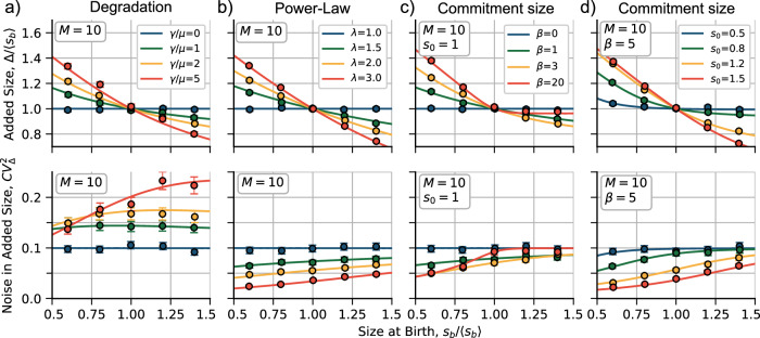
A negative slope is obtained in Δ vs sb as shown in Fig. 3a (top). The higher γ, the more pronounced the slope. This model reduces to the adder when γ ≪ μ. The noise signature of this model is presented in Fig. 3a (bottom) where, as the main property, we can see that for an increase γ, for fixed division steps M, it is expected a higher average .
Fig. 3. Trends on mean added size before division 〈Δ〉 (top) and its stochastic fluctuations (noise) (bottom) as functions of the size at birth sb.
a Trends considering the model of degradation for different values of the degradation rate γ relative to the growth rate μ in (13). b Predictions considering a division rate proportional to a power of size for different exponents λ in (14). c, d Predictions considering a commitment size with different values of the Hill function exponent β and the commitment size s0, respectively, in (15). The trend lines correspond to the numerical solutions of (2). Large dots are obtained from simulations. Error bars represent the 95%-confidence interval over 10000 simulated cycles. The constant k is set in each case such as 〈Δ〉 = 〈sb〉 = 1. Other parameters are shown inset.
Sizer-like via non-linear division rate
Following19, we consider a scenario where a molecule that triggers division is produced at a rate that depends on a power λ of cell size s, this is:
| 14 |
After substituting (14) in (2), plus the assumption of exponential growth, the division strategy exhibits a sizer-like behavior when λ > 1 (Fig. 3b top). For the particular case of λ = 1, the model reduces to the adder. Hence, the higher λ, the higher the slope of the relationship between Δ and sb. The power law on the production rate also affects the fluctuations of the added size : it lowers the average noise level for a given M and simultaneously increases the positive slope of versus sb (Fig. 3b bottom).
Sizer-like via a commitment size
Recent evidence suggests that cells may aim for a minimal size before starting division programs9. We will denote this minimal or commitment size by s0. We propose that it can be incorporated into our framework as
| 15 |
By replacing (15) with (2), we can see that the division strategy has a sizer-like behavior when β > 0 (Fig. 3c top). The parameter β can control the slope of the curve Δ versus sb for a fixed s0. It also influences the fluctuations of the added size : It reduces the average noise level for a given M, but increases the positive slope of versus sb (Fig. 3c bottom). This model encompasses the adder strategy as a special case when β = 0.
The commitment size s0 relative to the mean size at birth 〈sb〉 also affects the division strategy. For a fixed β, a low s0 ≪ sb mimics the adder, while a high s0 ≫ sb approximates the sizer (the strategy where the slope in Δ versus sb is −1). This is because cells born with a size below s0 follow a perfect sizer strategy, while cells born with size above s0 follow the adder. For small β, the adder strategy is recovered. For intermediate β, the transition from sizer to adder is smoother as sb increases.
Comparison: theory versus datasets
We have shown how to derive the PDF ρ(sd∣sb) from different models. Now, we want to estimate how accurate these distributions are relative to the data. We use the Akaike Information Criterion (AIC)39 to reward the fit of the models to the data while penalizing the number of free parameters. The adder model has one parameter (M), the degradation and power-law models have two (γ, M and λ, M respectively), and the commitment size model has three (s0, β and M). Taking into account all experiments, for each pair of data (sb, Δ) normalized by 〈sb〉 we numerically compute the size-at-division distribution given sb using (7). We find the parameters that maximize the likelihood function40 based on the data. Then we calculate the AIC for each model. The model with the lowest AIC is the most probable one, and we denote its AIC value by AICmin. To compare the relative probability of each model with the most probable one, we use the concept of relative likelihood41. For a model i with an AIC value of AICi, the relative likelihood p is given by:
| 16 |
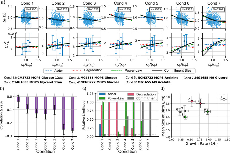
The AIC method is useful for estimating the accuracy of the models but is not easy to visualize since the match of the distributions is very similar. To gain a better intuitive understanding of how each model behaves, we can use the method of statistical moments. As shown in Fig. 3, this method involves plotting the division strategy (〈Δ〉 versus 〈sb〉) and the noise signature ( versus 〈sb〉) and comparing them with the data. From theory, we can obtain the moments directly: given a sb, they are calculated from the distribution using (8). From the data set, we visualize the moments given sb using quantile splitting. This method splits the data into a given number of quantiles and computes the statistics for each quantile separately. The points in Fig. 3 (five quantiles) represent the data from simulations, while Fig. 4a (also five quantiles) represents the experimental data. To study the division strategy, we plot and 〈Δ〉q for each quantile. The noise signature is obtained by plotting the variance for each quantile. Error bars indicate a 95% confidence interval using bootstrapping methods.
Fig. 4. Discriminating between models across different experimental conditions.
a Top: trends of the added size Δ vs the size at birth sb. Bottom: Noise signatures, quantified by stochastic fluctuations of the added size versus sb. The numerical prediction of the three models can be better discriminated: the degradation model (red dotted line), the power law (green dashed line), and the commitment size (black solid line). N represents the number of studied cell cycles. Cond represents the condition number. In the middle of the figure, more detailed labels are shown. Error bars represent the 95% CI using bootstrapping methods. b Comparison of the correlation function between Δ and sb for different conditions. The more negative this correlation is, the closer to sizer the division strategy is. c Relative likelihood of each model with respect to the model with the best AIC score using (16). The black dashed line represents the relative likelihood of 0.05. d Different conditions discriminated by the mean cell size at birth versus the mean growth rate. The color of the dots represents the most probable model, and the error bars represent the standard deviation of each variable.
Experiments
We analyzed two independent already published datasets of E. coli strains under different growth conditions4,19. Data were obtained from time-lapse microscopy images of confined single cells fed in a mother machine micro-fluidic device. References4,19 imaged slow-growing cells corresponding to steady growth conditions.
We measured the cell size using the cell length since the width is approximately constant. We normalize all lengths by the mean size at birth 〈sb〉. The theoretical division rate k was estimated, given the free parameters and setting the growth rate at , from the observed 〈Δ〉 with 〈sb〉 = 1. Besides simplify our computations, with this time and cell-size normalization, our idea is to obtain general results. Our conclusions should be scale-free because we only study dimensionless quantities such as the slope and correlation between Δ vs. sb and noise in added size . These properties of cell division emerge naturally given the model parameters. The experimental data and the data analysis scripts are available at42.
Results
Figure 4a shows the comparison between the data and the theory. First, we study the case where the division strategy is close to adder, that is, when Δ is independent of sb. This occurs for condition 1 (NCM3722 in MOPS with Glucose) since the 95% confidence interval for the correlation includes zero (Fig. 4b). For these conditions, the adder model has a relatively high AIC, but not as high as the other three models. The three models reach a similar likelihood, but the commitment size model is punished for its complexity. Both degradation and power-law models reach similar AIC scores, and it is not possible to discard one of those models with enough confidence.
For the other conditions, the division is sizer-like with statistical significance (Fig. 4c). In Fig. 4c, we observe that the degradation model has a minimum AIC (and therefore highest relative likelihood) for cells with a larger mean size at birth (conditions 2 and 3, MG1655 in MOPS with glycerol 11aa and MOPS with Glucose). For smaller strains, power-law and commitment show lower AICs. Power-law has the lowest AIC for conditions 4 and 5 (NCM3722 in MOPS glucose and MOPS arginine), and the commitment size model performs best for cell conditions with the most negative correlation Δ vs sb (conditions 6 and 7: MG1655 in M9 with Acetate and M9 with Glycerol).
To better visualize the performance of the models, we can compare the statistical moments with the binned moments of the data. In Fig. 4a, we observe that all of our proposed models capture the mean trend in Δ vs sb. However, they differ in noise signature ( vs sb). The degradation model predicts a low correlation between and sb, while the power-law model predicts an increasing function. The commitment size model behaves like the power law when the slope of Δ vs sb is small but predicts a higher slope of vs sb when the slope of Δ vs sb is large.
Discussion
In this paper, we propose a general model that includes different mechanisms of cell division regulation with experimental evidence. Each of them is a particular case. We found that each mechanism contributes differently to division depending on the growth condition. Our method can be used as a tool for the study of the origin of different division strategies not only in E. coli but also in other microorganisms. It includes complex observable variables such as the growth rate or the division-initiation size. We believe that our model can be generalized to other scenarios, such as cycle-stage-dependent growth rate or size-independent division transition rates, as seen in eukaryotic cells.
The transition from the adder to the sizer-like division strategies suggests the presence of a dual mechanism governing cell division: a primary mechanism that leads to the adder being the most important for larger cells, and a secondary mechanism that gives rise to the sizer-like is more visible in smaller cells. It is plausible that this secondary mechanism has often been overlooked in laboratory settings, where cells are commonly cultivated in nutrient-rich environments and are relatively large. However, this mechanism is of substantial importance, as it could elucidate cell survival and adaptation in real-world scenarios characterized by less-than-optimal or slower growth conditions.
Si et al.4 found experimental evidence that FtsZ degradation could be the basis for the sizer-like strategy. After inhibiting the production of ClpXP, an ATP-dependent protease43 that degrades FtsZ, E. coli cells that showed a sizer-like restored the adder division. Our investigation highlights the potency of the degradation model in explaining the sizer-like strategy, particularly when the relationship between added size and birth size is not excessively steep. However, for steeper slopes, alternative mechanisms align better with the data. We believe that the actual mechanism that explains sizer-like is a composition of degradation and commitment size since they are not exclusive.
The commitment size model posits a minimum size prerequisite for division initiation, which yields the sizer-like behavior. When the cell size at birth is relatively small, it often falls below this commitment size, needing to grow until reaching this threshold. Then the division machinery starts to build up. In contrast, when cells at birth exceed the commitment size, division starts immediately. With division starting just after birth, we expect large cells to follow the adder strategy. A visual representation of how the cell size plays a role in defining which model is more important is presented in Fig. 4d, where cells with a mean size at birth smaller than 2.3 μm (conditions 4–7) have a higher probability of being explained by a size-dependent division rate (either power law or commitment size models). Cells with a larger size (conditions 2 and 3) at birth are more likely to have a sizer-like by molecule degradation. Condition 1 has almost no correlation Δ versus sb and does not have a clear main contributor. In contrast, the average growth rate does not appear to define the main contributor to the division strategy.
This work shows that different models can fit the mean behavior, but the noise signature can be used to distinguish them. However, we acknowledge some limitations of these fluctuations-based methods. The added size distribution has multiple noise sources in addition to randomness related to the control mechanism; we can include others, such as instrumental precision: low camera resolutions, segmentation errors, and length as a size proxy that ignores fluctuations in cell width18. These uncertainties can add noise to the added size, but we believe that they cannot explain the high level of noise we observe and that the noise component of the division mechanism is high enough to neglect these instrumental sources.
The power-law rate model operates on the premise that the division rate has a stronger dependence on the size than the adder model. It is important to note that this model is heuristic, using nonlinear size dependency as an effective parameter to capture the intricacies of division. Thus, the power-law rate may approximate the Hill function rate of the commitment size model within a specific range. In contrast, the commitment size model, with more free parameters, can exhibit greater adaptability compared to the power-law model.
The notion of commitment size might relate to the initiation of chromosome replication at a fixed size per origin9,44,45. Although our model does not explicitly integrate chromosome replication, potential links between these variables remain open. Recent studies hint at coordination between division and replication in bacteria, resembling observations in more complex organisms35,46–48. Other variables that we did not study can contribute to the origin of the sizer-like. Recently, there is evidence about the dependence of growth rate and cell size at birth25,49, the correlation between lineage-related cells50 and randomness in growth rate51. We think that the addition of new free parameters makes the model more complex, and our fitting metric (the AIC score) punishes the complexity. Our aim is not to over-fit the model to each condition but to find the more general conclusion with the least number of assumptions.
The underlying biological mechanism enabling the cell to measure commitment size remains a subject of active debate. One perspective suggests tight synchronization between division and DNA replication, which triggers division when reaching a target size for replication initiation9,21,52. Another angle posits the existence of molecules acting as size proxies within cells, regulating division initiation24,53. Further experiments, as proposed in54,55, could provide additional information on the division strategy. Dynamic environments, where the division strategy changes dynamically, could shed light on these mechanisms. Other ways to achieve slow-growth conditions can also help us understand the validity of the models. These conditions can include decreasing growth temperature and growing in the presence of a mild concentration of antibiotics. Furthermore, exploring the division strategy with the clpX knockdown strain under different growth conditions, coupled with the use of AIC or other likelihood-based methods for data analysis, has the potential to provide a more comprehensive understanding of these phenomena.
Reporting summary
Further information on research design is available in the Nature Research Reporting Summary linked to this article.
Supplementary information
Acknowledgements
CN was supported by Colombian Science Ministry Convocatoria 647 para doctorados nacionales. A.S. acknowledges support from NIH-NIGMS via grant R35GM148351.
Author contributions
C.N. and C.V.G. developed the model and C.N. analyzed the data and designed the plots. C.V.G., J.M.P., and A.S. guided the research. All the authors contributed to the writing of the article.
Data availability
Dataset and code used for the data analysis discussed in this manuscript were gathered in a Zenodo repository with an MIT license at the following link: 10.5281/zenodo.3951080.
Competing interests
The authors declare no competing interests.
Footnotes
Publisher’s note Springer Nature remains neutral with regard to jurisdictional claims in published maps and institutional affiliations.
Supplementary information
The online version contains supplementary material available at 10.1038/s41540-024-00383-z.
References
- 1.Vargas-Garcia CA, Soltani M, Singh A. Conditions for cell size homeostasis: a stochastic hybrid system approach. IEEE Life Sci. Lett. 2016;2:47–50. doi: 10.1109/LLS.2016.2646383. [DOI] [Google Scholar]
- 2.Taheri-Araghi S, et al. Cell-size control and homeostasis in bacteria. Curr. Biol. 2015;25:385–391. doi: 10.1016/j.cub.2014.12.009. [DOI] [PMC free article] [PubMed] [Google Scholar]
- 3.Jun S, Taheri-Araghi S. Cell-size maintenance: universal strategy revealed. Trends Microbiol. 2015;23:4–6. doi: 10.1016/j.tim.2014.12.001. [DOI] [PubMed] [Google Scholar]
- 4.Si F, et al. Mechanistic origin of cell-size control and homeostasis in bacteria. Curr. Biol. 2019;29:1760–1770. doi: 10.1016/j.cub.2019.04.062. [DOI] [PMC free article] [PubMed] [Google Scholar]
- 5.Jun S, Si F, Pugatch R, Scott M. Fundamental principles in bacterial physiology—history, recent progress, and the future with focus on cell size control: a review. Rep. Prog. Phys. 2018;81:056601. doi: 10.1088/1361-6633/aaa628. [DOI] [PMC free article] [PubMed] [Google Scholar]
- 6.Campos M, et al. A constant size extension drives bacterial cell size homeostasis. Cell. 2014;159:1433–1446. doi: 10.1016/j.cell.2014.11.022. [DOI] [PMC free article] [PubMed] [Google Scholar]
- 7.Kar P, Tiruvadi-Krishnan S, Männik J, Männik J, Amir A. Using conditional independence tests to elucidate causal links in cell cycle regulation in Escherichia coli. Proc. Natl Acad. Sci. USA. 2023;120:e2214796120. doi: 10.1073/pnas.2214796120. [DOI] [PMC free article] [PubMed] [Google Scholar]
- 8.Männik J, Walker BE, Männik J. Cell cycle-dependent regulation of ftsz in Escherichia coli in slow growth conditions. Mol. Microbiol. 2018;110:1030–1044. doi: 10.1111/mmi.14135. [DOI] [PMC free article] [PubMed] [Google Scholar]
- 9.Wallden M, Fange D, Lundius EG, Baltekin Ö, Elf J. The synchronization of replication and division cycles in individual E. coli cells. Cell. 2016;166:729–739. doi: 10.1016/j.cell.2016.06.052. [DOI] [PubMed] [Google Scholar]
- 10.Priestman M, Thomas P, Robertson BD, Shahrezaei V. Mycobacteria modify their cell size control under sub-optimal carbon sources. Front. Cell Dev. Biol. 2017;5:64. doi: 10.3389/fcell.2017.00064. [DOI] [PMC free article] [PubMed] [Google Scholar]
- 11.Opalko HE, et al. Arf6 anchors cdr2 nodes at the cell cortex to control cell size at division. J. Cell Biol. 2021;221:e202109152. doi: 10.1083/jcb.202109152. [DOI] [PMC free article] [PubMed] [Google Scholar]
- 12.Erickson HP, Anderson DE, Osawa M. Ftsz in bacterial cytokinesis: cytoskeleton and force generator all in one. Microbiol. Mol. Biol. Rev. 2010;74:504–528. doi: 10.1128/MMBR.00021-10. [DOI] [PMC free article] [PubMed] [Google Scholar]
- 13.Jia C, Singh A, Grima R. Cell size distribution of lineage data: analytic results and parameter inference. Iscience. 2021;24:102220. doi: 10.1016/j.isci.2021.102220. [DOI] [PMC free article] [PubMed] [Google Scholar]
- 14.Ghusinga KR, Vargas-Garcia CA, Singh A. A mechanistic stochastic framework for regulating bacterial cell division. Sci. Rep. 2016;6:30229. doi: 10.1038/srep30229. [DOI] [PMC free article] [PubMed] [Google Scholar]
- 15.Basan M, et al. Inflating bacterial cells by increased protein synthesis. Mol. Syst. Biol. 2015;11:836. doi: 10.15252/msb.20156178. [DOI] [PMC free article] [PubMed] [Google Scholar]
- 16.Nieto C, Blanco SC, Vargas-García C, Singh A, Manuel PJ. Pyecolib: a python library for simulating stochastic cell size dynamics. Phys. Biol. 2023;20:045006. doi: 10.1088/1478-3975/acd897. [DOI] [PMC free article] [PubMed] [Google Scholar]
- 17.Serbanescu D, Ojkic N, Banerjee S. Cellular resource allocation strategies for cell size and shape control in bacteria. FEBS J. 2022;289:7891–7906. doi: 10.1111/febs.16234. [DOI] [PMC free article] [PubMed] [Google Scholar]
- 18.Nieto, C. et al. Coupling cell size regulation and proliferation dynamics of C. glutamicum reveals cell division based on surface area. bioRxiv10.1101/2023.12.26.573217 (2023).
- 19.Nieto C, Arias-Castro J, Sánchez C, Vargas-García C, Pedraza JM. Unification of cell division control strategies through continuous rate models. Phys. Rev. E. 2020;101:022401. doi: 10.1103/PhysRevE.101.022401. [DOI] [PubMed] [Google Scholar]
- 20.Sauls JT, Li D, Jun S. Adder and a coarse-grained approach to cell size homeostasis in bacteria. Curr. Opin. Cell Biol. 2016;38:38–44. doi: 10.1016/j.ceb.2016.02.004. [DOI] [PMC free article] [PubMed] [Google Scholar]
- 21.Knöppel A, Broström O, Gras K, Elf J, Fange D. Regulatory elements coordinating initiation of chromosome replication to the Escherichia coli cell cycle. Proc. Natl Acad. Sci. USA. 2023;120:e2213795120. doi: 10.1073/pnas.2213795120. [DOI] [PMC free article] [PubMed] [Google Scholar]
- 22.Boesen, T. et al. Robust control of replication initiation in the absence of dnaa-atp-dnaa-adp regulatory elements in Escherichia coli. bioRxiv10.1101/2022.09.08.507175 (2022).
- 23.Tiruvadi-Krishnan S, et al. Coupling between DNA replication, segregation, and the onset of constriction in Escherichia coli. Cell Rep. 2022;38:110539. doi: 10.1016/j.celrep.2022.110539. [DOI] [PMC free article] [PubMed] [Google Scholar]
- 24.Le Treut, G., Si, F., Li, D. & Jun, S. Quantitative examination of five stochastic cell-cycle and cell-size control models for Escherichia coli and Bacillus subtilis. Front. Microbiol. 3278 (2021). [DOI] [PMC free article] [PubMed]
- 25.Kohram M, Vashistha H, Leibler S, Xue B, Salman H. Bacterial growth control mechanisms inferred from multivariate statistical analysis of single-cell measurements. Curr. Biol. 2021;31:955–964. doi: 10.1016/j.cub.2020.11.063. [DOI] [PubMed] [Google Scholar]
- 26.Sekar K, et al. Synthesis and degradation of ftsz quantitatively predict the first cell division in starved bacteria. Mol. Syst. Biol. 2018;14:e8623. doi: 10.15252/msb.20188623. [DOI] [PMC free article] [PubMed] [Google Scholar]
- 27.Shi H, et al. Precise regulation of the relative rates of surface area and volume synthesis in bacterial cells growing in dynamic environments. Nat. Commun. 2021;12:1975. doi: 10.1038/s41467-021-22092-5. [DOI] [PMC free article] [PubMed] [Google Scholar]
- 28.Litsios A, et al. Differential scaling between g1 protein production and cell size dynamics promotes commitment to the cell division cycle in budding yeast. Nat. Cell Biol. 2019;21:1382–1392. doi: 10.1038/s41556-019-0413-3. [DOI] [PubMed] [Google Scholar]
- 29.Miller KE, Vargas-Garcia C, Singh A, Moseley JB. The fission yeast cell size control system integrates pathways measuring cell surface area, volume, and time. Curr. Biol. 2023;33:3312–3324. doi: 10.1016/j.cub.2023.06.054. [DOI] [PMC free article] [PubMed] [Google Scholar]
- 30.Nieto, C., Vargas-Garcia, C. A. & Singh, A. Statistical properties of dynamical models underlying cell size homeostasis. Tech. Rep., Center for Open Science (2023).
- 31.Harris LK, Theriot JA. Relative rates of surface and volume synthesis set bacterial cell size. Cell. 2016;165:1479–1492. doi: 10.1016/j.cell.2016.05.045. [DOI] [PMC free article] [PubMed] [Google Scholar]
- 32.Zhang Q, Zhang Z, Shi H. Cell size is coordinated with cell cycle by regulating initiator protein dnaa in E. coli. Biophys. J. 2020;119:2537–2557. doi: 10.1016/j.bpj.2020.10.034. [DOI] [PMC free article] [PubMed] [Google Scholar]
- 33.Speck C, Messer W. Mechanism of origin unwinding: sequential binding of dnaa to double-and single-stranded dna. EMBO J. 2001;20:1469–1476. doi: 10.1093/emboj/20.6.1469. [DOI] [PMC free article] [PubMed] [Google Scholar]
- 34.Schreiber G, Ron EZ, Glaser G. ppgpp-mediated regulation of dna replication and cell division in Escherichia coli. Curr. Microbiol. 1995;30:27–32. doi: 10.1007/BF00294520. [DOI] [PubMed] [Google Scholar]
- 35.Micali G, Grilli J, Osella M, Lagomarsino MC. Concurrent processes set E. coli cell division. Sci. Adv. 2018;4:eaau3324. doi: 10.1126/sciadv.aau3324. [DOI] [PMC free article] [PubMed] [Google Scholar]
- 36.Genthon A. Analytical cell size distribution: lineage-population bias and parameter inference. J. R. Soc. Interface. 2022;19:20220405. doi: 10.1098/rsif.2022.0405. [DOI] [PMC free article] [PubMed] [Google Scholar]
- 37.Nieto C, Vargas-Garcia C, Pedraza JM. Continuous rate modeling of bacterial stochastic size dynamics. Phys. Rev. E. 2021;104:044415. doi: 10.1103/PhysRevE.104.044415. [DOI] [PubMed] [Google Scholar]
- 38.Ghusinga KR, Dennehy JJ, Singh A. First-passage time approach to controlling noise in the timing of intracellular events. Proc. Natl Acad. Sci. USA. 2017;114:693–698. doi: 10.1073/pnas.1609012114. [DOI] [PMC free article] [PubMed] [Google Scholar]
- 39.Sakamoto, Y., Ishiguro, M. & Kitagawa, G. Akaike Information Criterion Statistics. (D. Reidel, Dordrecht, Boston, 1986) vol. 81, 26853.
- 40.Severini, T. A. Likelihood Methods in Statistics (Oxford University Press, 2000).
- 41.Burnham KP, Anderson DR. Multimodel inference: understanding aic and bic in model selection. Sociol. Methods Res. 2004;33:261–304. doi: 10.1177/0049124104268644. [DOI] [Google Scholar]
- 42.Nieto, C. Sizer-Like Division Analysis. 10.5281/zenodo.3951080 (2023).
- 43.Camberg JL, Hoskins JR, Wickner S. Clpxp protease degrades the cytoskeletal protein, ftsz, and modulates ftsz polymer dynamics. Proc. Natl Acad. Sci. USA. 2009;106:10614–10619. doi: 10.1073/pnas.0904886106. [DOI] [PMC free article] [PubMed] [Google Scholar]
- 44.Ho P-Y, Amir A. Simultaneous regulation of cell size and chromosome replication in bacteria. Front. Microbiol. 2015;6:662. doi: 10.3389/fmicb.2015.00662. [DOI] [PMC free article] [PubMed] [Google Scholar]
- 45.Chen J, Boyaci H, Campbell EA. Diverse and unified mechanisms of transcription initiation in bacteria. Nat. Rev. Microbiol. 2021;19:95–109. doi: 10.1038/s41579-020-00450-2. [DOI] [PMC free article] [PubMed] [Google Scholar]
- 46.Sun X-M, et al. Size-dependent increase in rna polymerase II initiation rates mediates gene expression scaling with cell size. Curr. Biol. 2020;30:1217–1230. doi: 10.1016/j.cub.2020.01.053. [DOI] [PubMed] [Google Scholar]
- 47.Kleckner NE, Chatzi K, White MA, Fisher JK, Stouf M. Coordination of growth, chromosome replication/segregation, and cell division in E. coli. Front. Microbiol. 2018;9:1469. doi: 10.3389/fmicb.2018.01469. [DOI] [PMC free article] [PubMed] [Google Scholar]
- 48.Ramkumar N, Baum B. Coupling changes in cell shape to chromosome segregation. Nat. Rev. Mol. Cell Biol. 2016;17:511–521. doi: 10.1038/nrm.2016.75. [DOI] [PubMed] [Google Scholar]
- 49.Nieto, C. et al. The role of division stochasticity on the robustness of bacterial size dynamics. bioRxiv10.1101/2022.07.27.501776 (2022).
- 50.ElGamel M, Vashistha H, Salman H, Mugler A. Multigenerational memory in bacterial size control. Phys. Rev. E. 2023;108:L032401. doi: 10.1103/PhysRevE.108.L032401. [DOI] [PubMed] [Google Scholar]
- 51.Modi S, Vargas-Garcia CA, Ghusinga KR, Singh A. Analysis of noise mechanisms in cell-size control. Biophys. J. 2017;112:2408–2418. doi: 10.1016/j.bpj.2017.04.050. [DOI] [PMC free article] [PubMed] [Google Scholar]
- 52.Zheng H, et al. General quantitative relations linking cell growth and the cell cycle in Escherichia coli. Nat. Microbiol. 2020;5:995–1001. doi: 10.1038/s41564-020-0717-x. [DOI] [PMC free article] [PubMed] [Google Scholar]
- 53.Berger M, Wolde PRT. Robust replication initiation from coupled homeostatic mechanisms. Nat. Commun. 2022;13:6556. doi: 10.1038/s41467-022-33886-6. [DOI] [PMC free article] [PubMed] [Google Scholar]
- 54.Bakshi S, et al. Tracking bacterial lineages in complex and dynamic environments with applications for growth control and persistence. Nat. Microbiol. 2021;6:783–791. doi: 10.1038/s41564-021-00900-4. [DOI] [PMC free article] [PubMed] [Google Scholar]
- 55.Nieto C, Vargas-García C, Pedraza JM, Singh A. Modeling cell size control under dynamic environments. IFAC-PapersOnLine. 2022;55:133–138. doi: 10.1016/j.ifacol.2023.01.061. [DOI] [Google Scholar]
Associated Data
This section collects any data citations, data availability statements, or supplementary materials included in this article.
Supplementary Materials
Data Availability Statement
Dataset and code used for the data analysis discussed in this manuscript were gathered in a Zenodo repository with an MIT license at the following link: 10.5281/zenodo.3951080.


