PLOS Neglected Tropical Diseases logoLink to PLOS Neglected Tropical Diseases
. 2024 May 17;18(5):e0011292. doi: 10.1371/journal.pntd.0011292

Factors associated with differential seropositivity to Leptospira interrogans and Leptospira kirschneri in a high transmission urban setting for leptospirosis in Brazil

Daiana de Oliveira 1,2, Hussein Khalil 3, Fabiana Almerinda G Palma 1, Roberta Santana 1, Nivison Nery Jr 1,2, Juan C Quintero-Vélez 4,5,6, Caio Graco Zeppelini 7, Gielson Almeida do Sacramento 2, Jaqueline S Cruz 2, Ricardo Lustosa 1, Igor Santana Ferreira 1, Ticiana Carvalho-Pereira 1, Peter J Diggle 8, Elsio A Wunder Jr 2,9,10, Albert I Ko 2,9, Yeimi Alzate Lopez 1, Mike Begon 11, Mitermayer G Reis 2,9,12, Federico Costa 1,2,9,*
Editor: Claudia Munoz-Zanzi13
PMCID: PMC11139309  PMID: 38758957

Abstract

Background

Leptospirosis is a zoonosis caused by pathogenic species of bacteria belonging to the genus Leptospira. Most studies infer the epidemiological patterns of a single serogroup or aggregate all serogroups to estimate overall seropositivity, thus not exploring the risks of exposure to distinct serogroups. The present study aims to delineate the demographic, socioeconomic and environmental factors associated with seropositivity of Leptospira serogroup Icterohaemorraghiae and serogroup Cynopteri in an urban high transmission setting for leptospirosis in Brazil.

Methods/Principal findings

We performed a cross-sectional serological study in five informal urban communities in the city of Salvador, Brazil. During the years 2018, 2020 2021, we recruited 2.808 residents and collected blood samples for serological analysis using microagglutination assays. We used a fixed-effect multinomial logistic regression model to identify risk factors associated with seropositivity for each serogroup. Seropositivity to Cynopteri increased with each year of age (OR 1.03; 95% CI 1.01–1.06) and was higher in those living in houses with unplastered walls (exposed brick) (OR 1.68; 95% CI 1.09–2.59) and where cats were present near the household (OR 2.00; 95% CI 1.03–3.88). Seropositivity to Icterohaemorrhagiae also increased with each year of age (OR 1.02; 95% CI 1.01–1.03) and was higher in males (OR 1.51; 95% CI 1.09–2.10), in those with work-related exposures (OR 1.71; 95% CI 1.10–2.66) or who had contact with sewage (OR 1.42; 95% CI 1.00–2.03). Spatial analysis showed differences in distribution of seropositivity to serogroups Icterohaemorrhagiae and Cynopteri within the five districts where study communities were situated.

Conclusions/Significance

Our data suggest distinct epidemiological patterns associated with the Icterohaemorrhagiae and Cynopteri serogroups in the urban environment at high risk for leptospirosis and with differences in spatial niches. We emphasize the need for studies that accurately identify the different pathogenic serogroups that circulate and infect residents of low-income areas.

Author summary

In this study, we investigated specific epidemiological patterns related to the Icterohaemorrhagiae and Cynopteri serogroups in urban environments at high risk for leptospirosis. Our results indicate that seropositivity for the Cynopteri serogroup increases with age. Seropositivity is also higher in places where houses have unplastered walls and where there are cats in the vicinity of the residences. Seropositivity for the Icterohaemorrhagiae serogroup also increases with age, and is more prevalent in men and in individuals exposed to work or who have contact with sewage. Spatial analysis reveals variations in the distribution of seropositivity for these serogroups within the five urban areas studied. We emphasize the importance of evaluating the epidemiological pattern for the different Leptospira serogroups, since each serogroup may be associated with different animal reservoirs. Our data suggest that the Icterohaemorrhagiae and Cynopteri serogroups present different epidemiological pattern, thus highlighting the need to understand the complex interactions between serogroups and reservoirs. These findings not only contribute to our understanding of the epidemiology of leptospirosis, but also have practical public health implications by helping to identify risk factors and thereby develop more effective preventive measures.

Introduction

Leptospirosis is a potentially severe infectious disease that affects animals (both domestic and wild) and humans [1]. The disease is caused by pathogenic species of the genus Leptospira and results in a wide array of clinical manifestations in humans [2]. Symptoms can range from asymptomatic infection and light fever to severe manifestations with risk of death [3]. Over a million cases occur globally every year, causing over sixty thousand deaths [4]. The majority of the disease burden occurs in developing nations, in local environments that lack adequate infrastructure and health services [5]. Human infection occurs through direct contact with urine or contaminated secretions from infected animals, or indirectly through contact with contaminated soil and water [1,6].

The genus Leptospira comprises 72 species and over 300 serovars, classified in more than 20 serogroups [710]. The microscopic agglutination test (MAT) is the reference serological assay for the diagnosis of leptospirosis. Despite its limitations, the MAT can provide the presumptive identity of the infecting serogroup, contributing for spatiotemporal epidemiological studies and reservoir identification when isolation of local strains is not available [11]. However, isolation or specific laboratory techniques like DNA sequence analysis are needed for definitive serotyping [12,13]. Some serovars can infect multiple animal species while others are more adapted to specific animal hosts [2]. This host preference might explain why some serovars/serogroups are more frequent in certain geographic areas. The diversity of reservoirs, serovars and serogroups, together with environmental and socio-behavioral determinants, poses a challenge to the characterization of the epidemiologic patterns of leptospirosis. These factors can influence the risk of transmission and consequently increase incidence and cause outbreaks [14].

Historically, tropical and subtropical nations have experienced seasonal leptospirosis incidence peaks associated with heavy rainfall and flooding events during the rainy season [1417]. The rapid, informal expansion of large urban centers, coupled with inadequate housing and sanitation, has resulted in environmental and social conditions that are highly favorable to disease hosts and pathogen transmission [18]. Over 33% of the world’s population, and around 28% if Brazil’s population live in urban poor communities [19] where, because of the deep social inequalities, there is a direct correlation between vulnerable environments, disaster risk and public health problems [20,21]. Historically, in the city of Salvador, where most of the population is of black and mixed race, people with lower income live in the peripheral areas of cities where access to basic rights is often absent and the prevalence of health problems such as leptospirosis is greater than in other areas of the city.

Studies performed in the past two decades in Salvador, Brazil, have reported Leptospira interrogans serogroup Icterohaemorrhagiae serovar Copenhageni (strain Fiocruz L1-130) as the serogroup responsible for over 95% of the clinical cases recorded throughout the city [14], as well as asymptomatic infections in residents of the poor urban community of Pau da Lima within Salvador [22,23]. Previous studies in the same community have shown that the animal reservoir of the serovar Copenhageni and serogroup Icterohaemorrhagiae is the brown rat (Rattus norvegicus) [24,25]. These studies identified the seroincidence of Leptospira kirschneri, serogroup Cynopteri (strain 3522C) as the second highest in residents of urban communities [26]. However, the animal reservoir of these serogroups and the determinants of its transmission remain unknown.

Investigating the Leptospira serologic profile of the residents in urban communities, accounting for serogroup-specific risk factors, will contribute to a better understanding of the risk determinants of urban infection and inform the development of more specific control measures. The present study aims to identify demographic, socioeconomic, and environmental factors associated with seropositivity of Leptospira interrogans Icterohaemorrhagiae (strain Fiocruz L1-130) and Leptospira kirschneri Cynopteri (strain 3522C) in residents of five urban low-income communities in Salvador, Brazil.

Methods

Ethics statement

The study was approved by the Ethics Committee of Fundação Oswaldo Cruz, Instituto Gonçalo Moniz (CAAE 45217415.4.0000.0040), the Ethics Committee for Research of Instituto de Saúde Coletiva (UFBA, 041 / 17–2.245.914.17–2.245.914) and the Institutional Review Board of Yale University (no. 2000031554). Those willing to participate signed an informed consent form and had a small blood sample drawn by a trained phlebotomist. Underage participants were enrolled with informed consent written of their legal guardian.

Study design

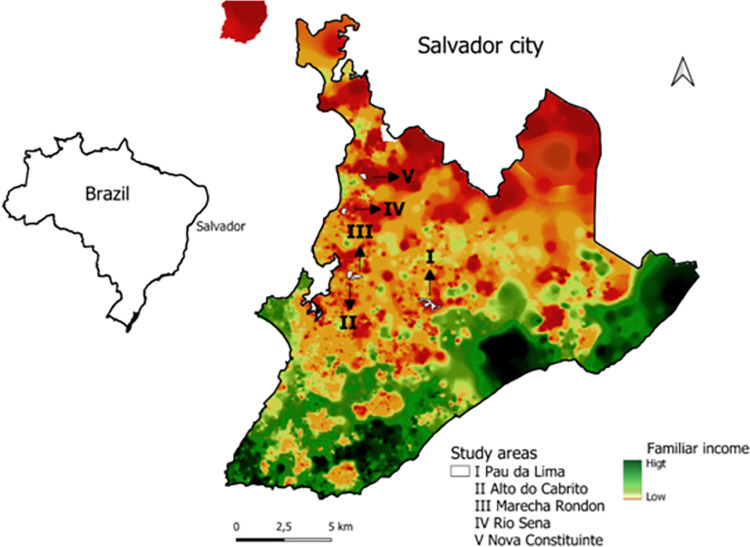
We performed a cross-sectional study in five urban poor communities: Pau da Lima (PL), Alto do Cabrito (AdC), Marechal Rondon (MR), Nova Constituinte (NC) and Rio Sena (RS) in the city of Salvador, Bahia, Brazil (Fig 1). In PL, we collected samples during the period from November 2020 to February 2021. Previous studies in the area indicated an annual leptospiral incidence of 37.8 infections per thousand [22]. In the four other communities, namely: AdC, MR, NC and RS, sampling occurred from April to September 2018. All five communities are considered low-income, with precarious sanitation and infrastructure.

Fig 1. Urban communities in the city of Salvador, Brazil.

Fig 1

Location of study sites (white) within the city. Distribution of family income, high (green) and low (orange). Shapefiles and data used for the base map were obtained from open and publicly accessible base of IBGE—Instituto Brasileiro de Geografia e Estatística [Brazilian Institute of Geography and Statistics]. Direct link to the base layer of the map: https://www.ibge.gov.br/geociencias/downloads-geociencias.html.

Eligible individuals were those ≥5 years of age, who slept 3 nights per week in a household. We applied a structured questionnaire to collect information on individual characteristics of the participant, domestic and peri-domestic environmental characteristics, exposure to sources of environmental contamination, and the presence of potential animal reservoirs.

Serological analysis was performed using the Microscopic Agglutination Test (MAT), the reference assay for diagnosis of human leptospirosis, as previously described [18]. MAT is based on dark-field microscopy detection of serum agglutination samples from an individual (antibodies) with live Leptospira antibodies. Samples were tested against a panel of seven antigens, including five reference strains (WHO Collaborative Laboratory for Leptospirosis, Royal Tropical Institute, Holland): L. kirschneri serogroups Cynopteri serovar Cynopteri strain 3522C and Grippothyphosa serovar Grippothyphosa strain Duyster; L. interrogans serogroups Canicola serovar Canicola strain H. Utrecht IV and Autumnalis serovar Autumnalis strain Akiyami A; and L. borgpetersenii serogroup Ballum serovar Ballum strain MUS 127. The panel also included two local clinical isolates: L. interrogans serogroup Icterohaemorrhagiae serovar Copenhageni strain Fiocruz L1-130 and L. santarosai serogroup Shermani strain LV3954 [22]. We performed association analyses only for serogroups Icterohaemorrhagiae strain Fiocruz L1-130 and Cynopteri strain 3522C, which were the most prevalent strains in our analyses, and presented adequate sample sizes for estimating associations.

Outcomes, exposures, and covariates

The outcome of positivity for leptospirosis was defined as seropositivity against serogroups Cynopteri or Icterohaemorrhagiae. The interpretation of MAT results for serogroup classification is limited due to the possibility of cross-reactivity between different serovars, lack of representative strains for the local circulating serogroup, and subjectivity of the interpretation of the agglutination [27]. A result was considered positive if 50% or more leptospires were agglutinated by MAT with a titer of ≥1:50. When agglutination was observed at a dilution of 1:100, the sample was titrated in serial two-fold dilutions to determine the highest positive titer [26]. In this study, the presumptive definition of the serogroup was established based on the serogroup that exhibited an antibody titer at least one dilution higher than any other serogroup. In cases where serogroups showed equal titers, the presumptive serogroup was classified as mixed and not considered in the final analysis. Additionally, samples that did not show significant reactivity, with an antibody titer below 1:50 in the tested serogroups, were considered negative.

The main explanatory variables at the individual-level were: age in years (as a quantitative variable); gender; ethnicity; highest education level achieved; receipt of the Federal government stimulus program (Bolsa Família); employment status; work involving contact with sewer/garbage/construction materials; use of protective boots; contact with mud in the last 12 months; walking barefoot outside; and cleaning sewer canals in the last 12 months. Additional environmental explanatory variables included: proximity of the household to open sewers; paved access to the household; household with a backyard; unplastered walls (a proxy for quality of construction); garbage accumulation near the household; and presence of domestic animals (cats, dogs, and chickens).

Spatial descriptive analysis

QGIS version 2.18.20 was used to construct a georeferenced database from satellite image WorldView-3 May 2017, at 31 cm resolution. The study team identified households within the study site and marked their positions onto hard copy 1:1,500 scale maps. The image WorldView-3 was acquired by the research project / Institute Gonçalo de Moniz—IGM—Fiocruz Bahia from the company Stamap, with disclosure permitted referencing the Copyrights of DigitalGlobe images. We used bivariate Kernel Density Estimation (KDE) [28], to evaluate possible territorial segregation of serogroups Icterohaemorrhagiae and Cynopteri. The KDE is defined as fhat(x) = n-1h-2 ∑k{(x-xi)/h}, where k(u) is the kernel function (a bivariate probability density), x denotes any location within the study-area, x1,…,xn are the data-locations and h is the bandwidth. The KDE delivers smoothed spatial distributions of the density of human cases of serogroups Icterohaemorrhagiae and Cynopteri, we set the bandwidth at 30 meters, based on the average distance typically traveled by rats per day [29], to give a possible exposure factor of households to each serogroup. To determine the smoothed, population-adjusted risk distribution for each of the serogroups Icterohaemorrhagiae and Cynopteri we then calculated the ratio between the KDE for households with positive cases and the KDE for all households evaluated. Boundary polygons and the data for the production of the Salvador income map were downloaded from the publicly accessible data base of IBGE—Instituto Brasileiro de Geografia e Estatística [Brazilian Institute of Geography and Statistics], using the Geosciences platform that can be accessed at https://www.ibge.gov.br/geociencias/downloads-geociencias.html. The maps were created by the authors using QGIS 2.18 software.

Statistical analysis

We performed a descriptive analysis using relative and absolute frequencies for qualitative variables and median and interquartile range for quantitative variables. A fixed-effect multinomial logistic regression model was used to estimate potential factors associated with seropositivity for each of the two serogroups, Icterohaemorrhagiae and Cynopteri. Variables considered in the multivariable analysis were those with p < 0.15 in single-factor analyses. The multivariable analysis used a stepwise method based on the purposeful selection of variables according to both statistical and research criteria (biological plausibility) [22,23]. Also, “area" was included in the final model as a five-level categorical fixed effect. The final model, constrained by the inclusion of variables selected on the grounds of biological plausibility was chosen to minimize Akaike’s Information Criteria (AIC). The linearity assumption was confirmed by visual inspection before the inclusion of each quantitative variables in bivariate and multivariate models. We then checked for high levels of correlation among the selected variables in the final model using a Variance Inflation Factor (VIF), but all VIF values were <5 and thus no previously selected variables were excluded. All statistical procedures were performed in R studio, using tidyverse, lme4 and nmet packages [3033]

Results

The study included 2,808 residents of five neighborhoods located in Salvador, Brazil (Fig 1); of which 1,512 were from Pau da Lima, 332 from Marechal Rondon, 367 from Alto do Cabrito, 304 from Nova Constituinte and 293 from Rio Sena (Table 1). The population studied was predominantly female (58%; 1,620/2,808) and self-declared black (51%; 1,422/2,808) (Table 1).

Table 1. Descriptive statistics for demography and socioenvironmental variables by area.

Characteristic N Total
N = 2,8081
Alto do Cabrito
N = 3671
Marechal Rondon
N = 3321
Nova Constituinte
N = 3041
Pau da Lima
N = 1,5121
Rio Sena
N = 2931
p-value2
Sociodemographic
Age (years) 2,81 <0.001
    0 to 16 666 (24) 59 (16) 48 (14) 72 (24) 395 (26) 92 (31)
    17 to 29 708 (25) 102 (28) 74 (22) 74 (24) 386 (26) 72 (25)
    30 to 44 721 (26) 97 (26) 84 (25) 87 (29) 388 (26) 65 (22)
    > 45 713 (25) 109 (30) 126 (38) 71 (23) 343 (23) 64 (22)
Sex 2,81 0.94
    Male 1,188 (42) 159 (43) 134 (40) 130 (43) 639 (42) 126 (43)
    Female 1,620 (58) 208 (57) 198 (60) 174 (57) 873 (58) 167 (57)
Ethnicity 2,77 <0.001
    Brown 1,171 (42) 146 (41) 148 (46) 115 (38) 635 (42) 127 (45)
    Black 1,422 (51) 191 (54) 151 (47) 169 (57) 781 (52) 130 (46)
    White 155 (5.6) 16 (4.5) 21 (6.6) 15 (5.0) 76 (5.0) 27 (9.5)
    Others 20 (0.7) 0 (0) 0 (0) 0 (0) 20 (1.3) 0 (0)
Education 2,81 <0.001
    5 or less 769 (27) 87 (24) 84 (25) 80 (26) 430 (28) 88 (30)
    5 to 9 919 (33) 94 (26) 103 (31) 87 (29) 515 (34) 120 (41)
    9 to 12 945 (34) 159 (43) 121 (36) 116 (38) 479 (32) 70 (24)
    Higher Education 61 (2.2) 17 (4.6) 7 (2.1) 10 (3.3) 25 (1.7) 2 (0.7)
    Never studied 114 (4.1) 10 (2.7) 17 (5.1) 11 (3.6) 63 (4.2) 13 (4.4)
Employment 2,81 982 (35) 127 (35) 105 (32) 98 (32) 568 (38) 84 (29) 0.016
Risk Actions
Risk occupation 2,81 215 (7.7) 24 (6.5) 37 (11) 15 (4.9) 121 (8.0) 18 (6.1) 0.029
Walk barefoot 2,81 1,046 (37) 141 (38) 105 (32) 101 (33) 561 (37) 138 (47) <0.001
Use of boots 2,81 559 (20) 60 (16) 59 (18) 58 (19) 341 (23) 41 (14) 0.002
Cleaned sewage 2,49 253 (10) 27 (8.2) 43 (14) 40 (15) 113 (8.5) 30 (12) <0.001
Sewage contact 2,81 600 (21) 48 (13) 97 (29) 56 (18) 332 (22) 67 (23) <0.001
Peridomiciliary
Open sewer 2,81 955 (34) 74 (20) 131 (39) 97 (32) 539 (36) 114 (39) <0.001
Access to the house is paved? 2,81 2,107 (75) 325 (89) 259 (78) 239 (79) 1,121 (74) 163 (56) <0.001
Wall plastered? 2,81 297 (11) 19 (5.2) 40 (12) 19 (6.2) 145 (9.6) 74 (25) <0.001
Presence of animals in the household
Cats 2,81 693 (25) 82 (22) 79 (24) 75 (25) 390 (26) 67 (23) 0.60
Dogs 2,81 1,092 (39) 154 (42) 108 (33) 140 (46) 544 (36) 146 (50) <0.001
Chickens 2,81 138 (4.9) 29 (7.9) 14 (4.2) 36 (12) 41 (2.7) 18 (6.1) <0.001
Leptospira seropositivity 2,81 0.97
    Cynopteri 45 (1.6) 6 (1.6) 7 (2.1) 3 (1.0) 23 (1.5) 6 (2.0)
    Icterohaemorrhagiae 208 (7.4) 24 (6.5) 24 (7.2) 23 (7.6) 114 (7.5) 23 (7.8)
Without evidence of seropositivity 2,555 (91) 337 (92) 301 (91) 278 (91) 1,375 (91) 264 (90)

1Mean (SD) or Frequency (%)

2Pearson’s Chi-squared test

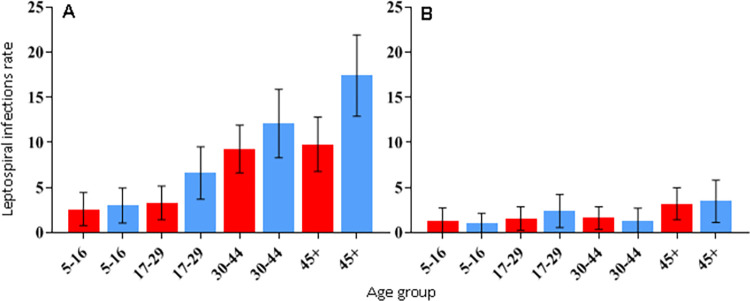
The overall unadjusted seroprevalence was 9% (253/2,808, 95% CI 8.0% - 10.1%). Of the 253 infected, 18% (45/253) were seropositive to serogroup Cynopteri, of which 43 (96%) reacted to titers of 50 to 400, while the remaining 2 (4%) reacted to titers of 800 to 6400. Also, 82% (208/253) were seropositive to serogroup Icterohaemorrhagiae, of which 190 (91%) reacted to titers of 50 to 400, while the remaining 18 (9%) reacted to titers of 800 to 1600. The seropositivity for the other tested serogroups (Canicola, Shermani, Autumnalis, Ballum, Grippotyphosa) did not exceed 2%, while the mixed result was 7% (S1 and S2 Tables). Seropositivity to serogroup Icterohaemorrhagiae was lowest in AdC (6.5%) and highest in RS (7.8%). Seroprevalence of Cynopteri ranged between 1.0% in NC and 2.1% in MR (Table 1). Seroprevalence of Icterohaemorrhagiae increased with age (OR 1.03 per year, 95% CI 1.02–1.04) as shown in S3 Table and described by age group in Fig 2. Females were less likely to be infected than males (OR 0.69, 95% CI 0.52–0.91, S3 Table). This pattern was not observed for serogroup Cynopteri (OR 1.07, 95% CI 0.59–1.99), which presented higher seroprevalence in the age group >45 years (Fig 2).

Fig 2.

Fig 2

Infection rates for Leptospira interrogans Icterohaemorrhagiae (A) and Leptospira kirschneri Cynopteri (B) by sex and age. Red bars: female. Blue bars: males. Whiskers: 95% CI.

The final multivariate multinomial analysis (Table 2), which includes data for residents > 18 years of age to investigate work-related exposures (n = 2,009), identified the following variables as significantly associated with seropositivity against Cynopteri: age in years (as a quantitative variable, OR 1.03, 95% CI 1.01–1.06), lives in house with unplastered walls (exposed brick, OR 1.68, 95% CI 1.09–2.59) and presence of cats near the household (OR 2.00, 95% CI 1.03–3.88). In addition, gender (males>females, OR 1.51, 95% CI 1.09–2.10), age in years (OR 1.02 per year, 95% CI 1.01–1.03), work-related exposures (OR 1.71, 95% CI 1.10–2.66) and contact with sewage (OR 1.42, 95% CI 1.00–2.03) were factors associated with seropositivity against Icterohaemorrhagiae.

Table 2. Multivariate multinomial regression of seropositivity for Leptospira kirschneri Cynopteri (3522C) and Leptospira interrogans Icterohaemorrhagiae (Fiocruz L1-130), (n = 2,009).

Note that there were no differences in seropositivity among neighborhoods.

Characteristics Cynopteri
(3522C)
Icterohaemorrhagiae
(Fiocruz L1-130)
OR 95% CI
Gender (male) - - 1.51 1.09–2.10
Age (years) 1.03 1.01–1.06 1.02 1.01–1.03
Work-related exposures - - 1.71 1.10–2.66
Contact with sewage - - 1.42 1.00–2.03
Lives in house with unplastered walls
(exposed brick)
1.68 1.09–2.59 - -
Neighborhood
Alto do Cabrito (reference)
Marechal Rondon 0.79 0.42–1.47 1.54 0.43–5.46
Nova Constituinte 0.98 0.51–1.88 0.97 0.21–4.44
Pau da Lima 1.19 0.74–1.90 1.39 0.46–4.15
Rio Sena 1.30 0.69–2.45 2.46 0.67–9.10
Cats near the household 2.00 1.03–3.88 - -

Acronyms: OR, odds ratio; 95% IC, Confidence intervals.

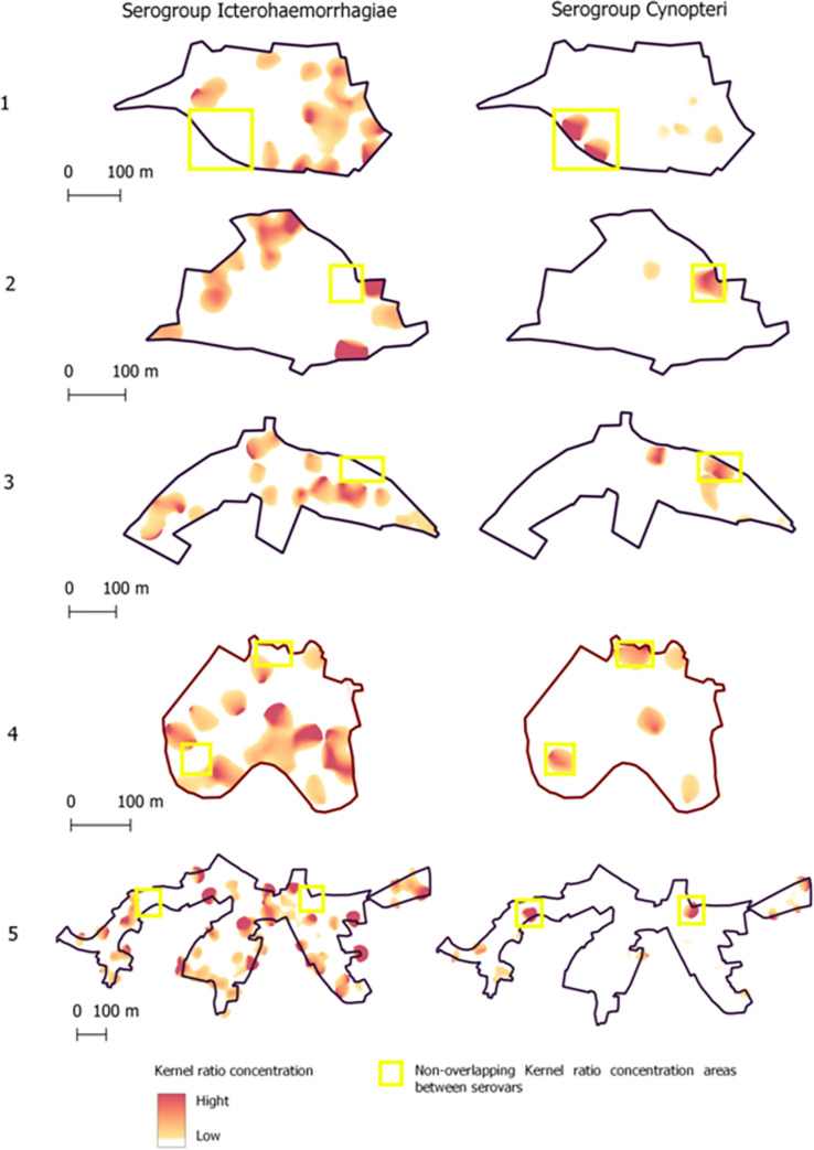
Fig 3 shows differences in the spatial patterns of seropositivity between serogroups Icterohaemorrhagiae and Cynopteri, where yellow rectangle indicate non-overlapping kernel ratio concentration areas between serogroups. The objective of this complementary analysis was to verify possible overlaps in the spatial distributions of both the two serogroups.

Fig 3. Spatial patterns of seropositivity to serogroups Icterohaemorrhagiae and Cynopteri (yellow rectangle) on five neighborhoods of Salvador, Bahia, Brazil.

Fig 3

The light red-to- hard red gradient represents increasing density in smoothing analyses which used 30 meters as the bandwidth.

Discussion

In this article, our MAT results indicated that L. interrogans serogroup Icterohaemorrhagiae is the most predominant in our areas of study in the city of Salvador, Brazil. In the local context, based on MAT, we found few circulating serogroups, and there was little evidence of cross-reaction (S1 and S2 Tables). Taken together, those results are in accordance with previous serosurveillance and ecological studies conducted in the same region [18,22,23], and supported by the high number of local clinical isolates obtained from humans and rats, where the majority of those were identified as serogroup Icterohaemorrhagiae [14,24]. However, we also found evidence of local exposure to L. kirschneri serogroup Cynopteri among residents of all the communities studied. Although MAT is a technique with limitations to precisely identify the infecting serogroup, the identification of individuals with high titers against the Cynopteri serogroup, considered pathogenic and whose reservoir in urban environments is unknown, represents an important epidemiological opportunity in attempting to understand the infection patterns of these two important serogroups of pathogenic Leptospira that seems to be circulating in this region.

We identified differences in the demographic and socioenvironmental factors that affect seropositivity for each serogroup. Residents exposed to serogroup Cynopteri had a different demographic profile from that of serogroup Icterohaemorrhagiae. We observed higher seroprevalence for serogroup Cynopteri in ages >45 years for both genders, suggesting that behavioral differences between men and women do not affect exposure, in contrast to what has been observed regarding overall seropositivity in previous studies. Men tend to be more engaged in outdoor activities that are considered environmental risk exposures (e.g.: sewer canal cleaning, construction work, debris and refuse management) that are known risk factors for seroprevalence [18,23]. However, for serogroup Icterohaemorrhagiae, we found that seropositivity increased with age, and that males were more likely to be seropositive. A similar relationship between age and exposure to Icterohaemorrhagiae has been observed in previous studies [18].

Occupation-related factors were associated with exposure to serogroup Icterohaemorrhagiae. Individuals who engaged in activities involving solid waste management, or contact with mud, floodwaters and/or sewage were more likely to be seropositive. This agrees with previous results from these and similar communities, where leptospirosis is associated with work-related exposures, including occupation in subsistence farming [3]. Further, inhabitants of low-income communities are commonly enrolled in informal employment, often close to their households. This finding highlights the role of the environment as an important source for transmission. We did not detect any independent associations between work-related risk exposures and seropositivity for Cynopteri. This negative finding may be a consequence of the lower number of individuals seropositive for Cynopteri.

Little is known about the animal and environmental reservoirs for serogroup Cynopteri, other than that the strain was first isolated from a bat kidney in Indonesia [34]. In our final model, we detected a positive association between anti-Cynopteri antibodies and the presence of cats in the household. Although occurrence of this serogroup has been reported in wild animals such as bats [35], there are also indications that cats can be exposed to Leptospira; in particular, higher seroprevalence of serogroup Cynopteri than other serogroups has been found in cats from Spain [36]. This suggests a need for future studies to accurately identify the different pathogenic serogroups that circulate in low-income areas and to evaluate the maintenance animal host of the Cynopteri serogroup in these urban environments.

Among the domestic and peri-domiciliary variables, the presence of open sewage near the home was associated with greater seropositivity for serogroup Icterohaemorrhagiae. This result is consistent with a previous study [22] in which “living near an open sewer” was a factor associated with seropositivity and suggests that environmental exposure is an important route of indirect transmission. It should also be noted that the urine of animals other than rats can release leptospires into the environment. This association has also been reported in other epidemiological studies [37,38]. A previous study in the same study area identified that, in addition to sewage, accumulations of rainwater on the ground can be a source of pathogenic Leptospira [39].

Infrastructure deficiencies in homes, for example living in a house with unplastered walls (exposed brick), have also been considered a source of transmission for repeated exposure to Leptospira [22]. Risk factors for peri-domiciliary exposure may therefore also be important for infection by serogroup Cynopteri. Structural precariousness in the household could be an indicator of low socioeconomic status, as suggested in another study [25].

The differences that we found in the spatial distribution of Icterohaemorrhagiae and Cynopteri (Fig 3) suggest that these two r serogroups occupy distinct niches within the urban environment. For example, our statistical analysis found that serogroup Icterohaemorrhagiae is associated with proximity to sewers and Cynopteri with the presence of cats.

There are limitations to this work that must be acknowledged. Firstly, whilst MAT is the standard test used in prevalence surveys and can, to some extent, help to indicate the presumptive serogroups circulating in a specific area [27] A positive result does not indicate that the individual necessarily has an active infection unless paired samples are taken [13]. Furthermore, as we collected data at a single point in time we cannot assess-incidence of infection events over time. Another important aspect to be considered is that, although we had previously tested a broader set of seven serogroups, we recognized the possible existence of other serogroups of Leptospira in the study population. However, our main focus was not on a comprehensive assessment of all serogroups present in each region, but on identifying the factors that could be associated with the seropositivity of the most highly prevalent serogroups of Leptospira in the populations under study. Additionally, we found a low prevalence of Cynopteri, making it difficult to understand its true epidemiological pattern. Future studies may be needed to deepen our understanding of the joint spatial distributions of multiple serogroups in relation to the abundance and mobility of rodents or felines, and proximity to households.

Conclusion

Our data suggest distinct epidemiological and spatial patterns associated with the Icterohaemorrhagiae and Cynopteri serogroups in the urban environment at high risk for leptospirosis. We emphasize the need for studies that accurately identify the different pathogenic serogroups that circulate and infect residents of low-income areas. Future studies need to further evaluate the role of cats in Leptospira transmission and identify the main maintenance animal host of the Cynopteri serogroup in urban environments.

Supporting information

S1 Table. Leptospira seropositivity by serogroup.

(XLSX)

pntd.0011292.s001.xlsx (11.2KB, xlsx)
S2 Table. Stratification Leptospira seropositivity.

(XLSX)

pntd.0011292.s002.xlsx (9.9KB, xlsx)
S3 Table. Bivariate models for seroprevalence of Leptospira kirschneri Cynopteri (3522C) and Leptospira interrogans Icterohaemorrhagiae (Fiocruz L1-130).

(DOCX)

pntd.0011292.s003.docx (20.1KB, docx)
S1 Database. Excel database containing the data used in the tables and figures.

(XLSX)

pntd.0011292.s004.xlsx (226.9KB, xlsx)

Acknowledgments

The authors would like to thank the individuals from the communities who participated in this study.

Data Availability

All relevant data are in the manuscript and its supporting information files.

Funding Statement

The study was funded by Medical Research Council (UK) Grant number: MR/P024084/1 to MB; Fundação de Amparo à Pesquisa do Estado da Bahia (BR) Grant number: 10206/2015, Wellcome Trust (UK) Grant number: 102330/Z/13/Z to FC; and National Institutes of Health (US) Grant numbers: R01 TW009504 and R01 AI121207 to AIK. The funders had no role in study design, data collection and analysis, decision to publish, or preparation of the manuscript.

References

  • 1.Ko AI, Goarant C, Picardeau M. Leptospira: the dawn of the molecular genetics era for an emerging zoonotic pathogen. Nat Rev Microbiol. 2009;7: 736–747. doi: 10.1038/nrmicro2208 [DOI] [PMC free article] [PubMed] [Google Scholar]
  • 2.Bharti AR, Nally JE, Ricaldi JN, Matthias MA, Diaz MM, Lovett MA, et al. Leptospirosis: A zoonotic disease of global importance. Lancet Infect Dis. 2003;3: 757–771. doi: 10.1016/s1473-3099(03)00830-2 [DOI] [PubMed] [Google Scholar]
  • 3.Mcbride AJA, Athanazio DA, Reis MG, Ko AI. Leptospirosis. Trop Travel Dis. 2005;18: 376–386. doi: 10.1097/01.qco.0000178824.05715.2c [DOI] [PubMed] [Google Scholar]
  • 4.Costa F, Hagan JE, Calcagno J, Kane M, Torgerson P, Martinez-Silveira MS, et al. Global morbidity and mortality of leptospirosis: a systematic review. PLoS Negl Trop Dis. 2015; 1–19. doi: 10.1371/journal.pntd.0003898 [DOI] [PMC free article] [PubMed] [Google Scholar]
  • 5.Goarant C. Leptospirosis: risk factors and management challenges in developing countries. Res Rep Trop Med. 2016;Volume 7: 49–62. doi: 10.2147/RRTM.S102543 [DOI] [PMC free article] [PubMed] [Google Scholar]
  • 6.Bierque E, Thibeaux R, Girault D, Soupé-Gilbert ME, Goarant C. A systematic review of Leptospira in water and soil environments. PLoS One. 2020;15: 1–22. doi: 10.1371/journal.pone.0227055 [DOI] [PMC free article] [PubMed] [Google Scholar]
  • 7.Picardeau M. Virulence of the zoonotic agent of leptospirosis: Still terra incognita? Nat Rev Microbiol. 2017;15: 297–307. doi: 10.1038/nrmicro.2017.5 [DOI] [PubMed] [Google Scholar]
  • 8.Vincent AT, Schiettekatte O, Goarant C, Neela VK, Bernet E, Thibeaux R, et al. Revisiting the taxonomy and evolution of pathogenicity of the genus Leptospira through the prism of genomics. PLoS Negl Trop Dis. 2019;13: e0007270. doi: 10.1371/journal.pntd.0007270 [DOI] [PMC free article] [PubMed] [Google Scholar]
  • 9.Casanovas-Massana A, Hamond C, Santos LA, de Oliveira D, Hacker KP, Balassiano I, et al. Leptospira yasudae sp. Nov. and Leptospira stimsonii sp. nov., two new species of the pathogenic group isolated from environmental sources. Int J Syst Evol Microbiol. 2019;70: 1450–1456. doi: 10.1099/ijsem.0.003480 [DOI] [PMC free article] [PubMed] [Google Scholar]
  • 10.dos Santos Ribeiro P, Carvalho NB, Aburjaile F, Sousa T, Veríssimo G, Gomes T, et al. Environmental Biofilms from an Urban Community in Salvador, Brazil, Shelter Previously Uncharacterized Saprophytic Leptospira. Microb Ecol. 2023;86: 2488–2501. doi: 10.1007/s00248-023-02253-3 [DOI] [PubMed] [Google Scholar]
  • 11.Diagnosis Picardeau M. and epidemiology of leptospirosis. Médecine Mal Infect. 2013;43: 1–9. doi: 10.1016/S0992-5945(17)30102-2 [DOI] [PubMed] [Google Scholar]
  • 12.Faine S, Adler B, Bolin C, Perolat P. Leptospira and Leptospirosis. 2a ed. MediSci. Melbourne, Australia; 1999. [Google Scholar]
  • 13.Levett PN. Leptospirosis. Clin Microbiol. 2001;14: 296–326. doi: 10.1128/CMR.14.2.296 [DOI] [PMC free article] [PubMed] [Google Scholar]
  • 14.Ko AI, Reis MG, Dourado CMR, Johnson WD Jr, Riley LW. Urban epidemic of severe leptospirosis in Brazil. Lancet. 1999;354: 820–825. Available from: http://www.ncbi.nlm.nih.gov/pubmed/10485724 [DOI] [PubMed] [Google Scholar]
  • 15.Hacker KP, Sacramento GA, Cruz JS, De Oliveira D, Nery N, Lindow JC, et al. Influence of rainfall on leptospira infection and disease in a tropical urban setting, Brazil. Emerg Infect Dis. 2020;26: 311–314. doi: 10.3201/eid2602.190102 [DOI] [PMC free article] [PubMed] [Google Scholar]
  • 16.Trevejo RT, Rigau-Pérez JG, Ashford DA, McClure EM, Jarquín-González C, Amador JJ, et al. Epidemic Leptospirosis Associated with Pulmonary Hemorrhage—Nicaragua, 1995. J Infect Dis. 1998;178: 1457–1463. doi: 10.1086/314424 [DOI] [PubMed] [Google Scholar]
  • 17.Amilasan AST, Ujiie M, Suzuki M, Salva E, Belo MCP, Koizumi N, et al. Outbreak of leptospirosis after flood, the Philippines, 2009. Emerg Infect Dis. 2012;18: 91–94. doi: 10.3201/eid1801.101892 [DOI] [PMC free article] [PubMed] [Google Scholar]
  • 18.Reis RB, Ribeiro GS, Felzemburgh RDM, Santana FS, Mohr S, Melendez AXTO, et al. Impact of environment and social gradient on Leptospira infection in urban slums. PLoS Negl Trop Dis. 2008;2: 11–18. doi: 10.1371/journal.pntd.0000228 [DOI] [PMC free article] [PubMed] [Google Scholar]
  • 19.UN-HABITAT. Streets as public spaces and drivers of urban prosperity. 2013. [Google Scholar]
  • 20.SANTOS M. A urbanização brasileira. Ed. Hucite. São Paulo; 1993. [Google Scholar]
  • 21.SPINK MJP. Viver em áreas de risco: reflexões sobre vulnerabilidades socioambientais. EDUC: Terc. São Paulo; 2018. [Google Scholar]
  • 22.Felzemburgh RDM, Ribeiro GS, Costa F, Reis RB, Hagan JE, Melendez AXTO, et al. Prospective Study of Leptospirosis Transmission in an Urban Slum Community: Role of Poor Environment in Repeated Exposures to the Leptospira Agent. PLoS Negl Trop Dis. 2014;8: 1–9. doi: 10.1371/journal.pntd.0002927 [DOI] [PMC free article] [PubMed] [Google Scholar]
  • 23.Hagan JE, Moraga P, Costa F, Capian N, Ribeiro GS, Wunder EA, et al. Spatiotemporal determinants of urban leptospirosis transmission: Four-year prospective cohort study of slum residents in Brazil. PLoS Negl Trop Dis. 2016;10: 1–16. doi: 10.1371/journal.pntd.0004275 [DOI] [PMC free article] [PubMed] [Google Scholar]
  • 24.Costa F, Porter FH, Rodrigues G, Farias H, de Faria MT, Wunder E a, et al. Infections by Leptospira interrogans, Seoul virus, and Bartonella spp. among Norway rats (Rattus norvegicus) from the urban slum environment in Brazil. Vector borne zoonotic Dis. 2014;14: 33–40. doi: 10.1089/vbz.2013.1378 [DOI] [PMC free article] [PubMed] [Google Scholar]
  • 25.Costa F, Ribeiro GS, Felzemburgh RDM, Santos N, Reis RB, Santos AC, et al. Influence of Household Rat Infestation on Leptospira Transmission in the Urban Slum Environment. PLoS Negl Trop Dis. 2014;8: 1–8. doi: 10.1371/journal.pntd.0003338 [DOI] [PMC free article] [PubMed] [Google Scholar]
  • 26.Khalil H, Santana R, de Oliveira D, Palma F, Lustosa R, Eyre MT, et al. Poverty, sanitation, and leptospira transmission pathways in residents from four Brazilian slums. PLoS Negl Trop Dis. 2021;15: 1–15. doi: 10.1371/journal.pntd.0009256 [DOI] [PMC free article] [PubMed] [Google Scholar]
  • 27.Levett PN. Usefulness of Serologic Analysis as a Predictor of the Infecting Serovar in Patients with Severe Leptospirosis. Clin Infect Dis. 2003;36: 447–452. doi: 10.1086/346208 [DOI] [PubMed] [Google Scholar]
  • 28.Silverman B. Estimativa de densidade para estatísticas e análise de dados. Chapman & Hall, Londres; 1986. [Google Scholar]
  • 29.Gómez Villafañe IE, Muschetto E, Busch M. Movements of Norway rats (Rattus norvegicus) in two poultry farms, Exaltación de la Cruz, Buenos Aires, Argentina. Mastozoología Neotrop. 2008;15: 203–208. [Google Scholar]
  • 30.RStudio: Integrated Development for R. RStudio, PBC, Boston M. RStudio Team. 2020. Available from: http://www.rstudio.com/
  • 31.Team RC. R: A language and environment for statistical computing. Foundation for Statistical Computing. Vienna, Austria; 2019. Available from: https://www.r-project.org/ [Google Scholar]
  • 32.Venables W, Ripley B. Modern Applied Statistics with S. New York; 2002. Available from: https://www.stats.ox.ac.uk/pub/MASS4/
  • 33.D B, M M, B B, S W. Fitting Linear Mixed-Effects Models Using lme4. J Stat Softw. 2015;67: 1–48. doi: 10.18637/jss.v067.i01 [DOI] [Google Scholar]
  • 34.Eribo B, Mingmongkolchai S, Yan T, Dubbs P, Nelson KE. Leptospire genomic diversity revealed by microarray-based comparative genomic hybridization. Appl Environ Microbiol. 2012;78: 3045–3050. doi: 10.1128/AEM.07465-11 [DOI] [PMC free article] [PubMed] [Google Scholar]
  • 35.Matthias MA, Campos KJ, Calderon M, Willig MR, Pacheco V, Gotuzzo E, et al. Diversity of bat-associated Leptospira in the peruvian amazon inferred by bayesian phylogenetic analysis of 16s ribosomal DNA sequences. Am Soc Trop Med Hyg Divers. 2005;73: 964–974. [PMC free article] [PubMed] [Google Scholar]
  • 36.Murillo A, Cuenca R, Serrano E, Marga G, Ahmed A, Cervantes S, et al. Leptospira detection in cats in Spain by serology and molecular techniques. Int J Environ Res Public Health. 2020;17. doi: 10.3390/ijerph17051600 [DOI] [PMC free article] [PubMed] [Google Scholar]
  • 37.Leal-Castellanos CB, García-Suárez R, González-Figueroa E, Fuentes-Allen JL, Escobedo-de la Peña J. Risk factors and the prevalence of leptospirosis infection in a rural community of Chiapas, Mexico. Epidemiol Infect. 2003;131: 1149–1156. doi: 10.1017/S0950268803001201 [DOI] [PMC free article] [PubMed] [Google Scholar]
  • 38.Escandón-Vargas K, Osorio L, Astudillo-Hernández M. Seroprevalence and factors associated with Leptospira infection in an urban district of Cali, Colombia. Cad Saude Publica. 2017;33: e00039216. doi: 10.1590/0102-311X00039216 [DOI] [PubMed] [Google Scholar]
  • 39.Casanovas-Massana A, Costa F, Riediger IN, Cunha M, Oliveira D de, Mota DC, et al. Spatial and temporal dynamics of pathogenic Leptospira in surface waters from the urban slum environment. Water Res. 2018;130: 176–184. doi: 10.1016/j.watres.2017.11.068 [DOI] [PMC free article] [PubMed] [Google Scholar]
PLoS Negl Trop Dis. doi: 10.1371/journal.pntd.0011292.r001

Decision Letter 0

Joseph M Vinetz, Claudia Munoz-Zanzi

24 Jul 2023

Dear Prof Costa,

Thank you very much for submitting your manuscript "Factors associated with differential seropositivity to Leptospira interrogans and Leptospira kirschneri in a high transmission urban setting for leptospirosis in Brazil" for consideration at PLOS Neglected Tropical Diseases. As with all papers reviewed by the journal, your manuscript was reviewed by members of the editorial board and by several independent reviewers. In light of the reviews (below this email), we would like to invite the resubmission of a significantly-revised version that takes into account the reviewers' comments.

In addition to addressing the reviewers' questions, in particular regarding methodological and results details in study design and analysis, please address the following:

MAT is the reference serological test but not a gold standard (and not “golden”) which implies perfect sensitivity and specificity. Please change to reference test.

Regarding the use of MAT as the primary method for the study. In Line 90, authors indicate that MAT can indicate presumptive infective serogroup. We generally use that statement as a word of “caution”, to convey that it is a far we should go in terms of attempting to interpret MAT results since MAT cannot identify serogroups and certainly not serovars. The short sentence in the introduction seems to imply that MAT is a tool validated for identifying serogroups and I believe this needs to be justified further in the context of this specific study and application. Presumptively means that there is a lot of room for misclassification which will affect results.

The use of MAT in the specific study in Salvador is fairly unique because of the long-term research and understanding of the local ecology indicating very few circulating strains and serogroups which would facilitate interpretation of MAT results. This would not be applicable to other locations with many circulating strains. Authors do explain the study is examining serologic profiles which is a valuable tool but I recommend expanding that introduction to clearly explain that MAT does not identify serogroups, that presumptive serogroups can be reported under certain assumptions, but it can be useful in some contexts such as population level examination of serologic profiles, in particular to detect temporal or spatial shifts in the profiles. Methods need to clearly specify the assumptions for interpretation of MAT and the discussion should include limitations due to potential MAT misclassifications and missing detection of other strains.

We cannot make any decision about publication until we have seen the revised manuscript and your response to the reviewers' comments. Your revised manuscript is also likely to be sent to reviewers for further evaluation.

When you are ready to resubmit, please upload the following:

[1] A letter containing a detailed list of your responses to the review comments and a description of the changes you have made in the manuscript. Please note while forming your response, if your article is accepted, you may have the opportunity to make the peer review history publicly available. The record will include editor decision letters (with reviews) and your responses to reviewer comments. If eligible, we will contact you to opt in or out.

[2] Two versions of the revised manuscript: one with either highlights or tracked changes denoting where the text has been changed; the other a clean version (uploaded as the manuscript file).

Important additional instructions are given below your reviewer comments.

Please prepare and submit your revised manuscript within 60 days. If you anticipate any delay, please let us know the expected resubmission date by replying to this email. Please note that revised manuscripts received after the 60-day due date may require evaluation and peer review similar to newly submitted manuscripts.

Thank you again for your submission. We hope that our editorial process has been constructive so far, and we welcome your feedback at any time. Please don't hesitate to contact us if you have any questions or comments.

Sincerely,

Claudia Munoz-Zanzi

Guest Editor

PLOS Neglected Tropical Diseases

Joseph Vinetz

Section Editor

PLOS Neglected Tropical Diseases

***********************

In addition to addressing the reviewers' questions, in particular regarding methodological and results details in study design and analysis, please address the following:

MAT is the reference serological test but not a gold standard (and not “golden”) which implies perfect sensitivity and specificity. Please change to reference test.

Regarding the use of MAT as the primary method for the study. In Line 90, authors indicate that MAT can indicate presumptive infective serogroup. We generally use that statement as a word of “caution”, to convey that it is a far we should go in terms of attempting to interpret MAT results since MAT cannot identify serogroups and certainly not serovars. The short sentence in the introduction seems to imply that MAT is a tool validated for identifying serogroups and I believe this needs to be justified further in the context of this specific study and application. Presumptively means that there is a lot of room for misclassification which will affect results.

The use of MAT in the specific study in Salvador is fairly unique because of the long-term research and understanding of the local ecology indicating very few circulating strains and serogroups which would facilitate interpretation of MAT results. This would not be applicable to other locations with many circulating strains. Authors do explain the study is examining serologic profiles which is a valuable tool but I recommend expanding that introduction to clearly explain that MAT does not identify serogroups, that presumptive serogroups can be reported under certain assumptions, but it can be useful in some contexts such as population level examination of serologic profiles, in particular to detect temporal or spatial shifts in the profiles. Methods need to clearly specify the assumptions for interpretation of MAT and the discussion should include limitations due to potential MAT misclassifications and missing detection of other strains.

Reviewer's Responses to Questions

Key Review Criteria Required for Acceptance?

As you describe the new analyses required for acceptance, please consider the following:

Methods

-Are the objectives of the study clearly articulated with a clear testable hypothesis stated?

-Is the study design appropriate to address the stated objectives?

-Is the population clearly described and appropriate for the hypothesis being tested?

-Is the sample size sufficient to ensure adequate power to address the hypothesis being tested?

-Were correct statistical analysis used to support conclusions?

-Are there concerns about ethical or regulatory requirements being met?

Reviewer #1: Overall, the study demonstrates a clear articulation of objectives, employs an appropriate study design, and describes a suitable population. The study recruited a large sample size and employed robust statistical analyses, which adequately support the claims.

Reviewer #2: Why was Pau da Lima surveyed so much later than the other neighborhoods? I do have some concerns about this given that it also makes up roughly half of the total sample size. Table 1 mostly alleviates these concerns, but this should be discussed especially given what occurred between 2018 and 2020, namely COVID-19. Are we sure that these are comparable populations.

Line 149: How was eligibility determined?

Line 149: What is meant by “free consent”? Is this informed consent? Also, were eligible individuals offered any incentive to participate?

Line 149: How were eligible individuals identified and approached, e.g. door-to-door visits?

Line 151: Please include a sample questionnaire as supplemental information or a citation if it was used previously.

Please include both the precise starting statistical model and the model after variable selection for both analyses. It is unclear to me which variables were initially included and which were excluded by the stepwise variable selection.

I have concerns about the initial screen of variables using bivariate analysis and a p<0.15 threshold. Variables that are not statistically significant in bivariate analysis can be in multivariate analysis. It would be better to include all biologically relevant variables and allow the stepwise variable selection to choose, assuming the model will converge with all variables.

The authors mention using a mixed-effect model, but don’t mention a random effect.

Please include the standard diagnostic plots for all statistical models as supplemental information so that the appropriateness of the models can be determined.

--------------------

Results

-Does the analysis presented match the analysis plan?

-Are the results clearly and completely presented?

-Are the figures (Tables, Images) of sufficient quality for clarity?

Reviewer #1: The analysis presented in the manuscript matches the analysis plan described in the methods section. The results are clearly and completely presented in the manuscript.

In Table 1, the study presents three outcomes for Leptospira seropositivity: "Icterohaemorrhagiae," "Cynopteri," and "No infection." However, the term "No infection" may not be the most appropriate label in this context. It implies the absence of infection by any serogroups, but the study only used two strains in the MAT to determine seropositivity. Therefore, there is a possibility that the samples could have been infected with serogroups not included in the MAT. It would be helpful if the author could clarify this point or acknowledge it as a potential limitation in the study.

Reviewer #2: In Table 1: Would “Wall material?” be better described as “Wall Plastered?”

Figure 1: This figure or the data used to create needs a citation.

Line 237: Where any individuals seropositive for both?

Line 243: Did any of the neighborhoods have significantly different seropositivity rates from the others before adjusting for other covariates?

Line 245: You state that seroprevalence of Icterohaemorrhagiae increased with age group, what precisely do you mean here? And what is the p-value associated with the associated test of significance?

Figure 2:

The axes need labels as do panel A and B.

Are these seropositivity rates as a percentage of total population?

The upper bound of the male 45+ group on the left plot was cut off

Please add labels showing which groups are significantly different from others

Please change gender to sex

Line 266: What model was used for each analysis? Did the analysis of age and sex shown previously include only those variables or other covariates? What variables were included in this analysis?

Line 272: What age group was the OR given for and what was the comparator group?

Table 2: What do the dashes denote? Are they variables that were not included in the final model?

--------------------

Conclusions

-Are the conclusions supported by the data presented?

-Are the limitations of analysis clearly described?

-Do the authors discuss how these data can be helpful to advance our understanding of the topic under study?

-Is public health relevance addressed?

Reviewer #1: Overall, the authors have presented a comprehensive analysis and discussion of the data, ensuring the conclusions are well-supported. They provide a clear understanding of how these findings can serve as a foundation for future research, emphasizing the significant public health implications of the study.

It would be helpful to expand and elaborate further on the limitation section in the discussion. This could involve addressing any potential biases or confounding factors that may have influenced the results. Additionally, limitations related to the study design, data collection methods, and generalizability of the findings should be acknowledged.

Reviewer #2: Line 305: If feel like the statement “suggesting that behavioral differences between men and women do not affect exposure” is too strong of a statement given that behavioral differences in men and women were not explored in the analysis. If there are specific covariates that are expected to vary between men and women, these should be discussed and evaluated.

Line 360: The authors only explored the spatial niches of seroprevalence, not of the reservoirs.

--------------------

Editorial and Data Presentation Modifications?

Use this section for editorial suggestions as well as relatively minor modifications of existing data that would enhance clarity. If the only modifications needed are minor and/or editorial, you may wish to recommend “Minor Revision” or “Accept”.

Reviewer #1: None.

Reviewer #2: Line 102: I’m not sure what you mean by an “informal accelerated expansion”.

Line 190: possible typo – should this read “as complementary analyses to evaluate”.

Line 191: I’m not sure what the last part of the sentence after “with” is referring to.

Line 319: possible typo – should “maybe because have a” be “maybe due to a”?

Line 354: suggested rewording – “which might not be possible to understand the true” might be better written as “might make it difficult to understand the true”

--------------------

Summary and General Comments

Use this section to provide overall comments, discuss strengths/weaknesses of the study, novelty, significance, general execution and scholarship. You may also include additional comments for the author, including concerns about dual publication, research ethics, or publication ethics. If requesting major revision, please articulate the new experiments that are needed.

Reviewer #1: The main claims of the paper are to identify the factors associated with seropositivity to Leptospira serogroup Icterohaemorrhagiae and serogroup Cynopteri in a high transmission urban setting for leptospirosis in city of Salvador, Brazil, and to analyze the spatial distribution patterns of these serogroups within the study area. These claims are novel and significant as they contribute to understanding the epidemiology of leptospirosis, specifically the risk factors and spatial distribution of different pathogenic serogroups in urban environments. While previous studies have investigated leptospirosis in similar contexts, this study's specific focus on these two serogroups and their associations with demographic, socioeconomic, and environmental factors adds novelty to the field.

Reviewer #2: I thank the author’s for their submission “Factors associated with differential seropositivity to Leptospira interrogans and Leptospira kirschneri in a high transmission urban setting for leptospirosis in Brazil”. Overall, I found the study well designed, the analysis appropriate, and the paper well written. I do believe that the paper could benefit from several revisions. Most importantly, I believe the methods would benefit from significantly more details about the models used as it not currently possible to replicate their results.

--------------------

PLOS authors have the option to publish the peer review history of their article (what does this mean?). If published, this will include your full peer review and any attached files.

If you choose “no”, your identity will remain anonymous but your review may still be made public.

Do you want your identity to be public for this peer review? For information about this choice, including consent withdrawal, please see our Privacy Policy.

Reviewer #1: No

Reviewer #2: No

Figure Files:

While revising your submission, please upload your figure files to the Preflight Analysis and Conversion Engine (PACE) digital diagnostic tool, https://pacev2.apexcova

PLoS Negl Trop Dis. doi: 10.1371/journal.pntd.0011292.r003

Decision Letter 1

Joseph M Vinetz, Claudia Munoz-Zanzi

19 Jan 2024

Dear Prof Costa,

Thank you very much for submitting your manuscript "Fatores associados à soropositividade diferencial para Leptospira interrogans e  Leptospira kirschneri em ambiente urbano de alta transmissão para leptospirose no Brasil." for consideration at PLOS Neglected Tropical Diseases. As with all papers reviewed by the journal, your manuscript was reviewed by members of the editorial board and by several independent reviewers. The reviewers appreciated the attention to an important topic. Based on the reviews, we are likely to accept this manuscript for publication, providing that you modify the manuscript according to the review recommendations.

Reviewer 3:

Please edit the submission form to eliminate non-English.

The manuscript continues to need English language corrections. For example, "unplaster" should be "unflustered." For example, "Our data suggests" should be changed to "our data suggest."Please go through the entire manuscript again to correct spelling, word choice/diction and grammar errors. This journal does not have copyeditors to do this task.

More importantly, this manuscript continues to appear to conflate serogroup seropositivity with actual infecting serovar. For example, in the abstract, "Our data suggests distinct epidemiological 73 patterns associated with serogroups

74 Icterohaemorrhagiae and Cynopteri within the high-risk urban environment for 75 leptospirosis and with differences of spatial niches." "Future studies must identify the different pathogenic serogroups circulating in low-income areas, and further evaluate the potential role of cats in the transmission of the serogroup Cynopteri in urban settings." What the field really needs is precise identification of the actual infecting Leptospira, either by obtaining an isolate for characterization or sequencing-based identification. Cats involved in leptospirosis transmission? Is this statement an error? It is presented as a conclusion. Perhaps "cats" are a confounder for another variable such as another transmitting species such as rodents. Cats are not thought to be important in leptospirosis ecology; no specific data are presented to support this assertion. As the authors state, "Little is known about the animal and environmental reservoirs for serogroup Cynopteri." This is true, but statistical associations should be treated in a much more circumspect way.

Generally speaking the manuscript can draw statistical conclusions based on seropositivity diagnosing leptospiral exposure but must refrain from drawing causal inference about the infecting strain, unless data are presented to confirm infecting strain.

The manuscript neither cites not discusses the following seminal manuscripts:

Levett PN. Leptospirosis. Clin Microbiol Rev. 2001 Apr;14(2):296-326. doi: 10.1128/CMR.14.2.296-326.2001. PMID: 11292640; PMCID: PMC88975.

Levett PN. Usefulness of serologic analysis as a predictor of the infecting serovar in patients with severe leptospirosis. Clin Infect Dis. 2003 Feb 15;36(4):447-52. doi: 10.1086/346208. Epub 2003 Jan 29. PMID: 12567302.

Abstract

The diagnosis of leptospirosis is often made using the microscopic agglutination test (MAT), in which live antigens representing >20 serogroups undergo reaction with patient serum samples to detect agglutinating antibodies. Data derived from this assay are often used to infer the identity of the infecting leptospiral serovar or serogroup; however, paradoxical reactions and cross-reactions between serogroups are common. To evaluate the usefulness of this approach, data on culture-proven cases of leptospirosis that occurred in Barbados from January 1980 through December 1998 were reviewed. A total of 151 isolates of 4 serovars were identified. The sensitivity of MAT for the prediction of the infecting serovar was determined. Overall, the predominant serogroup at a titer of >or=100 correctly predicted 46.4% of all serovars isolated. If a titer of >or=800 was used as the cutoff, sensitivity decreased slightly to 44.4%. The overall specificity for all serogroups was 64.8%. Serologic analysis appeared to be of little value for the identification of the infecting serovar in individual cases of leptospirosis in humans. Presumptive serogroup reactivity data should be used only to gain a broad idea of the serogroups present at the population level.

Please add a paragraph dedicated to delineating and discussing the limitations of the present work. It should start out something along the lines of "There are limitations to this work that should be considered in the context of the study design and data obtained....."

Information provided regarding serogroup Cynopteri is insufficient for the reader, given the important focus on this serogroup. Most publications citing Cynopteri cite L. interrogans server Cynopteri. What is the basis of L. kirschneri Cynopteri. A comprehensive but concise review of the original isolate of Cynopteri is important as well as information about the use of this serovar/serogroup in the literature.

Please prepare and submit your revised manuscript within 30 days. If you anticipate any delay, please let us know the expected resubmission date by replying to this email.

When you are ready to resubmit, please upload the following:

[1] A letter containing a detailed list of your responses to all review comments, and a description of the changes you have made in the manuscript.

Please note while forming your response, if your article is accepted, you may have the opportunity to make the peer review history publicly available. The record will include editor decision letters (with reviews) and your responses to reviewer comments. If eligible, we will contact you to opt in or out

[2] Two versions of the revised manuscript: one with either highlights or tracked changes denoting where the text has been changed; the other a clean version (uploaded as the manuscript file).

Important additional instructions are given below your reviewer comments.

Thank you again for your submission to our journal. We hope that our editorial process has been constructive so far, and we welcome your feedback at any time. Please don't hesitate to contact us if you have any questions or comments.

Sincerely,

Joseph M. Vinetz

Section Editor

PLOS Neglected Tropical Diseases

Joseph Vinetz

Section Editor

PLOS Neglected Tropical Diseases

***********************

Reviewer 3:

Please edit the submission form to eliminate non-English.

The manuscript continues to need English language corrections. For example, "unplaster" should be "unflustered." For example, "Our data suggests" should be changed to "our data suggest."Please go through the entire manuscript again to correct spelling, word choice/diction and grammar errors. This journal does not have copyeditors to do this task.

More importantly, this manuscript continues to appear to conflate serogroup seropositivity with actual infecting serovar. For example, in the abstract, "Our data suggests distinct epidemiological 73 patterns associated with serogroups

74 Icterohaemorrhagiae and Cynopteri within the high-risk urban environment for 75 leptospirosis and with differences of spatial niches." "Future studies must identify the different pathogenic serogroups circulating in low-income areas, and further evaluate the potential role of cats in the transmission of the serogroup Cynopteri in urban settings." What the field really needs is precise identification of the actual infecting Leptospira, either by obtaining an isolate for characterization or sequencing-based identification. Cats involved in leptospirosis transmission? Is this statement an error? It is presented as a conclusion. Perhaps "cats" are a confounder for another variable such as another transmitting species such as rodents. Cats are not thought to be important in leptospirosis ecology; no specific data are presented to support this assertion. As the authors state, "Little is known about the animal and environmental reservoirs for serogroup Cynopteri." This is true, but statistical associations should be treated in a much more circumspect way.

Generally speaking the manuscript can draw statistical conclusions based on seropositivity diagnosing leptospiral exposure but must refrain from drawing causal inference about the infecting strain, unless data are presented to confirm infecting strain.

The manuscript neither cites not discusses the following seminal manuscripts:

Levett PN. Leptospirosis. Clin Microbiol Rev. 2001 Apr;14(2):296-326. doi: 10.1128/CMR.14.2.296-326.2001. PMID: 11292640; PMCID: PMC88975.

Levett PN. Usefulness of serologic analysis as a predictor of the infecting serovar in patients with severe leptospirosis. Clin Infect Dis. 2003 Feb 15;36(4):447-52. doi: 10.1086/346208. Epub 2003 Jan 29. PMID: 12567302.

Abstract

The diagnosis of leptospirosis is often made using the microscopic agglutination test (MAT), in which live antigens representing >20 serogroups undergo reaction with patient serum samples to detect agglutinating antibodies. Data derived from this assay are often used to infer the identity of the infecting leptospiral serovar or serogroup; however, paradoxical reactions and cross-reactions between serogroups are common. To evaluate the usefulness of this approach, data on culture-proven cases of leptospirosis that occurred in Barbados from January 1980 through December 1998 were reviewed. A total of 151 isolates of 4 serovars were identified. The sensitivity of MAT for the prediction of the infecting serovar was determined. Overall, the predominant serogroup at a titer of >or=100 correctly predicted 46.4% of all serovars isolated. If a titer of >or=800 was used as the cutoff, sensitivity decreased slightly to 44.4%. The overall specificity for all serogroups was 64.8%. Serologic analysis appeared to be of little value for the identification of the infecting serovar in individual cases of leptospirosis in humans. Presumptive serogroup reactivity data should be used only to gain a broad idea of the serogroups present at the population level.

Please add a paragraph dedicated to delineating and discussing the limitations of the present work. It should start out something along the lines of "There are limitations to this work that should be considered in the context of the study design and data obtained....."

Information provided regarding serogroup Cynopteri is insufficient for the reader, given the important focus on this serogroup. Most publications citing Cynopteri cite L. interrogans server Cynopteri. What is the basis of L. kirschneri Cynopteri. A comprehensive but concise review of the original isolate of Cynopteri is important as well as information about the use of this serovar/serogroup in the literature.

Reviewer's Responses to Questions

Key Review Criteria Required for Acceptance?

As you describe the new analyses required for acceptance, please consider the following:

Methods

-Are the objectives of the study clearly articulated with a clear testable hypothesis stated?

-Is the study design appropriate to address the stated objectives?

-Is the population clearly described and appropriate for the hypothesis being tested?

-Is the sample size sufficient to ensure adequate power to address the hypothesis being tested?

-Were correct statistical analysis used to support conclusions?

-Are there concerns about ethical or regulatory requirements being met?

Reviewer #2: (No Response)

--------------------

Results

-Does the analysis presented match the analysis plan?

-Are the results clearly and completely presented?

-Are the figures (Tables, Images) of sufficient quality for clarity?

Reviewer #2: (No Response)

--------------------

Conclusions

-Are the conclusions supported by the data presented?

-Are the limitations of analysis clearly described?

-Do the authors discuss how these data can be helpful to advance our understanding of the topic under study?

-Is public health relevance addressed?

Reviewer #2: (No Response)

--------------------

Editorial and Data Presentation Modifications?

Use this section for editorial suggestions as well as relatively minor modifications of existing data that would enhance clarity. If the only modifications needed are minor and/or editorial, you may wish to recommend “Minor Revision” or “Accept”.

Reviewer #2: Line 113: extra space between and and the ,

--------------------

Summary and General Comments

Use this section to provide overall comments, discuss strengths/weaknesses of the study, novelty, significance, general execution and scholarship. You may also include additional comments for the author, including concerns about dual publication, research ethics, or publication ethics. If requesting major revision, please articulate the new experiments that are needed.

Reviewer #2: I thank the authors for their thoughtful feedback and edits. I am happy with the current version of the manuscript and believe it should be accepted for publication.

--------------------

PLOS authors have the option to publish the peer review history of their article (what does this mean?). If published, this will include your full peer review and any attached files.

If you choose “no”, your identity will remain anonymous but your review may still be made public.

Do you want your identity to be public for this peer review? For information about this choice, including consent withdrawal, please see our Privacy Policy.

Reviewer #2: No

Figure Files:

While revising your submission, please upload your figure files to the Preflight Analysis and Conversion Engine (PACE) digital diagnostic tool, https://pacev2.apexcovantage.com. PACE helps ensure that figures meet PLOS requirements. To use PACE, you must first register as a user. Then, login and navigate to the UPLOAD tab, where you will find detailed instructions on how to use the tool. If you encounter any issues or have any questions when using PACE, please email us at figures@plos.org.

Data Requirements:

Please note that, as a condition of publication, PLOS' data policy requires that you make available all data used to draw the conclusions outlined in your manuscript. Data must be deposited in an appropriate repository, included within the body of the manuscript, or uploaded as supporting information. This includes all numerical values that were used to generate graphs, histograms etc.. For an example see here: http://www.plosbiology.org/article/info%3Adoi%2F10.1371%2Fjournal.pbio.1001908#s5.

Reproducibility:

To enhance the reproducibility of your results, we recommend that you deposit your laboratory protocols in protocols.io, where a protocol can be assigned its own identifier (DOI) such that it can be cited independently in the future. Additionally, PLOS ONE offers an option to publish peer-reviewed clinical study protocols. Read more information on sharing protocols at https://plos.org/protocols?utm_medium=editorial-email&utm_source=authorletters&utm_campaign=protocols

References

Please review your reference list to ensure that it is complete and correct. If you have cited papers that have been retracted, please include the rationale for doing so in the manuscript text, or remove these references and replace them with relevant current references. Any changes to the reference list should be mentioned in the rebuttal letter that accompanies your revised manuscript. If you need to cite a retracted article, indicate the article's retracted status in the References list and also include a citation and full reference for the retraction notice.

PLoS Negl Trop Dis. doi: 10.1371/journal.pntd.0011292.r005

Decision Letter 2

Joseph M Vinetz, Claudia Munoz-Zanzi

28 Mar 2024

Dear Prof Costa,

Thank you very much for submitting your manuscript "Factors associated with differential seropositivity to Leptospira interrogans and Leptospira kirschneri in a high transmission urban setting for leptospirosis in Brazil." for consideration at PLOS Neglected Tropical Diseases. As with all papers reviewed by the journal, your manuscript was reviewed by members of the editorial board and by several independent reviewers. The reviewers appreciated the attention to an important topic. Based on the reviews, we are likely to accept this manuscript for publication, providing that you modify the manuscript according to the review recommendations.

It has been increasingly recognized that MAT has limited value for classification. It is widely used and plays an important role in sero-epidemiological studies and serologic surveillance; however, as stated in previous reviews caution is needed when making claims about strain identification and causality. I think the current language in this revised version still emphasizes causality without giving proper context. The local disease ecology may be suitable, better than in other locations, due to may be few circulating strains, but this is not immediately represented in the text.

I believe that if the definition used and assumptions made are properly described and justified in the Methods section, the rest of the analysis is appropriate. Then, in the discussion section, this limitation can be reiterated and the current statement expanded to discuss the impact on the results.

Specific changes:

Line 111: The microscopic agglutination test (MAT) is the reference serological assay for the diagnosis of leptospirosis. This enables identification of the strain involved in the infection. It can also help to indicate the serogroups circulating in a specific area or region, thereby supporting spatiotemporal epidemiological studies and reservoir identification [11].

This sentence explicitly states that MAT is used for strain identification, which is not the case. Regarding serogroup identification, “identification of presumptive serogroup” has been used more recently as a way to represent the uncertainty in such classification.

Line 183: Samples were tested against a panel of seven antigens, including five reference strains (WHO Collaborative Laboratory for Leptospirosis, Royal Tropical Institute, Holland): L. kirschneri serovars Cynopteri strain 3522C and Grippothyphosa strain Duyster; L. interrogans serovars Canicola strain H. Utrecht IV and Autumnalis strain Akiyami A; and L. borUTHgpetersenii serovar Ballum strain MUS 127.

State also the serogroups for those strains since this is what MAT resulting are indicating/approximating.

Line 196: The outcome of seropositive for leptospirosis was defined as seropositivity against serogroups Cynopteri and/or Icterohaemorrhagiae. Line 200: Seropositivity for a specific serogroup was defined as the one with the highest titer.

Explain here the limitation of using MAT for serogroup classification, that it may only give an idea of the presumptive serogroup, and how MAT results were interpreted to produce the outcome for analysis. For example: highest titer, at least 2 titers higher than the titer for any other serogroup, how it was classified if the highest titer was to more than one serogroup, describe how “negative” (the comparison group) was defined, etc.

Line 315: In this paper, we found that L. interrogans serogroup Icterohaemorrhagiae was the main serogroup responsible for leptospirosis cases in Salvador.

This statement needs to be toned down regarding finding that serogroup Icterohaemorrhagiae was the main infecting serogroup. Statement needs to be consistent with the limitation of MAT to identify serogroups. If in the local context of expected few circulating serogroups and the interpretation of results (for example: few individuals had titers to other serogroups, there was little evidence of cross-reactions, etc. – these results are not shown), then authors can elaborate a bit more about the relative importance of the investigated serogroups.

Please prepare and submit your revised manuscript within 30 days. If you anticipate any delay, please let us know the expected resubmission date by replying to this email.

When you are ready to resubmit, please upload the following:

[1] A letter containing a detailed list of your responses to all review comments, and a description of the changes you have made in the manuscript.

Please note while forming your response, if your article is accepted, you may have the opportunity to make the peer review history publicly available. The record will include editor decision letters (with reviews) and your responses to reviewer comments. If eligible, we will contact you to opt in or out

[2] Two versions of the revised manuscript: one with either highlights or tracked changes denoting where the text has been changed; the other a clean version (uploaded as the manuscript file).

Important additional instructions are given below your reviewer comments.

Thank you again for your submission to our journal. We hope that our editorial process has been constructive so far, and we welcome your feedback at any time. Please don't hesitate to contact us if you have any questions or comments.

Sincerely,

Claudia Munoz-Zanzi

Guest Editor

PLOS Neglected Tropical Diseases

Joseph Vinetz

Section Editor

PLOS Neglected Tropical Diseases

***********************

It has been increasingly recognized that MAT has limited value for classification. It is widely used and plays an important role in sero-epidemiological studies and serologic surveillance; however, as stated in previous reviews caution is needed when making claims about strain identification and causality. I think the current language in this revised version still emphasizes causality without giving proper context. The local disease ecology may be suitable, better than in other locations, due to may be few circulating strains, but this is not immediately represented in the text.

I believe that if the definition used and assumptions made are properly described and justified in the Methods section, the rest of the analysis is appropriate. Then, in the discussion section, this limitation can be reiterated and the current statement expanded to discuss the impact on the results.

Specific changes:

Line 111: The microscopic agglutination test (MAT) is the reference serological assay for the diagnosis of leptospirosis. This enables identification of the strain involved in the infection. It can also help to indicate the serogroups circulating in a specific area or region, thereby supporting spatiotemporal epidemiological studies and reservoir identification [11].

This sentence explicitly states that MAT is used for strain identification, which is not the case. Regarding serogroup identification, “identification of presumptive serogroup” has been used more recently as a way to represent the uncertainty in such classification.

Line 183: Samples were tested against a panel of seven antigens, including five reference strains (WHO Collaborative Laboratory for Leptospirosis, Royal Tropical Institute, Holland): L. kirschneri serovars Cynopteri strain 3522C and Grippothyphosa strain Duyster; L. interrogans serovars Canicola strain H. Utrecht IV and Autumnalis strain Akiyami A; and L. borUTHgpetersenii serovar Ballum strain MUS 127.

State also the serogroups for those strains since this is what MAT resulting are indicating/approximating.

Line 196: The outcome of seropositive for leptospirosis was defined as seropositivity against serogroups Cynopteri and/or Icterohaemorrhagiae. Line 200: Seropositivity for a specific serogroup was defined as the one with the highest titer.

Explain here the limitation of using MAT for serogroup classification, that it may only give an idea of the presumptive serogroup, and how MAT results were interpreted to produce the outcome for analysis. For example: highest titer, at least 2 titers higher than the titer for any other serogroup, how it was classified if the highest titer was to more than one serogroup, describe how “negative” (the comparison group) was defined, etc.

Line 315: In this paper, we found that L. interrogans serogroup Icterohaemorrhagiae was the main serogroup responsible for leptospirosis cases in Salvador.

This statement needs to be toned down regarding finding that serogroup Icterohaemorrhagiae was the main infecting serogroup. Statement needs to be consistent with the limitation of MAT to identify serogroups. If in the local context of expected few circulating serogroups and the interpretation of results (for example: few individuals had titers to other serogroups, there was little evidence of cross-reactions, etc. – these results are not shown), then authors can elaborate a bit more about the relative importance of the investigated serogroups.

Figure Files:

While revising your submission, please upload your figure files to the Preflight Analysis and Conversion Engine (PACE) digital diagnostic tool, https://pacev2.apexcovantage.com. PACE helps ensure that figures meet PLOS requirements. To use PACE, you must first register as a user. Then, login and navigate to the UPLOAD tab, where you will find detailed instructions on how to use the tool. If you encounter any issues or have any questions when using PACE, please email us at figures@plos.org.

Data Requirements:

Please note that, as a condition of publication, PLOS' data policy requires that you make available all data used to draw the conclusions outlined in your manuscript. Data must be deposited in an appropriate repository, included within the body of the manuscript, or uploaded as supporting information. This includes all numerical values that were used to generate graphs, histograms etc.. For an example see here: http://www.plosbiology.org/article/info%3Adoi%2F10.1371%2Fjournal.pbio.1001908#s5.

Reproducibility:

To enhance the reproducibility of your results, we recommend that you deposit your laboratory protocols in protocols.io, where a protocol can be assigned its own identifier (DOI) such that it can be cited independently in the future. Additionally, PLOS ONE offers an option to publish peer-reviewed clinical study protocols. Read more information on sharing protocols at https://plos.org/protocols?utm_medium=editorial-email&utm_source=authorletters&utm_campaign=protocols

References

Please review your reference list to ensure that it is complete and correct. If you have cited papers that have been retracted, please include the rationale for doing so in the manuscript text, or remove these references and replace them with relevant current references. Any changes to the reference list should be mentioned in the rebuttal letter that accompanies your revised manuscript. If you need to cite a retracted article, indicate the article's retracted status in the References list and also include a citation and full reference for the retraction notice.

PLoS Negl Trop Dis. doi: 10.1371/journal.pntd.0011292.r007

Decision Letter 3

Joseph M Vinetz, Claudia Munoz-Zanzi

2 May 2024

Dear Prof Costa,

We are pleased to inform you that your manuscript 'Factors associated with differential seropositivity to Leptospira interrogans and Leptospira kirschneri in a high transmission urban setting for leptospirosis in Brazil.' has been provisionally accepted for publication in PLOS Neglected Tropical Diseases.

Before your manuscript can be formally accepted you will need to complete some formatting changes, which you will receive in a follow up email. A member of our team will be in touch with a set of requests.

Please note that your manuscript will not be scheduled for publication until you have made the required changes, so a swift response is appreciated.

IMPORTANT: The editorial review process is now complete. PLOS will only permit corrections to spelling, formatting or significant scientific errors from this point onwards. Requests for major changes, or any which affect the scientific understanding of your work, will cause delays to the publication date of your manuscript.

Should you, your institution's press office or the journal office choose to press release your paper, you will automatically be opted out of early publication. We ask that you notify us now if you or your institution is planning to press release the article. All press must be co-ordinated with PLOS.

Thank you again for supporting Open Access publishing; we are looking forward to publishing your work in PLOS Neglected Tropical Diseases.

Best regards,

Claudia Munoz-Zanzi

Guest Editor

PLOS Neglected Tropical Diseases

Joseph Vinetz

Section Editor

PLOS Neglected Tropical Diseases

***********************************************************

PLoS Negl Trop Dis. doi: 10.1371/journal.pntd.0011292.r008

Acceptance letter

Joseph M Vinetz, Claudia Munoz-Zanzi

10 May 2024

Dear Prof Costa,

We are delighted to inform you that your manuscript, "Factors associated with differential seropositivity to Leptospira interrogans and Leptospira kirschneri in a high transmission urban setting for leptospirosis in Brazil," has been formally accepted for publication in PLOS Neglected Tropical Diseases.

We have now passed your article onto the PLOS Production Department who will complete the rest of the publication process. All authors will receive a confirmation email upon publication.

The corresponding author will soon be receiving a typeset proof for review, to ensure errors have not been introduced during production. Please review the PDF proof of your manuscript carefully, as this is the last chance to correct any scientific or type-setting errors. Please note that major changes, or those which affect the scientific understanding of the work, will likely cause delays to the publication date of your manuscript. Note: Proofs for Front Matter articles (Editorial, Viewpoint, Symposium, Review, etc...) are generated on a different schedule and may not be made available as quickly.

Soon after your final files are uploaded, the early version of your manuscript will be published online unless you opted out of this process. The date of the early version will be your article's publication date. The final article will be published to the same URL, and all versions of the paper will be accessible to readers.

Thank you again for supporting open-access publishing; we are looking forward to publishing your work in PLOS Neglected Tropical Diseases.

Best regards,

Shaden Kamhawi

co-Editor-in-Chief

PLOS Neglected Tropical Diseases

Paul Brindley

co-Editor-in-Chief

PLOS Neglected Tropical Diseases

Associated Data

    This section collects any data citations, data availability statements, or supplementary materials included in this article.

    Supplementary Materials

    S1 Table. Leptospira seropositivity by serogroup.

    (XLSX)

    pntd.0011292.s001.xlsx (11.2KB, xlsx)
    S2 Table. Stratification Leptospira seropositivity.

    (XLSX)

    pntd.0011292.s002.xlsx (9.9KB, xlsx)
    S3 Table. Bivariate models for seroprevalence of Leptospira kirschneri Cynopteri (3522C) and Leptospira interrogans Icterohaemorrhagiae (Fiocruz L1-130).

    (DOCX)

    pntd.0011292.s003.docx (20.1KB, docx)
    S1 Database. Excel database containing the data used in the tables and figures.

    (XLSX)

    pntd.0011292.s004.xlsx (226.9KB, xlsx)
    Attachment

    Submitted filename: Response to Reviewer_DSO.docx

    pntd.0011292.s005.docx (70.2KB, docx)
    Attachment

    Submitted filename: Response to Reviewer_Final_DSO_v2.pdf

    pntd.0011292.s006.pdf (176KB, pdf)
    Attachment

    Submitted filename: Response to Reviewer_Final_DSO_v6.pdf

    pntd.0011292.s007.pdf (92.9KB, pdf)

    Data Availability Statement

    All relevant data are in the manuscript and its supporting information files.


    Articles from PLOS Neglected Tropical Diseases are provided here courtesy of PLOS

    RESOURCES