



Abstract
Type 1 diabetes mellitus (T1DM) is an autoimmune condition that results in the destruction of insulin-secreting β cells of the islets of Langerhans. Allogeneic islet transplantation could be a successful treatment for T1DM; however, it is limited by the need for effective, permanent immunosuppression to prevent graft rejection. Upon transplantation, islets are rejected through non-specific, alloantigen specific, and recurring autoimmune pathways. Immunosuppressive agents used for islet transplantation are generally successful in inhibiting alloantigen rejection, but they are suboptimal in hindering non-specific and autoimmune pathways. In this review, we summarize the challenges with cellular immunological rejection and therapeutics used for islet transplantation. We highlight agents that target these three immune rejection pathways and how to package them for controlled, local delivery via biomaterials. Exploring macro-, micro-, and nano-scale immunomodulatory biomaterial platforms, we summarize their advantages, challenges, and future directions. We hypothesize that understanding their key features will help identify effective platforms to prevent islet graft rejection. Outcomes can further be translated to other cellular therapies beyond T1DM.
Keywords: Controlled release, Immunosuppression, Islet transplantation, Allograft, Autoimmunity, Immune rejection, Biomaterials, Allogeneic
Graphical Abstract

1. Introduction
1.1. Type 1 Diabetes
Type 1 diabetes mellitus (T1DM) is an autoimmune disease in which the patient’s insulin-secreting β cells, housed in the islets of Langerhans within the pancreas, are targeted and destroyed by the patient’s immune system. A polygenetic disorder, T1DM is the most common chronic, childhood diseases [1]; however, recent data has shown an increasing number of new cases occurring in adulthood [2]. While there are many theories on what initiates T1DM, there are several key players involved in autoimmune rejection [3–5]. First, antigen presenting cells (APCs), which process and present β cell autoantigens, are implicated in disease progression [3]. In addition, β cell reactive CD4+ helper T cells and CD8+ cytotoxic T cells are found within damaged islets in pancreatic sections from individuals with T1DM [6]. Autoreactive B cells are also relevant, as islet autoantibodies are a highly predictive T1DM biomarker [7]. Finally, localized islet inflammation, generated either by effector T cell trafficking to the site or the islets themselves, further propogates immunological effector pathways [4].
At some point in disease progression, too many β cells are destroyed to support sufficient endogenous insulin production, which leads to glycemic dysregulation and the manifestation of clinical symptoms [1]. Current treatment methods for this disease include exogenous insulin injections with frequent blood glucose measurements; yet, this requires high patient compliance and fails to replicate physiological insulin secretion [8]. To provide a more durable and physiological treatment option for people living with T1DM, β cell transplantation is currently being explored.
2. Clinical islet transplantation & cell sourcing
The most common cell source used for the treatment of T1DM is pancreatic islets. Clinical islet transplantation (CIT) involves isolating cadaveric, allogeneic donor islets and infusing them into the hepatic portal vein of the patient’s liver. There, the cell clusters become lodged into the microcapillaries and secrete insulin into the bloodstream in response to glucose [9]. Reliance on allogeneic sources and multiple donors for these cells means that cell supply issues are a significant restriction for broader use [10]. In addition, because these allogeneic islets are recognized by the patient’s immune system and can be rejected via allo- and auto-rejection pathways, an intense immunosuppressive protocol is required [11]. This chronic, systemic immunosuppression imposes further limitations to patient eligibility due to associated risks and negative side effects, such as an increased susceptibility to infection, organ damage, and off target impacts [12,13]. Additionally, secondary islet infusions are often necessary to achieve insulin independence, which can further exacerbate immunological rejection [14,15]. Results from recent trials have indicated that a robust islet dose, as well as broad immunosuppression, was needed for favorable five year outcomes [16]. Given these challenges, alternate cell sourcing and immune-evasive strategies are needed to elevate efficacy and accessibility.
A potential solution to the islet donor shortage problem is utilizing a stem cell source to derived β-like cells (sBC) [17]. One regenerative medicine company, ViaCyte, initiated their first clinical trial in 2014 using pancreatic endoderm cells derived from pluripotent embryonic stem cells (termed PEC-01) and seeded them inside a microencapsulation subcutaneous device (termed VC-01). The product was well tolerated by the patients in the clinical trial; however, cell survival was poor due to aggressive foreign body responses [18,19]. To mitigate this reaction, ViaCyte added large pores to support host intra-device vascularization (termed VC-02). In a follow-up clinical trial, engraftment and insulin expression was observed in 63% of the patients up to a year post-transplantation; however, insulin independence was not achieved and systemic immunosuppression was required [18,20]. Vertex, a biopharmaceutical company that acquired ViaCyte earlier this year, subsequently initiated clinical trials for their product, VX-880, which utilized a different protocol to generate sBC and used the CIT intra-portal implant site with systemic immunosuppression in a device-free approach [21]. Vertex has reported that all six recipients of this product exhibited positive markers of endogenous β cell function (i.e., positive c-peptide, decreased HbA1c, improved glucose time-in-range via glucose monitoring, and decreased exogenous insulin use), although results are still early [22,23]. While establishing the potential of large-scale stem cell sources for the treatment of T1DM, both approaches still require a robust drug regimen to suppress rejection. Of note, the combination of stem cell derived sources and systemic immunosuppressants raises serious safety concerns [24–27].
An alternate solution to decrease reliance on systemic immunosuppression is using gene editing techniques to modify the immunogenicity of the cell. This can be achieved by knocking out specific cell surface antigens and/or increasing expression of immunosuppressive molecules on the cell surface [28]. Silencing major histocompatibility complex (MHC) expression of HLA class 1 and 2 proteins on the transplanted cells is one promising approach to suppress alloantigen directed rejection, as it would prevent antigen presentation to CD8+ and CD4+ T cells [28]. Additional edits are needed, however, as the loss of MHC expression can lead to cellular clearance via alternative pathways. Other edits have been introduced, such as the upregulation of CD47 to prevent macrophage phagocytosis [29]. Combining these two approaches, suppressed immune rejection and long-term survival of cell-edited grafts in fully immunocompetent allogeneic mouse models was observed [30]. Translating this editing approach to primary human islets showed successful islet engraftment with a lack of allogeneic and autoimmune rejection in mouse models for up to 30 days [31]. Despite successful engraftment and reduced immunogenicity, gene editing strategies to remove HLA do not fully eliminate the immunological barrier, as minor histocompatibility antigens on the donor cells can still initiate allogeneic rejection via indirect APC presentation [32]. Additionally, islet autoantigens, such as insulin and glutamic acid decarboxylase 65 (GAD65), cannot be knocked out without a loss of islet function, so β cells could still be rejected through autoimmune pathways [33,34]. Another concern is the elevated risk of cellular transformation into malignancy when using both stem cells and gene editing [32]. Therefore, additional or alternative immunosuppressive approaches may still be necessary in β cell transplantation to enhance protection and/or balance risks.
3. Immunological rejection of β cell transplants
3.1. Non-specific antigen response
Without immunosuppression, transplanted allogeneic islets or β cells are rejected through a combination of innate (non-specific) and adaptive (specific) immune responses (Fig. 1). Due to the nature of the transplant procedure, both generalized tissue injury and cellular damage contribute to the initiation of the innate rejection cascade (Fig. 1A) [35]. Within the foreign home of the hepatic portal vein, islets are in direct contact with blood and are stressed and damaged by the transplant procedure; in response, cells release damage-associated molecular patterns (DAMPs) and express inflammatory agents, such as tissue factor (TF), which initiates the instant blood mediated inflammatory reaction (IBMIR) [36]. IBMIR involves the binding of blood proteins and platelets to the stressed islet surface, triggering thrombosis, inflammation, and the coagulation pathway [37–39]. IBMIR also increases pro-inflammatory cytokine production of IL-6, IL-8, IL-10, and MCP-1, which accelerates immune cell recruitment [40].
Fig. 1.

Key cellular players involved in the progression of immunological rejection of transplanted allogeneic cells and common therapeutics (A) Non-specific antigen rejection involves the release of damage associated molecular patterns (DAMPs) and alloantigens, upregulation of tissue factor (TF) expression, and blood platelet binding to initiate the instant blood mediated inflammatory cascade (IBMIR). Neutrophils recognize DAMPs and alloantigens, recruit NK cells to induce islet apoptosis, and recruit antigen presenting cells (APCs) to phagocytose, process shed cellular debris, and travel to local draining lymph nodes. (B) Upon traveling to the lymph nodes, APCs present alloantigens and co-stimulatory signals primarily to T cells. CD4+ T cells help activate B and CD8+ T cell effector functions. B cells expand and mature to secrete donor specific antibodies, which tag the donor cells for attack and phagocytosis. CD8+ T cell clones expand, mature, and exit the lymph node to induce their cytotoxic effect on donor islets. (C) Shedding of autoantigens initiates the reactivation and clonal expansion of β cell specific memory T and B cells, which then execute their effector functions to destroy the β cells. (D) Common therapeutics currently being studied for their immunoprotective effect of transplanted cellular grafts include biologics and small molecule drugs that target key immune cells involved in the three immunological rejection pathways.
Neutrophils are the first immune cells arriving at the graft site in response to these danger signals. Neutrophil cells initiate phagocytosis, release cytotoxic signals to degrade damaged cells, and recruit other immune cells, e.g., monocytes/macrophages, dendritic cells, and natural killer (NK) cells [11,41]. Once at the scene, host NK cells recognize the foreign donor cells as “non-self” via lack of self-MHC class 1 ligands and kill the foreign cells through perforin and granzyme secretion [42,43]. NK cells also amplify inflammation and secrete IFN-γ to further activate monocyte-derived macrophages [11,35]. Of note, recent evidence on the existence of distinct tissue-resident NK cell populations suggests that these cells can play a different role in rejection, when compared to conventional NK cells [44,45]. Subsequently, macrophages phagocytose cellular fragments and release additional inflammatory cytokines to amplify immune cell chemotaxis [46]. Globally, these early non-specific immune events have a profound impact on graft survival, with studies indicating over a 60 % loss of the engrafted islets within the first few days post-transplantation [47]. This robust innate response also supports antigen specific adaptive immune responses. For example, after phagocytosing foreign cellular debris, macrophages express the foreign antigen on their MHC class 2 ligand and present it to CD4+ T lymphocytes [35,46]. In addition, dendritic cells, which are also APCs, process and present foreign antigens and migrate to secondary lymphoid organs to present to complementary T cells [11,35].
3.2. Alloantigen specific response
Graft rejection via alloantigen specific responses includes recognition, activation and clonal expansion, and effector phases (Fig. 1B). Allorecognition can occur through direct, indirect, or semi-direct mechanisms. Beginning with the direct mechanism, donor cells are directly recognized by their alloantigens, which are typically related to MHC incompatibility. The most immunogenic cells within the transplanted tissue are donor APCs, as they can present alloantigens to T cells via both MHC class 1 and 2 expression, but endothelial cells can also play a role [48,49]. Alternatively, the indirect recognition pathway involves the uptake of alloantigens by recipient APCs, which then process and present the antigens via MHC class 2 [48]. Lastly, the semi-direct pathway of allorecognition involves a transfer of “intact” alloantigens between cells. In this pathway, an MHC-alloantigen complex from a donor APC is presented to and uptaken by a recipient APC [50]. Alternatively, the MHC-alloantigen complex can be released by the donor APC, transported via exosome, and captured by the recipient APC. Contrary to the indirect pathway, this alloantigen is not processed by the APC. Instead, the alloantigen is presented as an “intact” allo-peptide on the APC surface [48].
Antigen recognition itself, however, is insufficient to initiate effector T cell function, as immunological pathways have several checkpoints to prevent inadvertent immune activation. For CD8+ T cells, T cell receptor (TCR) recognition of antigens presented via the MHC class 1 ligand must be followed by binding between their CD28 (CD152) ligand with the complementary CD80/86 receptor on the APC. CD4+ T cells, on the other hand, are activated via TCR binding to APC MHC class 2, along with a co-stimulation signal between CD40 ligand (CD40L, CD154) and CD40. Typically, these activation pathways are heavily regulated, but alterations in state, either due to inflammation or autoimmunity, can lead to dysregulation. Once activated, T cells undergo clonal expansion and maturation. Of note, there are additional signals and pathways that induce a diverse portfolio of T cell phenotypes and functions [51–53]. Generally, the resulting effector CD8+ cytotoxic T cells migrate to the graft site, where they bind directly to the foreign cells and release perforins to impart donor cell death. Alternatively, effector CD4+ helper T cells serve to support the clonal expansion and maturation of both CD8+ T cells and B cells. The combinatory effect of the alloantigen binding to the B cell receptor and help from the CD4+ T cells activates B cells [11]. These effector B cells not only secrete antibodies against the donor cells [54], but also act as APCs for CD4+ T cell activation and expansion [55]. For the adaptive immune arm, alloreactive memory cells are also generated to support efficient reactivation in the event of re-exposure [56].
3.3. Recurring Autoimmunity
In addition to allogeneic-mediated rejection, transplanted β cells are distinctly susceptible to recurring autoimmunity (Fig. 1C) [50]. While still poorly understood, this smoldering autoimmune response is thought to be attributed to the presence of autoreactive memory T and B cells against β cell-specific autoantigens, such as insulin, GAD65, and islet amyloid peptide [7,56,57]. β cell-specific memory T and B cells have the ability to react more quickly to re-exposure, as they are already primed against these antigens and have a reduced requirement for co-stimulation when compared to naïve T cells [50,58,59]. Evidence to date implicates CD4+ T cells as the main driver of this autoimmune response [7]. Thus, as opposed to allogeneic-mediated rejection, recurring autoimmunity primarily occurs through the indirect pathway, meaning these immune cells are less susceptible to standard immunosuppressive protocols [59–61].
3.4. Regulatory immune cells
While the goal of the immune system is to protect from foreign cells and pathogens, a specific subset of immune cells serve a regulatory role to prevent destruction of self-constituents [62]. APCs, such as macrophages and dendritic cells, and B and T lymphocytes all have regulatory subpopulations that function to suppress effector responses. As such, they help maintain autoimmune peripheral tolerance [63–65]. Dysregulation of these cell populations results in a lack of immunity surveillance and eventually the development of an autoimmune disorder [63,65]. Engagement of these cellular subsets can be important in T1DM cell transplant approaches, as therapeutics targeted at expanding these regulatory cell populations could not only serve as a promising treatment to minimize immune rejection of transplanted grafts, but may also modulate autoimmune processes responsible for the underlying T1DM condition [66].
4. Immunosuppression for CIT
Continuous and systemic immunosuppressive therapy is required for solid organ transplantation, as well as in CIT, to dampen graft rejection. Selected immunosuppressive agents for this therapy target both non-specific and antigen specific immune rejection pathways and are delivered in two stages, an induction phase followed by a maintenance phase [67]. The induction phase involves broad immunosuppressants with a potent antibody cocktail with the goal of globally, but temporarily, depleting the patient’s immune cells to reduce acute rejection [68,69]. This is followed by a maintenance phase, which is used to prevent chronic rejection and maintain graft acceptance long-term while still preserving some broader immune cell function [68,69]. Corticosteroids are widely used for both the induction and maintenance phases in solid organ transplants due to their anti-inflammatory and immunosuppressive properties [70]; however, at effective systemic dosages, they are toxic to β cells [71,72]. The Edmonton protocol, developed in the 1990s, outlined an immunosuppressive regimen that avoided corticosteroid use, which was uniquely successful in establishing insulin independence post-CIT [69,71,73]. This protocol uses a specific selection of multiple drugs with distinct mechanisms of action to minimize the required dosage and subsequent toxicity of the individual agents [74]. The Edmonton protocol not only avoids corticosteroids, but uses a low-dose calcineurin inhibitor to balance immunosuppression with decreased β cell toxicity [71,75]. Despite its success, long-term insulin independence in patients is still challenged by multiple detrimental implications associated with chronic immunosuppression, such as off target effects and toxicity [21,36,71]. Therefore, many new agents are currently being explored in different combinations for use in islet graft immunoprotection (Fig. 1D). Agents showing promise in extending graft function include biologics and immunosuppressants [76].
4.1. Small molecule immunosuppressive agents
Small molecule immunosuppressants are chemically synthesized, stable compounds that are typically administered orally [77]. Small molecule agents are most commonly used in the maintenance phase of an immunosuppressive regimen to prolong graft tolerance and minimize immune rejection, due to their long-term stability and decreased side effects when compared to biologics [68,69,77]. These small molecule immunosuppressants can be separated into five broad categories based on their mechanism of action: calcineurin inhibitors, mTOR inhibitors, antiproliferative agents, sphingosine-1-phosphate receptor modulators, and corticosteroids.
4.1.1. Calcineurin inhibitors
Calcineurin is an intracellular protein phosphatase required for T cell cytokine production and subsequent activation, thus inhibition leads to broad immunosuppression [68]. Two common calcineurin inhibitors used for immunosuppression are cyclosporine and tacrolimus [68]. Cyclosporine targets the calcineurin/NFAT pathway to inhibit naïve T cell activation and block the transcription of cytokine genes for IL-2 and IL-4 production [78]. While IL-2 depletion hinders CD4+ and CD8+ T cell expansion, its undesired effect on impairing regulatory T cell function should also be noted [79]. This agent is most commonly delivered via oral administration, despite its poor oral bioavailability; it is metabolized by the cytochrome P450 enzyme in the gut and liver, and is mostly biliary excreted [80]. While cyclosporine was a common calcineurin inhibitor choice beginning in the late 1970s, tacrolimus (FK506) quickly replaced it in the late 1990s [69,81]. Despite a similar mechanism of action, tacrolimus lowered acute islet rejection rates and increased graft survival rates compared to cyclosporine; it was also efficacious at much lower concentrations (~100x), although the reason for this remains unclear [81–83]. Tacrolimus is administered, metabolized, and cleared in the same fashion as cyclosporine [81,82]. Unlike cyclosporine which binds to cyclophilin A, tacrolimus binds to FK binding proteins before inhibiting calcineurin [83]. Despite their effectiveness at inhibiting T cell activation and IL-2 production, high concentrations of calcineurin inhibitors can impart detrimental side effects such as nephrotoxicity, diabetogenicity, and inhibition of insulin secretion and islet vascularization post-transplantation [84,85]. However, as shown with the Edmonton protocol, these agents can be successful at lower concentrations in combination with other immunosuppressive agents, such as mTOR inhibitors and anti-IL-2 antibodies [69,73,75].
4.1.2. mTOR inhibitors
Mammalian target of rapamycin (mTOR) inhibitors are commonly administered in conjunction with calcineurin inhibitors in the maintenance phase of immunosuppression [75]. mTOR is an intracellular kinase that is critical to cell proliferation, angiogenesis, and metabolism [86]. Rapamycin, also referred to as sirolimus, is an mTOR inhibitor that, like tacrolimus, forms a complex with FK binding proteins, but inhibits mTOR instead of calcineurin [86]. While tacrolimus and cyclosporine influence early T lymphocyte activation, rapamycin impedes the second phase [87]. This inhibition results in restricted lymphocyte proliferation; rapamycin impedes proliferation by arresting the cells in the G1-S phases of the cell cycle and disrupting IL-2 cytokine signaling [87]. Rapamycin is delivered orally, metabolized by CYP3A enzymes and P-glycoproteins in the liver, and excreted via the feces [86]. A consideration when using rapamycin for islet transplantation is the elevated risk of β cell toxicity and suppression of angiogenesis [88–90]. However, rapamycin has been shown to work successfully with tacrolimus in the maintenance phase to protect islet grafts from rejection for up to five years, as shown in CIT trials [69,73].
4.1.3. Antiproliferative agents
In clinical trials, antiproliferative agents work synergistically with calcineurin inhibitors for solid organ and islet transplants [91–94]. One anti-proliferative agent, mycophenolic acid (MPA), is a non-competitive inhibitor of inosine monophosphate dehydrogenase (IMPDH), which is an enzyme involved in purine synthesis [95]. MPA is able to specifically impede B and T cell proliferation and clonal expansion because lymphocytes rely almost exclusively on purine synthesis to proliferate and lack the salvage or alternative pathways retained by other cells [96–99]. MPA is the active form of the prodrug mycophenolate mofetil, where mycophenolate mofetil is hydrolyzed into MPA in the blood and tissues [94]. Within the blood, MPA readily binds to human serum albumin, which limits its bioavailability; thus, only unbound MPA can interact with its target [100,101]. Unlike the calcineurin inhibitors, MPA is absorbed readily via oral administration, is metabolized into inactive glucuronide in the liver, and is excreted renally [102]. Commonly delivered in the maintenance phase, MPA is most frequently used after transplantation in combination with tacrolimus to prevent rejection; both have been shown to work synergistically due to their differing mechanisms of action [92,103,104]. Azathioprine is another anti-proliferative agent successfully used in liver, heart, and kidney transplants [105]. This agent is a prodrug of mercaptopurine that functions to inhibit purine synthesis and subsequent lymphocyte proliferation, similar to MPA [106]. Upon oral or intravenous administration, absorption in the gut, and metabolism in the liver, azathioprine is broken down into metabolites that are incorporated into replicating DNA before halting the process [105,106]. Then, the agent is renally excreted, like MPA [105]. Azathioprine is not commonly used for islet transplantation, as the bioavailability of this agent is highly variable between individuals. It is also associated with severe side effects, such as hepatotoxicity, with chronic use [106]. Additionally, because azathioprine is incorporated into DNA strands, there is a risk of chromosomal breaks and genetic mutations [102].
4.1.4. Sphingosine-1-phosphate receptor modulators
Another subtype of small molecule immunosuppressants are sphingosine-1-phosphate receptor modulators, such as fingolimod (FTY-720). Unlike calcineurin inhibitors, mTOR inhibitors, and anti-proliferative agents, which inhibit T and B cell activation and expansion, fingolimod sequesters naïve lymphocytes in the lymph nodes and prevents homing of these cells towards the allogeneic target [74]. Originally developed for the treatment of multiple sclerosis, systemic fingolimod administration has shown promise in extending islet allograft survival in a mouse model [107]. Additionally, evidence has shown that fingolimod can enhance graft vascularization [108]. Fingolimod is a prodrug that is phosphorylated upon oral administration to become its active form: fingolimod-phosphate. Fingolimod-phosphate activates lymphocyte sphingosine-1-phosphate before downregulating the receptor to prevent naïve and memory T and B cells from exiting the lymph nodes [109,110]. Fingolimod exhibits efficient oral bioavailability, is metabolized by cytochrome P450 enzymes like the calcineurin inhibitors, and is mostly excreted in the urine [111].
4.1.5. Corticosteroids
Corticosteroids have been broadly successful in both the induction and maintenance phases of solid organ transplantation; one common and potent corticosteroid is dexamethasone [68,70,112]. Its mechanism of action involves intracellular binding to the glucocorticoid receptor to suppress innate cell migration and lymphocyte clonal expansion [113]. Unlike other small molecule drugs that only focus on suppressing adaptive lymphocytes, dexamethasone inhibits leukocyte migration and macrophage phagocytosis in the innate arm of the immune system, in addition to suppressing T cell activity; although, it does have less of an influence on humoral immunity [112–114]. Based on its broad suppressive effects, this agent is typically used to minimize acute inflammation at the time of surgery and early in the maintenance phase [84,112,115]. Dexamethasone is commonly administered either orally or intravenously, is metabolized by CYP3A enzymes in the liver, and is mostly excreted in the urine [113]. Although successful in solid organ transplantation, high dosages and cumulative duration of dexamethasone can contribute to an increased risk of side effects such as hyperglycemia, hypertension, and other metabolic disorders [68,84,112]. Considering the intensified vulnerability of islets upon transplantation, effective systemic dosages of dexamethasone cause β cell toxicity [116]; thus, this agent is not typically used in CIT anti-rejection regimens.
4.2. Biological agents
An alternative or complementary approach to small molecule immunosuppressants are biologics, which can impart broad spectrum impacts such as immune cell inhibition and/or killing, as well as anti-inflammatory effects [76]. Biologics are complex, unstable molecules, such as antibodies and cytokines, and are derived from living cells [77]. Biologics are commonly used in the induction phase of the immunosuppressive regimen, as this step requires potent lymphocyte depletion and dysfunction; yet, some have been used in the maintenance phase, as well [117]. The most common biologics can be broadly split into three categories: T cell depleting agents, co-stimulation inhibitors, and anti-inflammatory and pro-regulatory agents.
4.2.1. T cell depleting agents
Biologics in the form of antibodies can be used to deplete immune cell populations by targeting cells for antibody-dependent cytolysis. Common agents used for immune cell depletion for CIT include specially engineered anti-thymocyte globulin (ATG), anti-CD3, and anti-CD52; these are typically administered via intravenous injection and are metabolized into smaller fragments by proteases [118]. ATG, sourced from rabbit or horse plasma inoculated with human thymocytes, is a cocktail of polyclonal antibodies that not only target multiple immune cell markers (e.g., CD3, CD4, CD8, CD11) but also HLA and other tissue antigens [119]; it has shown to be a highly effective component of the CIT immunosuppressant protocol [120]. Anti-CD3, used as a T cell depleting agent in organ transplantation and CIT, has newer variants specifically engineered to not impact resting T cells but to induce apoptosis in activated cells via anergy [71,121]. A common anti-CD3 variant, teplizumab, has not only shown efficacy in CIT but in delaying disease progression in T1DM patients [122]. Anti-CD52, alemtuzumab, is another antibody that targets a general T and B cell antigen and has shown strong immunoprotective effects in CIT [123,124].
Antibodies can also be used to specifically target apoptotic pathways in activated T cells that are typically used to induce cell death post-infection. For example, upregulated CD95(Fas), expressed on activated T lymphocytes, is targeted by FasL, expressed by T regulatory cells, to initiate activation-induced cell death [125]. An anti-CD95 antibody could mimic this FasL pathway, binding to CD95 and inducing T cell apoptosis [126]; however, the systemic delivery of anti-CD95 has shown high toxicity in preclinical models due to off target effects [127]. Another approach is targeting a second mechanism of T cell apoptosis by suppressing key cytokine signaling. For example, daclizumab blocks CD25, which is a key receptor of IL-2 signaling. Blocking this receptor results in the suppression of IL-2 pathways, including activated T cell survival [128]. Daclizumab was successfully used in the induction phase for CIT in early Edmonton protocols [73].
Given the critical role of APCs and T lymphocytes in initiating immune rejection, antibodies that target and deplete these cells can be highly effective; however, widespread and potent T cell depletion can induce significant side effects, as T cells may not fully recover from depletion. Thus, these agents are typically limited for use in the induction phase.
4.2.2. Co-stimulation inhibitors
Antibodies have also been leveraged to inhibit immune cell activation at various stages of the immune response, including APC activation, T cell priming, and T cell survival [129]. As previously outlined, immunological pathways have several checkpoints that require co-stimulation to fully initiate immune responses. Co-stimulation (or checkpoint) inhibitors are biologics that seek to block those co-stimulatory signals, thereby stopping the progression of immune activation. This approach is advantageous in that it can reduce T cell responsiveness in lieu of T cell depletion; however, co-stimulatory inhibitors can still induce T cell anergy and subsequent cell death [71]. One example of this approach is interfering with the co-stimulatory signal between APCs and CD4+ T cells, specifically CD40 and CD154, to suppress CD4+ T cell activation. Bleselumab is an anti-CD40 antibody used in whole organ transplants that has prolonged allogeneic islet survival in nonhuman primate models [130–132]. Alternatively, APC mediated activation of naïve CD4+ T cells, and to a lesser extent B cells, can be impaired by blocking CD154 (CD40L) [129]. While studies have found anti-CD154 antibodies more effective than anti-CD40 in protecting allogeneic islet grafts in nonhuman primates [131,133], fatal thrombotic events were observed in anti-CD154 clinical trials due to CD154 expression on blood platelets [134,135]. Thus, co-stimulation inhibitors with high specificity but broad cellular expression are challenged in their systemic translation. The generation of cytotoxic CD8+ T cells could also be interrupted by blocking co-stimulatory activation pathways via anti-CD80/86 and anti-CD152 (CTLA-4-Ig) [129]. Belatacept, an anti-CD80/86 antibody, was the first antibody used chronically in the maintenance phase for solid organ immunosuppression and is now used in islet transplantation [56,81]. Similarly, anti-CD152 antibodies inhibit CD8+ T cell binding with the CD80/86 ligand on APCs, which also blocks downstream T cell activation, proliferation, and IL-2 production [121]. These antibodies are all delivered via intravenous injection and are broken down by intracellular catabolism; a delivery method that is challenging for long-term delivery during the maintenance phase [136,137]. Another key caveat in translating co-stimulatory blockers towards a desired immunosuppressive effect is the complexity of co-stimulatory marker expression and their variable effects on different T cell subtypes. This can lead to unpredictable impacts, whereby an agent can instigate inhibitory effects on one T cell, but impart stimulatory effects on another [138]. The variable response of antibodies targeting co-stimulatory pathways can be further challenged by their systemic delivery.
4.2.3. Anti-inflammatory and pro-regulatory cell agents
Other than co-stimulation inhibitors and T cell depleting agents, biologics such as anti-inflammatory cytokines can also be useful in suppressing immune responses. A few examples of these include IL-2, TGF-β1, and IL-33. IL-2 has long been considered a promoter of T cell proliferation and effector function; however, recent evidence indicates that low-dose IL-2 concentrations promotes T regulatory cell (Treg) maturation and suppresses immune rejection [139]. Also, IL-2 variants that selectively target Treg cell expansion are currently being investigated [140–142]. TGF-β1 is similar to IL-2 in that it induces CD4+ T cell differentiation into Treg cells and suppresses effector T cell function [143–145]. It can also regulate the maturation and activation of APCs, impede antigen presentation, and induce peripheral T cell immune tolerance [144,146,147]. Lastly, IL-33, like IL-2, has pro-inflammatory and anti-inflammatory properties [148]. One study showed that IL-33 derived from epithelial cells stimulated inflammation, whereas IL-33 from myeloid APCs promoted Treg expansion and suppressed inflammation [149]. Despite these advantages, cytokines are limited in their use due to their short half-lives when injected intravenously, their need for customized levels to induce desired effects, and their off target impacts due to broadly expressed receptors (e.g. TGF-β1 instigated fibrosis) [144,150,151].
4.2.4. Challenges with systemic delivery of immunomodulatory agents
While anti-rejection therapy has provided an avenue for the support of organ and cellular transplantation, their systemic administration poses many obstacles (Fig. 2A). Following their systemic delivery via oral or intravenous administration, immunosuppressive agents are subjected to challenges associated with absorption, distribution, metabolism, and excretion. Liver metabolism is a common sink for most agents, whereby their bioavailability is greatly diminished and agents are excreted before they are able to produce a desired effect [152,153]. The remaining drug load is further reduced by metabolism and excretion via the gut and kidneys. By the time the agent reaches its intended target cells, only a small fraction of the original drug load remains. Therefore, elevated therapeutic dosages are required in systemic delivery to produce an intended effect. Because of this, off target adverse effects are likely to occur in distal organs [154]. Further, because of the high dosing frequency necessary for successful systemic immunosuppression, patient compliance is critical to mitigate rejection [76]. An additional concern is the presence of islets within the liver microvasculature. Agents in the blood can impart toxic effects on the transplanted islets before their high concentrations are reduced by the first hepatic pass [155,156]. Overall, this intense systemic immunosuppressive regimen must be sustained for the life of the implant, which leaves the patient continuously susceptible to elevated risk of infectious diseases and cancer [11,157]. Given these challenges, different strategies to facilitate the local delivery of immunosuppressive agents are needed.
Fig. 2.
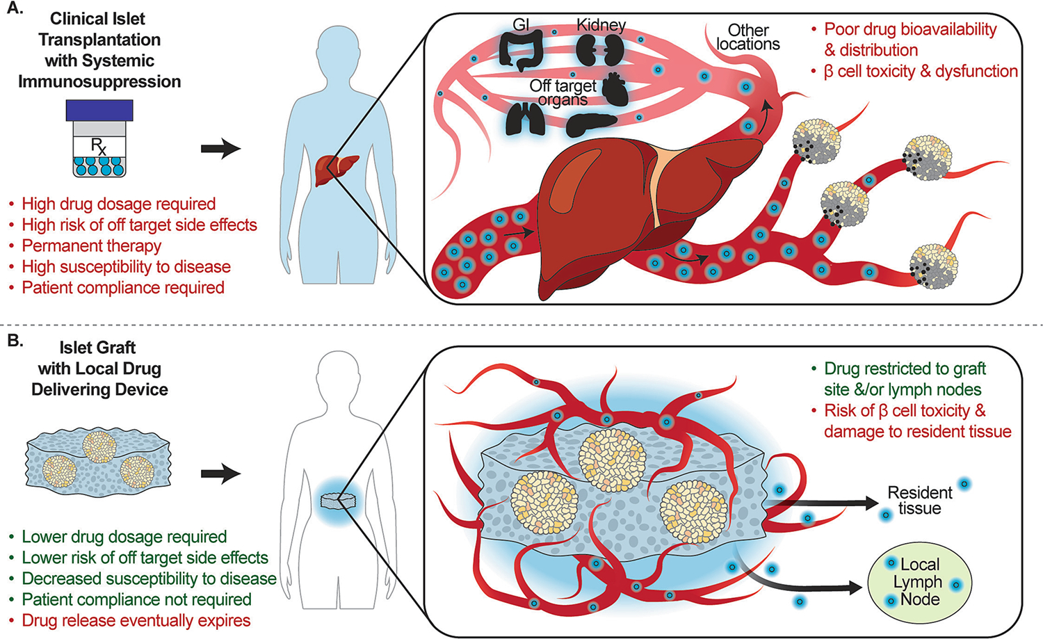
Advantages and disadvantages of systemic versus local delivery of immunosuppressants for the protection of allogeneic islet transplants. (A) Systemic immunosuppression, while potent for clinical islet transplantation, has many disadvantages, as noted. Upon administration, most agents have poor bioavailability and distribution in systemic circulation, leading to high clearance and off target effects to other organs. Because islets are transplanted in the liver, high drug dosages can also contribute to β cell toxicity before their concentrations are reduced after first-pass metabolism. (B) Local delivery of immunosuppressants can localize the agent directly to the transplant site. This provides distinct advantages, as noted. While patient compliance is not required with an implantable device, the amount of drug in the device is finite and eventually expires. Within the graft, the drug is typically restricted to the implant site and/or the local draining lymph nodes, thereby imparting a more targeted effect.
5. Local delivery of immunosuppressants
Local drug delivery involves administering the agent directly where it is needed, thereby avoiding or reducing broad and systemic impacts (Fig. 2B). Restricting immunosuppressants to the graft minimizes the required dosage to produce a suppressive effect and increases drug bioavailability and absorption at the site and within nearby draining lymph nodes [158,159]. Thus, this minimizes patient risk to off target effects [158]. Concerns with local delivery, however, are potential impacts to the local tissue and the transplanted islets. Given the susceptibility of islets to drug-induced dysregulation, careful dosage modulation and spatial control is needed to prevent deleterious effects [158]. In addition, the ultimate goal of local drug delivery for cellular transplantation is to deliver a cocktail of agents that achieves local immune tolerance. In other words, the ideal scenario would be to generate a localized environment with a lack of reactivity to donor alloantigens while retaining the capacity of the immune system to target other foreign antigens [50,157]. Some common strategies to deliver therapeutics locally include daily injections, infusion pumps, and drug-loaded biomaterials. Herein, we will discuss local drug delivery strategies using biomaterial platforms.
6. Biomaterial-based delivery of local immunosuppressants
Biomaterials have been used in numerous applications for the controlled, local delivery of therapeutic agents [160–164]. There are many engineering parameters that can be tailored to optimize biomaterial-based drug delivery. The drug type and its targeted cell population are two important things to consider. For example, entrapping a biologic agent inside an organic polymer is commonly challenged by their harsh fabrication techniques, e.g., intense heat, pH changes, and oxidative exposure, which can inactivate the biologic; thus, a water-based material fabricated using biologically compatible processes is more suitable [165]. Drug solubility also plays a role in the encapsulation efficiency within different materials [166], e.g., a hydrophobic drug typically encapsulates more readily and homogenously within a hydrophobic material [167]. Material selection and the agent’s mechanism of release from that material can affect the initial burst and duration of drug release [158,168,169]. Using a non-degradable polymer, for example, to encapsulate a small molecule drug may exhibit a more sustained release in comparison to the same drug encapsulated within a degradable material [166,169]. Additional considerations involve the desired target of the drug, including their targeted cell populations and their respective locations, as well as the location of the implant. For example, if the goal is to induce cytotoxicity of effector T cells at the graft site, a more localized release profile and material design would be selected. However, if the goal is to localize the agent within the lymph nodes that drain from the implant site, therapeutics encapsulated in a nanoparticle format may be more beneficial than in a macro or microscale device, as the former exhibits more favorable mobility characteristics [166]. Additionally, as noted earlier, some drugs may need highly tailored release rates to avoid toxicity, while others may impart a different mechanism of action when delivered locally [152,170,171]. The drug’s activity is another parameter. Some prodrugs require metabolic processing by the liver to exert their effect, thus they are likely not compatible with local delivery.
Despite this flexibility in modulating drug release based on the device parameters, there is typically a finite amount of drug in the biomaterial itself; thus, eventually the drug will run out and biomaterial replacement/refill or systemic drug administration will be necessary. Methods to circumvent this issue are being explored. One approach would be creating refillable reservoirs; this was demonstrated recently using a device designed for the local release of CTLA-4-Ig and anti-lymphocyte serum that could be refilled subcutaneously [172–174]. Another approach is to target the agents to the site. For example, one group engineered an azido-modified biomaterial that used click chemistry to target systemically administered tacrolimus, rapamycin, and MPA prodrugs to the material at the graft site [175]. Overall, there are several design parameters to define and consider for local drug delivery. To summarize these various approaches, we categorized these drug-delivery material designs according to their scale: macroscale (>500 μm); microscale (500 μm – 500 nm); or nanoscale (<500 nm) (Fig. 3).
Fig. 3.

Summary of current drug delivering devices engineered for the immunoprotection of cellular transplants categorized by scale (A) Macroscale drug delivery devices, such as porous scaffolds and hydrogels, are site retained and can house both the allogeneic islets and drug source in one location. (B) Microscale drug delivery devices hold the therapeutic agent only, can be injected with islets, and are retained at the graft site for immunoprotection of the allogeneic cells. (C) Nanoscale drug delivery devices are unique in that they can travel to local lymph nodes to directly inhibit immune rejection of the transplanted allogeneic islets. A subset of nanoparticles are liposomes and micelles, which are particularly useful for incorporating hydrophilic or hydrophobic agents, respectively. Without immune intervention via drug delivering devices, allogeneic islets can be rejected through non-specific, alloantigen specific, and recurring autoimmune rejection pathways.
6.1. Macroscale devices
Macroscale devices are commonly used for controlled drug delivery [160,163,175–177]. This scale has many advantages; they are site retained, serve a dual purpose in housing both drugs and the cell source in a three-dimensional space, have high control over spatial drug release, can hold a large amount of drug, and have flexibility in drug loading methods to tailor release (Fig. 3A). Because they remain at the implant location, macroscale devices confine most of the drug source in one location to orchestrate interactions between the host and transplanted cells [178,179]. This is ideal for therapeutics whose mechanism of action occurs at the graft site, such as anti-inflammatory agents. For example, one group fabricated a macroscale device loaded with ketoprofen, an anti-inflammatory agent, which downregulated pro-inflammatory markers and upregulated anti-inflammatory genes on macrophages in vitro [180]. Further, large scale platforms are capable of housing and delivering the most amount of drug, meaning they are most suitable for agents that need to be released over a long period of time (i.e., maintenance phase drugs). For example, one group encapsulated tacrolimus in their macroscale device for an allogeneic skin transplant and observed extended release with ~ 35% of the agent still left in the material on day 33 [163]. In addition to these advantages, a macroscale biomaterial provides larger control over the spatial release of the drugs, e.g. therapeutics could be loaded on the outside versus the core of the device. To demonstrate this, one group loaded anti-CD3 or anti-CD95 monoclonal antibodies into macroscale spheres (~2 mm diameter). These spheres were loaded into the core or periphery of different devices (6 – 10 mm) before loading islets into the remaining pores of the implant, resulting in reduced autoimmune responses to the transplanted islets [181]. Lastly, large scale devices have the flexibility of incorporating therapeutics into the device in different formats; agents have been loaded into macroscale devices monolithically, in discreet microbeads, coated on the device’s surface, and numerous other methods to tailor their drug release profiles. For example, one group coated a graphene-based macroscale device (~2 mm length) with dexamethasone to house and protect islets and mesenchymal stem cell units in vivo; however dexamethasone release lasted just 14 days [182]. Alternatively, our group loaded this same agent inside a macroscale scaffold device (~10 mm diameter), either as a monolithic or microbead format, where dexamethasone was integrated into microbeads and the beads were then loaded into the bulk scaffold material. The microbead scaffolds exhibited a more sustained, mono-phasic release profile compared to the bi-phasic release profile of the monolithic scaffolds, but all formats released the drug over 30 days [183]. Despite the many advantages of macroscale devices, a single implant drug source can create an outsized drug release gradient, which can impair surrounding tissue or be toxic to the housed cells due their proximity to the drug source. Therefore, the careful modulation of drug release kinetics from the macroscale devices is needed when using agents that can be problematic to β cell function and/or viability at high concentrations.
The most common formats of macroscale devices are porous scaffolds and hydrogels. While both are retained at the implant site, hydrogels are comprised of an aggregate network of polymer chains that can be injected or pre-gelled prior to implantation, whereas porous scaffolds are typically pre-formed 3D structures with macro or microscale porosity [169]. Hydrogels pose a unique advantage in that they are injectable, water-based, typically gel in situ, and can fill the implantation space. Injectability of a biomaterial is advantageous, as it is a less invasive implant procedure compared to a preformed device. It also allows for ease in the homogenous integration of cells and the drug. For instance, one group engineered a cyclosporine-loaded hydrogel that was injectable and slowed the proliferation and IL-2 production of T cells in vitro over 14 days [177]. Injectability may be a disadvantage, however, when delivering the drug-loaded material with cells, as the cells could be sheared and damaged during the injection procedure. Although not as common, hydrogels can be pre-fabricated in vitro, such as in the generation of a 3D-printed dexamethasone-eluting hydrogel [184]. Due to their high water retention capacity, hydrophilic agents are commonly used for hydrogel-based drug delivery [169]. Hydrogels are ideal for antibodies or cytokines, which are highly unstable, hydrophilic, and require gentler fabrication techniques. For example, Garcia’s team used a polyethylene glycol (PEG) hydrogel to successfully encapsulate an IL-2 analog and deliver FasL presenting microgels. With the drug and islet-loaded hydrogel, they achieved local release of the immunomodulatory proteins; however, significant extension of allograft survival was not observed when compared to non-drug controls [185]. Despite these advantages, the injectability of hydrogels limits their material selection and fabrication techniques. If injectable, the chosen material must be flowable to fit through the needle and crosslink using cell compatible methods. For example, one group engineered a silk hydrogel to house IL-4 and dexamethasone with islets, which could crosslink without an external stimulus at physiological-like conditions and preserve islet functionality in vitro [186,187]. Further, there can be initial drug loss depending on the time it takes for the material to gel at physiological conditions.
As opposed to hydrogels, scaffolds have enhanced versatility in material choices and fabrication techniques, as their 3D structures are developed before implantation [178]. Typically, scaffolds possess a rigid structure with micro- or macroscale pores where cells are added post-fabrication. Because porous scaffolds have a wider selection of material and fabrication choices, both hydrophilic and hydrophobic drugs can be encapsulated inside. For instance, Shea’s group delivered hydrophilic TGF-β1 and IL-33 cytokines along with islets in a poly(lactide-co-glycolide) (PLGA) porous scaffold (~3 mm diameter) and observed a decrease in pro-inflammatory cytokines and delayed allogeneic islet rejection in a diabetic mouse model [148,188]. Alternatively, our group loaded hydrophobic fingolimod into polydimethylsiloxane (PDMS), porous scaffolds, which exhibited controlled release of the agent over 30 days; however, fingolimod was detrimental to islet function in vivo [170]. Porous scaffolds can be fabricated in vitro using many different techniques, such as 3D-printing, salt leaching, or electrospinning. For these more intense fabrication processes, small molecule agents are typically better candidates for integration. One group mixed a small molecule agent, methotrexate, with polylactic acid, melted it, and 3D-printed the mixture using an extrusion-based technique. This fabrication method exposed the drug to high temperatures (220 °C) in order to 3D-print the drug-loaded material into the desired scaffold geometry, but still retained the drug’s anti-tumor activity [160]. Alternatively, drugs could be loaded within reservoirs for multi-drug release. For example, Grattoni’s group 3D-printed a macro-device, termed NICHE, which housed islets in a central core surrounded by a refillable drug reservoir. After pre-vascularizing the core for six weeks and filling the drug reservoir with an immunosuppressant cocktail (CTLA-4-Ig, anti-lymphocyte serum, ATG, and/or anti-CD154) three days before allogeneic islet transplantation, normoglycemia was achieved long-term (>150 days) in an immunocompetent, diabetic rat model; short-term islet engraftment was also achieved (17 days) in a nonhuman primate model [173,174]. Despite this flexibility in using different fabrication methods, optimizing a method to load the agents into the scaffold material to evoke a particular release profile can be challenging.
6.2. Microscale devices
Microparticles, small spheres typically about 500 nm – 500 μm in diameter that are loaded with a drug, provide an alternative format for controlled drug delivery [189]. The advantages of the microparticle-based systems are site retention, injectability, discreet spatial depots, increased surface to volume ratio, material choice flexibility, and ease in modulation of multiple drug combinations (Fig. 3B) [189]. Microparticles can be fabricated using numerous material types and house a various portfolio of drugs. For example, they can encapsulate lipophilic drugs, such as rapamycin and tacrolimus, as well as hydrophilic drugs, such as proteins and peptides. PLGA is a common material used for local drug delivery via microspheres, with examples of delivery of hydrophilic TGF-β1 or hydrophobic dexamethasone for islet transplantation [144,190]. Although not applied for islet transplantation, one team incorporated hydrophilic oligonucleotides, complementary to CD40, CD80, and CD86, into polyvinylpyrrolidone and PEG co-polymer microspheres (~2.5 μm) for the immunomodulation of autoimmune pathways; this approach could be translated to protect islet transplants [191]. Another desired feature of microparticles is their increased surface to volume ratio when compared with macroscale devices. That is, they can release a large amount of drug initially upon transplantation; however, this prompt burst release and large surface to volume ratio could be considered disadvantageous as the drug release will be exhausted more quickly than a macroscale device [192]. For example, one group engineered tacrolimus-loaded PLGA microspheres (~5 μm) and after co-administration with rat islets into a diabetic mouse model, these microspheres successfully impeded T cell activation and prolonged islet survival in vivo compared to non-drug and soluble drug controls. Yet, in vitro drug release studies revealed that 80% of the total amount of encapsulated drug was released within the first 25 days [193]. Leveraging their injectability feature, microparticles can also be easily combined with cells to create a graft with spatially discreet drug sources. To demonstrate this, one group encapsulated rapamycin, inside polycaprolactone and PLGA microparticles (~100 μm) and co-injected them with allogeneic islets in the anterior chamber of the eye. These discreet rapamycin sources protected the islets from rejection over the 30-day study at a much lower dosage than systemic delivery [194]. Further, having spatially discreet drug sources opens the opportunity to deliver multiple microparticle capsules containing different agents. While not specific to islet transplantation, one group fabricated phagocytosable (~2 μm) and non-phagocytosable (~30 μm) PLGA microparticles, which individually encapsulated either rapamycin, all-trans retinoic acid (inhibits anti-inflammatory cytokine production in APCs), TGF-β1, or IL-10. In different combinations, these microparticles reduced co-stimulatory molecule expression, such as MHC class 2, CD80, and CD86, and suppressed dendritic cell-mediated proliferation of allogeneic T cells compared to single type microparticles [195]. This platform could be translated to allogeneic islet transplantation in the future to minimize T cell proliferation and rejection. Another approach used a combination of three different PLGA-based drug delivering microparticles to suppress allograft limb rejection in a murine model. The study found that the triple combination of rapamycin, IL-2, and TGF-β releasing particles, injected near the implant site, produced the most protective effect [196]. Because microparticles are site retained, like macroscale devices, agents that target cells at the graft site are also suitable for this format. For example, Pepper’s group delivered dexamethasone-loaded PLGA microparticles (~50 μm), along with systemic CTLA-4-Ig, and observed increased conversion of CD4+ T cells to IL-10 secreting Treg cells, thereby inhibiting CD8+ T cell proliferation. This local dexamethasone delivery system also decreased pro-inflammatory cytokine production within the graft and improved immune tolerance of transplanted allogeneic islets [197]. Additionally, another group encapsulated curcumin, an antioxidant and anti-inflammatory agent, inside PLGA microspheres (3 – 5 μm) [198,199]. While not assessed in vivo, these microspheres did mitigate islet apoptosis and oxidative stress-induced cell death in hypoxic in vitro conditions [199]. Despite these advantages, the scale of microparticles limits their capacity to co-house drugs and cells. Additionally, in comparison to macroscale devices, microparticle retrieval is more challenging, due to their size and numerosity.
6.3. Nanoscale devices
Nanoscale drug delivery devices, such as liposomes, micelles, and nanoparticles, provide a unique way for immunosuppressants to be transported to their intended target. Advantages of nanoscale devices include implantation flexibility, high targeting specificity, and non-site retaining features (Fig. 3C). Nanoscale devices are flexible in that they can be integrated with cells into the implant site, implanted after the cells have already engrafted, or injected at multiple points and/or locations over time. For example, one group fabricated rapamycin-loaded PEG-polypropylene sulfide polymersomes (~100 nm) and injected them subcutaneously in mouse models pre- and post-allogeneic islet transplantation into the portal vein [152]. Typical oral administration of rapamycin inhibits T cell activation and can have detrimental effects to β cells [90,200]. Delivering these polymersomes in a distal location and at different time points enabled rapamycin to drain into brachial lymph nodes and be uptaken by APCs to induce CD4+ T cell anergy, essentially reengineering the drug’s mechanism of action [152]. This implantation flexibility was due to the fact that nanoparticles were not site retained and could drain from the implant location to the local lymph node(s). Thus, nanoscale materials are ideal for agents whose mechanism of action primarily occurs in the lymph nodes, such as fingolimod. In addition to these advantages, nanoscale devices have higher targeting specificity when compared with macro- and microscale devices. Nanoparticle size is a key parameter in driving particle mobility [157]. Larger nanoparticles, i.e., greater than 200 nm, are typically phagocytosed by graft tissue resident macrophages or dendritic cells, whereas smaller nanoparticles, i.e., between 20 and 200 nm, usually travel into neighboring lymph nodes [201,202]. For example, one group encapsulated MPA inside PLGA nanoparticles (~170 nm) and found that, upon intraperitoneal injection in a mouse skin allograft model, most of the nanoparticles trafficked to the spleen by 6 h [203]. Another group used slightly larger nanoparticles (~190 nm) made from a PLGA and PEG copolymer to encapsulate genistein, a naturally occurring anti-inflammatory agent [204,205]. The goal of the approach was to target delivery of the anti-inflammatory agent to the local islet capillary endothelial cells, which was shown to inhibit leukocyte adhesion in vitro [204]. Additional studies are needed to validate this targeting in vivo. This phenomenon of nanoparticle phagocytosis opens the opportunity for drugs to be contained in the particles and only execute their mechanism of action once they have been phagocytosed. For example, one group engineered PEG-PLGA nanoparticles (~138 nm) containing an auto-antigen peptide, a CRISPR-Cas9 plasmid, and three guide RNAs to inhibit dendritic cell co-stimulatory molecules (CD80, CD86, and CD40) [206]. While the nanoparticles were not used for an islet transplantation, the authors used a diabetic mouse model to show that, upon intravenous infusion, these nanoparticles were phagocytosed by dendritic cells and could deliver the three RNAs intracellularly, resulting in auto-antigen specific Treg expansion and prevention of islet autoimmune rejection in the pancreas [206].
Besides being convenient for implantation, targeting specific cell populations, and the capacity for distal mobility, certain types of nanocarriers are more efficient at encapsulating different agents. Liposomes and micelles are nanocarriers composed of lipids [76]. Micelles are advantageous for encapsulating hydrophobic drug molecules because they are comprised of a lipid monolayer with a hydrophilic surface and a hydrophobic core. Alternatively, liposomes utilize a lipid bilayer with a hydrophilic surface and core [76]. For example, one group successfully encapsulated hydrophobic tacrolimus inside pol(ε-caprolactone) + PEG + pol(ε-caprolactone) micelles (~20 nm) [207]. Alternatively, another group used liposomes (~120 – 160 nm) to encapsulate the hydrophilic agent, clodronate, and measured increased macrophage apoptosis and lowered pro-inflammatory cytokine concentrations in rheumatoid arthritis patients [208]. Both liposomal-based approaches could be beneficial for protecting islets post-transplantation. While simple to formulate and generally nontoxic, liposomes and micelles can have short circulation half-lives due to rapid uptake via monocytes and macrophages [209,210].
Despite their many advantages, nanoscale drug delivery is not an ideal format when seeking to target cells in the implant space, as they can migrate from the delivery site. As such, their use for delivering anti-inflammatory agents may not be favorable. Although the ability to travel can be considered advantageous, it does introduce the risk of traveling to unwanted locations, such as the blood brain barrier or lungs, before releasing their housed agents [153,211,212]. As size dictates their ability to travel, unwanted protein and cell absorption to the nanoscale device surface can alter their function and mobility [153]. Lastly, the nanoscale results in the least amount of total drug delivered, when compared to macro- and microscale devices, which is not ideal for agents that require extended release.
6.4. Islet encapsulation with local immunosuppression
A common method employed for suppressing immune attack of pancreatic islets is polymeric encapsulation. In this approach, islets are enveloped within a permselective material, which serves to support nutrient and insulin exchange but block direct cell contact. While polymeric encapsulation can be an effective approach for suppressing direct cell mediated attack, complementary local or systemic immunosuppression is necessary to minimize indirect mechanisms of rejection. This is because encapsulation only prevents direct interactions and killing between host CD8+ T cells and the donor cells, whereas cytokines and antibodies secreted from donor cells can still impart rejection and fibrotic responses via indirect T cell activation from host APC presentation of shed donor antigens [213]. Knowing this, one group encapsulated xenogeneic islets inside alginate microparticles with PEG plus rapamycin chains bound to the surface to suppress local immune activation and deleterious foreign body responses [214]. While the authors did observe a significant decrease in fibrotic cell infiltration, there was not a significant difference in islet efficacy within rapamycin or non-rapamycin alginate microbeads in chemically induced diabetic mice [214]. Another group encapsulated either curcumin or dexamethasone along with xenogeneic islets inside alginate capsules before transplanting in chemically induced diabetic mice [215]. While the curcumin capsules decreased fibrotic capsule formation compared to non-drug and dexamethasone capsules, hyperglycemia was observed in all groups after 30 days [215]. To further limit the interactions between transplanted islets and the host’s immune system, immunoisolation devices can be useful; however, the cell cargo can become hypoxic and exacerbation of fibrotic capsule formation is still a concern [216]. Although islet encapsulation could be additive to local drug delivering systems, it has lingering issues such as an elevated risk for foreign body response, insufficient nutritional delivery, and poor islet functionality that hinders its clinical translation [217].
7. Methods and challenges in local drug delivery
7.1. Methods for assessing efficacy of local immunosuppressant delivering devices
While the in vitro evaluation of drug kinetics and cellular impacts are key assessments needed prior to translation to preclinical models, in vivo trials for local drug delivering devices are critical in determining effectiveness and catching unintended effects. To fully capture both efficacy and broad impacts, there is a portfolio of laboratory techniques that are useful. One important screen is capturing the in vivo kinetics of drug release and delivery, as the implant environment can significantly change these parameters due to alterations in the microenvironment, drug drainage/processing, and material-host interactions (e.g., foreign body responses, altered degradation). As such, it is useful to quantify local release and broad systemic impacts. For this, serum and local tissues can be temporally collected, with levels compared to therapeutic systemic doses. For example, one group collected the nearby tissue, homogenized it, and quantified the amount of drug inside using high-performance liquid chromatography (HPLC) [197].
Post-transplantation explant analysis is key for understanding the device and drug effects on the host. Imaging techniques, such as immunohistochemistry and digital spatial profiling, are suitable for visualizing the spatial distribution of immune cells within the graft. Standard paraffin embedded explant analysis was used in one application to evaluate local regulatory effects, with staining for CD4+, CD25+, and FoxP3+ T cells implicating elevated Treg cells within their graft [197]. Another group stained the implant for F4/80, NK1.1, and CD8+ cells to visualize macrophages, NK cells, and cytotoxic T cells, respectively [188]. A newer technology, digital spatial profiling, allows for immune profiling at the protein or RNA level on formalin-fixed, paraffin embedded samples [218,219]. The technology relies on antibody or RNA staining probes that are tagged with oligonucleotides to spatially resolve proteins or RNA of interest in the sample [220]. This method can quantify immune cell types at distinct spatial locations in the explanted graft in comparison to normal tissue, which could be particularly insightful when interrogating the spatial effects of a drug on immune cell populations in the microenvironment. Another way to analyze intragraft pro-inflammatory cytokines or immune cell populations could be homogenizing the tissue explant and quantifying cytokines using an ELISA or staining and quantifying immune cell populations using flow cytometry [197]. Overall, these techniques are useful for spatially analyzing immune cell populations to determine how well the local drug delivering device suppresses its intended cell targets.
Besides the graft itself, neighboring tissues, such as the spleen, blood, and local draining lymph nodes, could also provide valuable information about the immunomodulatory effects of the localized drug delivery approach. One valuable tool could be a pharmacodynamic in vitro assay to measure immune cell response to immunosuppressive drugs [221]. The assay involves purifying and activating desired immune cells from blood or lymphoid organs, embedding them in a hydrogel mixture, and exposing them to a concentration gradient of an immunosuppressant of interest [221]. This could be informative when assessing, for example, T cell proliferation after exposure to a tacrolimus delivering device in vivo. An in vitro pharmacodynamic assay could also be helpful before transplantation, during the device optimization period, when determining an appropriate drug loading concentration. Another tool for assessing local immune tolerance of the graft is an in vitro mixed lymphocyte reaction (MLR). An MLR is an in vitro co-culture of donor lymphocytes (stimulators) with recipient lymphocytes (responders) to assess the proliferation and/or effector phenotype of the responder cells. For instance, one group completed an islet allograft study in C57BL/6 mice and used MLR to quantify immune tolerance to islets with and without FasL on their surface [222]. Lymphocytes from the spleen and draining lymph nodes of the recipient mice were harvested and assessed for CD4+ and CD8+ T cell proliferation in response to donor and third-party stimulators. Third-party stimulators were used as a positive control, while the donor stimulators assessed systemic (spleen) or local (draining lymph nodes) immune competency. This assay captured information about how the FasL-expressing islets established local immune tolerance without compromising the mice’s systemic immune responsiveness [222]. As a more robust in vivo model, skin graft tests or transplanting islets in distal locations using donor or third party tissues/cells can assess global versus local immune tolerance in the recipient [223]. Of note, screening in both small and larger preclinical models is needed, as pharmacokinetics, drug cross reactivity, and toxicity effects can alter as you progress from murine to more clinically similar models. These challenges further support the need for more accurate benchtop methods that can mimic complex human-centric pharmacokinetic challenges.
7.2. Global challenges
Despite the many different local drug delivery devices being developed for islet transplantation, there are still many remaining global challenges. One common problem is the burst release of the housed therapeutic agent upon implantation. In biomaterials, this is typically caused by factors such as an initial gradient between a high drug concentration in the material and the surrounding tissue, fabrication techniques where a large amount of drug is bound close to the surface, and, in the case of injectable hydrogels, high loss of drug before the material can gel in vivo [224,225]. This is not only deleterious for durable drug release kinetics, but the sensitivity of islets and host cells to immunosuppressive agents can alter positive islet engraftment by either impairing insulin sensitivity to glucose and/or dampening healthy host cell infiltration and vascularization [90,170,226–228]. This issue is of particular concern if the device and islets are co-localized. Numerous immunosuppressive drugs, e.g., rapamycin and tacrolimus, can alter insulin secretion dynamics [90,156,228]. For instance, one group showed that tacrolimus suppressed glucose-stimulated insulin secretion of isolated rat islets by ~ 32% at a therapeutic concentration [228]. Another group showed that systemic administration of tacrolimus and rapamycin impaired insulin secretion in mouse models transplanted with human islets [156]. Efforts to minimize burst release from local drug delivering devices involve incorporating crystalized drugs into the platform [229]. Furthermore, immunosuppressants can alter host migration to the site and impair vascularization. Rapid vascularization of islets is crucial for successful engraftment, as islets secrete hormones directly into the bloodstream. Selecting therapeutics that hinder or inhibit vascularization of the graft, such as dexamethasone or rapamycin, may delay graft function or ultimately debilitate the islets entirely [88,89,183]. For example, one group showed that systemic rapamycin inhibited vascularization of implanted hernia meshes in hamster models compared to cyclosporine [89]. Engineering a device that could minimize burst release without inhibiting vascularization and still maintain therapeutic efficacy for inducing immune tolerance would help local drug delivering devices come closer to clinical translation.
8. Conclusions
In conclusion, islet transplantation for T1DM is largely hindered by the need for permanent and systemic immunosuppression. Without immunosuppression, transplanted islets are rejected through a combination of non-specific, alloantigen, and autoantigen specific immune rejection pathways. Combinatory therapeutic approaches using agents with distinct mechanisms of action are successfully employed for CIT; however, this systemic administration evokes negative side effects and issues. The local delivery of these agents using drug delivering biomaterials can alleviate these issues and protect islets from immune rejection. Current biomaterials used for local drug delivery and islet transplantation range from nano- to macroscales. Future work should focus on leveraging engineering design parameters to customize biomaterial release platforms to release a portfolio of drugs that can modulate the multitude of immune responses to allogeneic islet transplants. In addition, emerging novel cell sources, such as stem cell derived β cells and engineered tissues, can be combined with these approaches to further improve efficacy and accessibility.
Acknowledgments
The authors are grateful for funding from the US National Institutes of Health grants DK126413 and DK122638, as well as from JDRF grants 3-SRA-2021-1033-S-B and 2-SRA-2021-1024-S-B. TR Lansberry is supported by the NIH NIDDK T32 Interdisciplinary Graduate Program in Type 1 Diabetes and Biomedical Engineering training program (DK108736).
Footnotes
Declaration of competing interest
The authors declare that they have no known competing financial interests or personal relationships that could have appeared to influence the work reported in this paper.
Data availability
No data was used for the research described in the article.
References
- [1].Atkinson MA, Eisenbarth GS, Michels AW, Type 1 diabetes, Lancet 383 (2014) 69–82, 10.1016/S0140-6736(13)60591-7. [DOI] [PMC free article] [PubMed] [Google Scholar]
- [2].Leslie RD, Evans-Molina C, Freund-Brown J, Buzzetti R, Dabelea D, Gillespie KM, Goland R, Jones AG, Kacher M, Phillips LS, Rolandsson O, Wardian JL, Dunne JL, Adult-onset type 1 diabetes: current understanding and challenges, Diabetes Care. 44 (2021) 2449–2456, 10.2337/dc21-0770. [DOI] [PMC free article] [PubMed] [Google Scholar]
- [3].Yoon J-W, Jun H-S, Autoimmune Destruction of pancreatic β Cells, Am. J. Ther. 12 (2005) 580–591, 10.1097/01.mjt.0000178767.67857.63. [DOI] [PubMed] [Google Scholar]
- [4].Roep BO, Thomaidou S, van Tienhoven R, Zaldumbide A, Type 1 diabetes mellitus as a disease of the β-cell (do not blame the immune system?), Nat. Rev. Endocrinol. 17 (2021) 150–161, 10.1038/s41574-020-00443-4. [DOI] [PMC free article] [PubMed] [Google Scholar]
- [5].Bresson D, Von Herrath M, Mechanisms underlying type I diabetes, Drug Discov. Today Dis. Mech. 1 (2004) 321–327, 10.1016/j.ddmec.2004.11.015. [DOI] [Google Scholar]
- [6].Pathak V, Pathak NM, O’Neill CL, Guduric-Fuchs J, Medina RJ, Therapies for type 1 diabetes: current scenario and future perspectives, Clin. Med. Insights Endocrinol. Diabetes. 12 (2019), 10.1177/1179551419844521. [DOI] [PMC free article] [PubMed] [Google Scholar]
- [7].Burrack AL, Martinov T, Fife BT, T cell-mediated beta cell destruction: autoimmunity and alloimmunity in the context of type 1 diabetes, Front. Endocrinol. (lausanne) 8 (2017) 1–15, 10.3389/fendo.2017.00343. [DOI] [PMC free article] [PubMed] [Google Scholar]
- [8].Quizon MJ, García AJ, Engineering β Cell replacement therapies for type 1 diabetes: biomaterial advances and considerations for macroscale constructs, Annu. Rev. Pathol. Mech. Dis. 17 (2021) 485–513, 10.1146/annurev-pathol-042320-094846. [DOI] [PubMed] [Google Scholar]
- [9].Rickels MR, Paul Robertson R, Pancreatic islet transplantation in humans: Recent progress and future directions, Endocr. Rev. 40 (2019) 631–668, 10.1210/er.2018-00154. [DOI] [PMC free article] [PubMed] [Google Scholar]
- [10].Anazawa T, Okajima H, Masui T, Uemoto S, Current state and future evolution of pancreatic islet transplantation, Ann. Gastroenterol. Surg. 3 (2019) 34–42, 10.1002/ags3.12214. [DOI] [PMC free article] [PubMed] [Google Scholar]
- [11].Frei AW, Stabler CL, Engineering immune responses to allografts, in: Kirschner CM, Brennan AB (Eds.), Bio-Inspired Mater. Biomed, Eng., John Wiley & Sons Ltd, 2014, pp. 327–355, 10.1002/9781118843499.ch17. [DOI] [Google Scholar]
- [12].Shapiro AMJ, Pokrywczynska M, Ricordi C, Clinical pancreatic islet transplantation, Nat. Rev. Endocrinol. 13 (2017) 268–277, 10.1038/nrendo.2016.178. [DOI] [PubMed] [Google Scholar]
- [13].Qin T, Smink AM, de Vos P, Enhancing longevity of immunoisolated pancreatic islet grafts by modifying both the intracapsular and extracapsular environment, Acta Biomater. (2023), 10.1016/j.actbio.2023.06.038. [DOI] [PubMed] [Google Scholar]
- [14].Gamble A, Pepper AR, Bruni A, Shapiro AMJ, The journey of islet cell transplantation and future development, Islets. 10 (2018) 80–94, 10.1080/19382014.2018.1428511. [DOI] [PMC free article] [PubMed] [Google Scholar]
- [15].Forbes S, Flatt AJ, Bennett D, Crookston R, Pimkova M, Birtles L, Pernet A, Wood RC, Burling K, Barker P, Counter C, Lumb A, Choudhary P, Rutter MK, Rosenthal M, Sutherland A, Casey J, Johnson P, Shaw JAM, The impact of islet mass, number of transplants, and time between transplants on graft function in a national islet transplant program, Am. J. Transplant. 22 (2022) 154–164, 10.1111/ajt.16785. [DOI] [PMC free article] [PubMed] [Google Scholar]
- [16].Hering BJ, Ballou CM, Bellin MD, Payne EH, Kandeel F, Witkowski P, Alejandro R, Rickels MR, Barton FB, Factors associated with favourable 5 year outcomes in islet transplant alone recipients with type 1 diabetes complicated by severe hypoglycaemia in the collaborative islet transplant registry, Diabetologia 66 (2023) 163–173, 10.1007/s00125-022-05804-4. [DOI] [PMC free article] [PubMed] [Google Scholar]
- [17].Matsumoto S, Clinical allogeneic and autologous islet cell transplantation: Update, Diabetes Metab. J. 35 (2011) 199–206, 10.4093/dmj.2011.35.3.199. [DOI] [PMC free article] [PubMed] [Google Scholar]
- [18].Ramzy A, Thompson DM, Ward-Hartstonge KA, Ivison S, Cook L, Garcia RV, Loyal J, Kim PTW, Warnock GL, Levings MK, Kieffer TJ, Implanted pluripotent stem-cell-derived pancreatic endoderm cells secrete glucose-responsive C-peptide in patients with type 1 diabetes, Cell Stem Cell. 28 (2021) 2047–2061.e5, 10.1016/j.stem.2021.10.003. [DOI] [PubMed] [Google Scholar]
- [19].Henry RR, Pettus J, Wilensky J, Shapiro AMJ, Senior PA, Roep B, Wang R, Kroon EJ, Scott M, D’Amour K, Foyt HL, Initial clinical evaluation of VC-01TM Combination Product—A stem cell-derived islet replacement for type 1 diabetes (T1D), Diabetes. 67 (2018), 10.2337/db18-138-OR. [DOI] [Google Scholar]
- [20].Shapiro AMJ, Thompson D, Donner TW, Bellin MD, Hsueh W, Pettus J, Wilensky J, Daniels M, Wang RM, Brandon EP, Jaiman MS, Kroon EJ, D’Amour KA, Foyt HL, Insulin expression and C-peptide in type 1 diabetes subjects implanted with stem cell-derived pancreatic endoderm cells in an encapsulation device, Cell Reports Med. 2 (2021) 100466, 10.1016/j.xcrm.2021.100466. [DOI] [PMC free article] [PubMed] [Google Scholar]
- [21].Shapiro AMJ, Verhoeff K, A spectacular year for islet and stem cell transplantation, Nat. Rev. Endocrinol. 19 (2023) 68–69, 10.1038/s41574-022-00790-4. [DOI] [PubMed] [Google Scholar]
- [22].Dolgin E, Diabetes cell therapies take evasive action, Nat. Biotechnol. 40 (2022) 291–295, 10.1038/s41587-022-01246-w. [DOI] [PubMed] [Google Scholar]
- [23].Vertex Presents Positive VX-880 Results From Ongoing Phase 1/2 Study in Type 1 Diabetes at the American Diabetes Association 83rd Scientific Sessions | Vertex Pharmaceuticals, Boston, 2023. https://investors.vrtx.com/news-releases/news-release-details/vertex-presents-positive-vx-880-results-ongoing-phase-12-study. [Google Scholar]
- [24].Hentze H, Soong PL, Wang ST, Phillips BW, Putti TC, Dunn NR, Teratoma formation by human embryonic stem cells: Evaluation of essential parameters for future safety studies, Stem Cell Res. 2 (2009) 198–210, 10.1016/j.scr.2009.02.002. [DOI] [PubMed] [Google Scholar]
- [25].Gutierrez-Aranda I, Ramos-Mejia V, Bueno C, Munoz-Lopez M, Real PJ, Mácia A, Sanchez L, Ligero G, Garcia-Parez JL, Menendez P, Human induced pluripotent stem cells develop teratoma more efficiently and faster than human embryonic stem cells regardless the site of injection, Stem Cells 28 (2010) 1568–1570, 10.1002/stem.471. [DOI] [PMC free article] [PubMed] [Google Scholar]
- [26].Aldahmash A, Atteya M, Elsafadi M, Al-Nbaheen M, Al-Mubarak HA, Vishnubalaji R, Al-Roalle A, Al-Harbi S, Manikandan M, Matthaei KI, Mahmood A, Teratoma formation in immunocompetent mice after syngeneic and allogeneic implantation of germline capable mouse embryonic stem cells, asian pacific, J Cancer Prev. 14 (2013) 5705–5711, 10.7314/APJCP.2013.14.10.5705. [DOI] [PubMed] [Google Scholar]
- [27].Bedel A, Beliveau F, Lamrissi-Garcia I, Rousseau B, Moranvillier I, Rucheton B, Guyonnet-Dupérat V, Cardinaud B, de Verneuil H, Moreau-Gaudry F, Dabernat S, Preventing pluripotent cell teratoma in regenerative medicine applied to hematology disorders, Stem. Cells Transl. Med. 6 (2017) 382–393, 10.5966/sctm.2016-0201. [DOI] [PMC free article] [PubMed] [Google Scholar]
- [28].Lanza R, Russell DW, Nagy A, Engineering universal cells that evade immune detection, Nat. Rev. Immunol. 19 (2019) 723–733, 10.1038/s41577-019-0200-1. [DOI] [PubMed] [Google Scholar]
- [29].Hayat SMG, Bianconi V, Pirro M, Jaafari MR, Hatamipour M, Sahebkar A, CD47: role in the immune system and application to cancer therapy, Cell. Oncol. 43 (2020) 19–30, 10.1007/s13402-019-00469-5. [DOI] [PMC free article] [PubMed] [Google Scholar]
- [30].Deuse T, Hu X, Gravina A, Wang D, Tediashvili G, De C, Thayer WO, Wahl A, Garcia JV, Reichenspurner H, Davis MM, Lanier LL, Schrepfer S, Hypoimmunogenic derivatives of induced pluripotent stem cells evade immune rejection in fully immunocompetent allogeneic recipients, Nat. Biotechnol. 37 (2019) 252–258, 10.1038/s41587-019-0016-3. [DOI] [PMC free article] [PubMed] [Google Scholar]
- [31].Hu X, Gattis C, Olroyd AG, Friera AM, White K, Young C, Basco R, Lamba M, Wells F, Ankala R, Dowdle WE, Lin A, Egenberger K, Rukstalis JM, Millman JR, Connolly AJ, Deuse T, Schrepfer S, Human hypoimmune primary pancreatic islets avoid rejection and autoimmunity and alleviate diabetes in allogeneic humanized mice, Sci. Transl. Med. 15 (2023) 1–17, 10.1126/scitranslmed.adg5794. [DOI] [PubMed] [Google Scholar]
- [32].Meissner TB, Schulze HS, Dale SM, Immune editing: overcoming immune barriers in stem cell transplantation, Curr. Stem Cell Reports. 8 (2022) 206–218, 10.1007/s40778-022-00221-0. [DOI] [PMC free article] [PubMed] [Google Scholar]
- [33].Roep BO, Peakman M, Antigen targets of type 1 diabetes autoimmunity, Cold Spring Harb. Perspect. Med. 2 (2012) a007781–a, 10.1101/cshperspect.a007781. [DOI] [PMC free article] [PubMed] [Google Scholar]
- [34].Kash S, Condie B, Baekkeskov S, Glutamate decarboxylase and GABA in pancreatic islets: lessons from knock-out mice, Horm. Metab. Res. 31 (1999) 340–344, 10.1055/s-2007-978750. [DOI] [PubMed] [Google Scholar]
- [35].LaRosa DF, Rahman AH, Turka LA, The innate immune system in allograft rejection and tolerance, J. Immunol. 178 (2007) 7503–7509, 10.4049/jimmunol.178.12.7503. [DOI] [PMC free article] [PubMed] [Google Scholar]
- [36].Marfil-Garza BA, Shapiro AMJ, Kin T, Clinical islet transplantation: current progress and new frontiers, J. Hepatobiliary. Pancreat. Sci. 28 (2021) 243–254, 10.1002/jhbp.891. [DOI] [PubMed] [Google Scholar]
- [37].Johansson H, Lukinius A, Moberg L, Lundgren T, Berne C, Foss A, Felldin M, Källen R, Salmela K, Tibell A, Tufveson G, Ekdahl KN, Elgue G, Korsgren O, Nilsson B, Tissue factor produced by the endocrine cells of the islets of langerhans is associated with a negative outcome of clinical islet transplantation, Diabetes 54 (2005) 1755–1762, 10.2337/diabetes.54.6.1755. [DOI] [PubMed] [Google Scholar]
- [38].Moberg L, Johansson H, Lukinius A, Berne C, Foss A, Källen R, Østraat Ø, Salmela K, Tibell A, Tufveson G, Elgue G, Ekdahl KN, Korsgren O, Nilsson B, Production of tissue factor by pancreatic islet cells as a trigger of detrimental thrombotic reactions in clinical islet transplantation, Lancet. 360 (2002) 2039–2045, 10.1016/S0140-6736(02)12020-4. [DOI] [PubMed] [Google Scholar]
- [39].Peppelenbosch MP, Spek CA, Type I diabetes: a role for tissue factor in pancreatic islet transplantation? Lancet. 360 (2002) 1999–2000, 10.1016/S0140-6736(02)12033-2. [DOI] [PubMed] [Google Scholar]
- [40].Foster GA, García AJ, Bio-synthetic materials for immunomodulation of islet transplants, Adv. Drug Deliv. Rev. 114 (2017) 266–271, 10.1016/j.addr.2017.05.012. [DOI] [PMC free article] [PubMed] [Google Scholar]
- [41].Mantovani A, Cassatella MA, Costantini C, Jaillon S, Neutrophils in the activation and regulation of innate and adaptive immunity, Nat. Rev. Immunol. 11 (2011) 519–531, 10.1038/nri3024. [DOI] [PubMed] [Google Scholar]
- [42].Sackett SD, Kaplan SJ, Mitchell SA, Brown ME, Burrack AL, Grey S, Huangfu D, Odorico J, Genetic engineering of immune evasive stem cell-derived islets, Transpl. Int. 35 (2022) 1–11, 10.3389/ti.2022.10817. [DOI] [PMC free article] [PubMed] [Google Scholar]
- [43].Benichou G, Yamada Y, Aoyama A, Madsen JC, Natural killer cells in rejection and tolerance of solid organ allografts, Curr. Opin. Organ Transplant. 16 (2011) 47–53, 10.1097/MOT.0b013e32834254cf. [DOI] [PMC free article] [PubMed] [Google Scholar]
- [44].Sojka DK, Plougastel-Douglas B, Yang L, Pak-Wittel MA, Artyomov MN, Ivanova Y, Zhong C, Chase JM, Rothman PB, Yu J, Riley JK, Zhu J, Tian Z, Yokoyama WM, Tissue-resident natural killer (NK) cells are cell lineages distinct from thymic and conventional splenic NK cells, Elife. 3 (2014) 1–21, 10.7554/eLife.01659. [DOI] [PMC free article] [PubMed] [Google Scholar]
- [45].Peng H, Sun R, Liver-resident NK cells and their potential functions, Cell. Mol. Immunol. 14 (2017) 890–894, 10.1038/cmi.2017.72. [DOI] [PMC free article] [PubMed] [Google Scholar]
- [46].Martin KE, García AJ, Macrophage phenotypes in tissue repair and the foreign body response: Implications for biomaterial-based regenerative medicine strategies, Acta Biomater. 133 (2021) 4–16, 10.1016/j.actbio.2021.03.038. [DOI] [PMC free article] [PubMed] [Google Scholar]
- [47].Biarnés M, Montolio M, Nacher V, Raurell M, Soler J, Montanya E, β-Cell death and mass in syngeneically transplanted islets exposed to short- and long-term hyperglycemia, Diabetes. 51 (2002) 66–72, 10.2337/diabetes.51.1.66. [DOI] [PubMed] [Google Scholar]
- [48].Siu JHY, Surendrakumar V, Richards JA, Pettigrew GJ, T cell allorecognition pathways in solid organ transplantation, Front. Immunol. 9 (2018) 1–14, 10.3389/fimmu.2018.02548. [DOI] [PMC free article] [PubMed] [Google Scholar]
- [49].Al-Lamki RS, Bradley JR, Pober JS, Endothelial cells in allograft rejection, Transplantation. 86 (2008) 1340–1348, 10.1097/TP.0b013e3181891d8b. [DOI] [PMC free article] [PubMed] [Google Scholar]
- [50].Wang X, Brown NK, Wang B, Shariati K, Wang K, Fuchs S, Melero-Martin JM, Ma M, Local immunomodulatory strategies to prevent allo-rejection in transplantation of insulin-producing cells, Adv. Sci. 8 (2021) 2003708, 10.1002/advs.202003708. [DOI] [PMC free article] [PubMed] [Google Scholar]
- [51].Buchholz VR, Schumacher TNM, Busch DH, T Cell Fate at the Single-Cell Level, Annu. Rev. Immunol. 34 (2016) 65–92, 10.1146/annurev-immunol-032414-112014. [DOI] [PubMed] [Google Scholar]
- [52].Gerriets VA, Rathmell JC, Metabolic pathways in T cell fate and function, Trends Immunol. 33 (2012) 168–173, 10.1016/j.it.2012.01.010. [DOI] [PMC free article] [PubMed] [Google Scholar]
- [53].Hosokawa H, Rothenberg EV, How transcription factors drive choice of the T cell fate, Nat. Rev. Immunol. 21 (2021) 162–176, 10.1038/s41577-020-00426-6. [DOI] [PMC free article] [PubMed] [Google Scholar]
- [54].Adusei KM, Ngo TB, Sadtler K, T lymphocytes as critical mediators in tissue regeneration, fibrosis, and the foreign body response, Acta Biomater. 133 (2021) 17–33, 10.1016/j.actbio.2021.04.023. [DOI] [PubMed] [Google Scholar]
- [55].Silveira PA, Grey ST, B cells in the spotlight: innocent bystanders or major players in the pathogenesis of type 1 diabetes, Trends Endocrinol. Metab. 17 (2006) 128–135, 10.1016/j.tem.2006.03.006. [DOI] [PubMed] [Google Scholar]
- [56].Samojlik MM, Stabler CL, Designing biomaterials for the modulation of allogeneic and autoimmune responses to cellular implants in Type 1 Diabetes, Acta Biomater. 133 (2021) 87–101, 10.1016/j.actbio.2021.05.039. [DOI] [PMC free article] [PubMed] [Google Scholar]
- [57].Burrack AL, Landry LG, Siebert J, Coulombe M, Gill RG, Nakayama M, Simultaneous recognition of allogeneic MHC and cognate autoantigen by autoreactive T cells in transplant rejection, J. Immunol. 200 (2018) 1504–1512, 10.4049/jimmunol.1700856. [DOI] [PMC free article] [PubMed] [Google Scholar]
- [58].Pennock ND, White JT, Cross EW, Cheney EE, Tamburini BA, Kedl RM, T cell responses: Naïve to memory and everything in between, Adv. Physiol. Educ. 37 (2013) 273–283, 10.1152/advan.00066.2013. [DOI] [PMC free article] [PubMed] [Google Scholar]
- [59].Espinosa JR, Samy KP, Kirk AD, Memory T cells in organ transplantation: progress and challenges, Nat. Rev. Nephrol. 12 (2016) 339–347, 10.1038/nrneph.2016.9. [DOI] [PMC free article] [PubMed] [Google Scholar]
- [60].Pearl JP, Parris J, Hale DA, Hoffmann SC, Bernstein WB, McCoy KL, Swanson SJ, Mannon RB, Roederer M, Kirk AD, Immunocompetent T-cells with a memory-like phenotype are the dominant cell type following antibody-mediated T-cell depletion, Am. J. Transplant. 5 (2005) 465–474, 10.1111/j.1600-6143.2005.00759.x. [DOI] [PubMed] [Google Scholar]
- [61].Neujahr DC, Chen C, Huang X, Markmann JF, Cobbold S, Waldmann H, Sayegh MH, Hancock WW, Turka LA, Accelerated memory cell homeostasis during T cell depletion and approaches to overcome it, J. Immunol. 176 (2006) 4632–4639, 10.4049/jimmunol.176.8.4632. [DOI] [PubMed] [Google Scholar]
- [62].Wing JB, Sakaguchi S, Regulatory immune cells, Clin. Immunol, Elsevier; (2019) 261–271.e1, 10.1016/B978-0-7020-6896-6.00018-1. [DOI] [Google Scholar]
- [63].Kondělková KKK, Vokurková D, Krejsek J, Borská L, Fiala Z, Ctirad A, Andrýs C, Regulatory T cells (Treg) and Their Roles in Immune System with Respect to Immunopathological Disorders, Acta Medica (Hradec Kral. Czech Republic). 53 (2010) 73–77. 10.14712/18059694.2016.63. [DOI] [PubMed] [Google Scholar]
- [64].Li XC, Turka LA, An update on regulatory T cells in transplant tolerance and rejection, Nat. Rev. Nephrol. 6 (2010) 577–583, 10.1038/nrneph.2010.101. [DOI] [PubMed] [Google Scholar]
- [65].Wood KJ, Bushell A, Hester J, Regulatory immune cells in transplantation, Nat. Rev. Immunol. 12 (2012) 417–430, 10.1038/nri3227. [DOI] [PubMed] [Google Scholar]
- [66].Raffin C, Vo LT, Bluestone JA, Treg cell-based therapies: challenges and perspectives, Nat. Rev. Immunol. 20 (2020) 158–172, 10.1038/s41577-019-0232-6. [DOI] [PMC free article] [PubMed] [Google Scholar]
- [67].Hartono C, Muthukumar T, Suthanthiran M, Immunosuppressive drug therapy, Cold Spring Harb. Perspect. Med. 3 (2013), 10.1101/cshperspect.a015487. [DOI] [PMC free article] [PubMed] [Google Scholar]
- [68].Jasiak NM, Park JM, Immunosuppression in solid-organ transplantation essentials and practical tips, Crit. Care Nurs. q. 39 (2016) 227–240, 10.1097/CNQ.0000000000000117. [DOI] [PubMed] [Google Scholar]
- [69].Shapiro AMJ, Lakey JRT, Ryan EA, Korbutt GS, Toth E, Warnock GL, Kneteman NM, Rajotte RV, Islet transplantation in seven patients with type 1 diabetes mellitus using a glucocorticoid-free immunosuppressive regimen, N. Engl. J. Med. 343 (2000) 230–238, 10.1056/nejm200007273430401. [DOI] [PubMed] [Google Scholar]
- [70].Dashti-Khavidaki S, Saidi R, Lu H, Current status of glucocorticoid usage in solid organ transplantation, World J. Transplant. 11 (2021) 443–465, 10.5500/wjt.v11.i11.443. [DOI] [PMC free article] [PubMed] [Google Scholar]
- [71].Gala-Lopez B, Pepper AR, Shapiro AMJ, Biologic agents in islet transplantation, Curr. Diab. Rep. 13 (2013) 713–722, 10.1007/s11892-013-0414-8. [DOI] [PubMed] [Google Scholar]
- [72].Pepper AR, Bruni A, Shapiro AMJ, Clinical islet transplantation: is the future finally now? Curr. Opin. Organ Transplant. 23 (2018) 10.1097/MOT.0000000000000546. [DOI] [PubMed] [Google Scholar]
- [73].Van Belle T, Von Herrath M, Immunosuppression in islet transplantation, J. Clin. Invest. 118 (2008) 1625–1628, 10.1172/JCI35639. [DOI] [PMC free article] [PubMed] [Google Scholar]
- [74].Pinschewer DD, Ochsenbein AF, Odermatt B, Brinkmann V, Hengartner H, Zinkernagel RM, FTY720 immunosuppression impairs effector t cell peripheral homing without affecting induction, expansion, and memory, J. Immunol. 164 (2000) 5761–5770, 10.4049/jimmunol.164.11.5761. [DOI] [PubMed] [Google Scholar]
- [75].Rother KI, Harlan DM, Challenges facing islet transplantation for the treatment of type 1 diabetes mellitus, J. Clin. Invest. 114 (2004) 877–883, 10.1172/JCI23235. [DOI] [PMC free article] [PubMed] [Google Scholar]
- [76].Anggelia MR, Huang R-W, Cheng H-Y, Lin C-H, Lin C-H, Implantable immunosuppressant delivery to prevent rejection in transplantation, Int. J. Mol. Sci. 23 (2022) 1592, 10.3390/ijms23031592. [DOI] [PMC free article] [PubMed] [Google Scholar]
- [77].Makurvet FD, Biologics vs. small molecules: drug costs and patient access, Med. Drug Discov. 9 (2021) 100075, 10.1016/j.medidd.2020.100075. [DOI] [Google Scholar]
- [78].Matsuda S, Koyasu S, Mechanisms of action of cyclosporine, Immunopharmacology. 47 (2000) 119–125, 10.1016/S0162-3109(00)00192-2. [DOI] [PubMed] [Google Scholar]
- [79].Spolski R, Li P, Leonard WJ, Biology and regulation of IL-2: from molecular mechanisms to human therapy, Nat. Rev. Immunol. 18 (2018) 648–659, 10.1038/s41577-018-0046-y. [DOI] [PubMed] [Google Scholar]
- [80].Ptachcinski RJ, Venkataramanan R, Burckart GJ, Clinical pharmacokinetics of cyclosporin, Clin. Pharmacokinet. 11 (1986) 107–132, 10.2165/00003088-198611020-00002. [DOI] [PubMed] [Google Scholar]
- [81].Pilch NA, Bowman LJ, Taber DJ, Immunosuppression trends in solid organ transplantation: the future of individualization, monitoring, and management, Pharmacotherapy. 41 (2021) 119–131, 10.1002/phar.2481. [DOI] [PMC free article] [PubMed] [Google Scholar]
- [82].Wallemacq PE, Verbeeck RK, Comparative clinical pharmacokinetics of tacrolimus in paediatric and adult patients, Clin. Pharmacokinet. 40 (2001) 283–295, 10.2165/00003088-200140040-00004. [DOI] [PubMed] [Google Scholar]
- [83].Barbarino JM, Staatz CE, Venkataramanan R, Klein TE, Altman RB, PharmGKB summary, Pharmacogenet, Genomics. 23 (2013) 563–585, 10.1097/FPC.0b013e328364db84. [DOI] [PMC free article] [PubMed] [Google Scholar]
- [84].Wu S, Wang L, Fang Y, Huang H, You X, Wu J, Advances in encapsulation and delivery strategies for islet transplantation, Adv. Healthc. Mater. 10 (2021) 2100965, 10.1002/adhm.202100965. [DOI] [PubMed] [Google Scholar]
- [85].Nishimura R, Nishioka S, Fujisawa I, Shiku H, Shimada M, Sekiguchi S, Fujimori K, Ushiyama A, Matsue T, Ohuchi N, Satomi S, Goto M, Tacrolimus inhibits the revascularization of isolated pancreatic islets, PLoS One. 8 (2013) e56799. [DOI] [PMC free article] [PubMed] [Google Scholar]
- [86].Hartford CM, Ratain MJ, Rapamycin: Something old, something new, sometimes borrowed and now renewed, Clin. Pharmacol. Ther. 82 (2007) 381–388, 10.1038/sj.clpt.6100317. [DOI] [PubMed] [Google Scholar]
- [87].Dumont FJ, Su Q, Mechanism of action of the immunosuppressant rapamycin, Life Sci. 58 (1995) 373–395, 10.1016/0024-3205(95)02233-3. [DOI] [PubMed] [Google Scholar]
- [88].Wang M, Xu Y, Wen GZ, Wang Q, Yuan SM, Rapamycin suppresses angiogenesis and lymphangiogenesis in melanoma by downregulating VEGF-A/VEGFR-2 and VEGF-C/VEGFR-3 expression, Onco. Targets. Ther. 12 (2019) 4643–4654, 10.2147/OTT.S205160. [DOI] [PMC free article] [PubMed] [Google Scholar]
- [89].Laschke MW, Häufel JM, Roller J, Schorr H, Menger MD, Rapamycin, but not cyclosporine A, inhibits vascularization and incorporation of implanted surgical meshes, Transpl. Int. 22 (2009) 654–662, 10.1111/j.1432-2277.2009.00841.x. [DOI] [PubMed] [Google Scholar]
- [90].Barlow AD, Nicholson ML, Herbert TP, Evidence for rapamycin toxicity in pancreatic β-Cells and a review of the underlying molecular mechanisms, Diabetes. 62 (2013) 2674–2682, 10.2337/db13-0106. [DOI] [PMC free article] [PubMed] [Google Scholar]
- [91].Ojo AO, Meier-Kriesche HU, Hanson JA, Leichtman AB, Cibrik D, Magee JC, Wolfe RA, Agodoa LY, Kaplan B, Mycophenolate mofetil reduces late renal allograft loss independent of acute rejection, Transplantation. 69 (2000) 2405–2409, 10.1097/00007890-200006150-00033. [DOI] [PubMed] [Google Scholar]
- [92].Johnson C, Ahsan N, Gonwa T, Halloran P, Stegall M, Hardy M, Metzger R, Shield C, Rocher L, Scandling J, Sorensen J, Mulloy L, Light J, Corwin C, Danovitch G, Wachs M, VanVeldhuisen P, Salm K, Tolzman D, Fitzsimmons WE, Randomized trial of tacrolimus (Prograf) in combination with azathioprine or mychophenolate mofetil versus cyclosporine (Neoral) with mycophenolate mofetil after cadaveric kidney transplantation, Transplantation 69 (2000) 834–841, 10.1097/00007890-200003150-00028. [DOI] [PubMed] [Google Scholar]
- [93].Kaplan B, West P, Neeley H, Martellotto J, Iqbal R, Gangemi A, Hatipoglu B, Benedetti E, Oberholzer J, Use of low dose tacrolimus, mycophenolate mofetil and maintenance IL-2 receptor blockade in an islet transplant recipient, Clin. Transplant. 22 (2007) 250–253, 10.1111/j.1399-0012.2007.00757.x. [DOI] [PubMed] [Google Scholar]
- [94].Jacobson PA, Green KG, Hering BJ, Mycophenolate mofetil in islet cell transplant: Variable pharmacokinetics but good correlation between total and unbound concentrations, J. Clin. Pharmacol. 45 (2005) 901–909, 10.1177/0091270005278599. [DOI] [PubMed] [Google Scholar]
- [95].Lamba V, Sangkuhl K, Sanghavi K, Fish A, Altman RB, Klein TE, PharmGKB summary: mycophenolic acid pathway, Pharmacog. Genomics 24 (2014) 73–79, 10.1097/FPC.0000000000000010. [DOI] [PMC free article] [PubMed] [Google Scholar]
- [96].Eugui EM, Almquist SJ, Muller CD, Allison AC, Lymphocyte-selective cytostatic and immunosuppressive effects of mycophenolic acid in vitro: role of deoxyguanosine nucleotide depletion, Scand. J. Immunol. 33 (1991) 161–173, 10.1111/j.1365-3083.1991.tb03746.x. [DOI] [PubMed] [Google Scholar]
- [97].Allison AC, Hovi T, Watts RWE, Webster ADB, The Role of de novo Purine Synthesis in Lymphocyte Transformation, in, Purine Pyrimidine Metab. (1977) 207–224, 10.1002/9780470720301.ch13. [DOI] [PubMed] [Google Scholar]
- [98].Ransom JT, Mechanism of Action of Mycophenolate Mofetil, Ther. Drug Monit. 17 (1995) 681–684, 10.1097/00007691-199512000-00023. [DOI] [PubMed] [Google Scholar]
- [99].Gummert JF, Barten MJ, Sherwood SW, Van Gelder T, Morris RE, Pharmacodynamics of immunosuppression by mycophenolic acid: Inhibition of both lymphocyte proliferation and activation correlates with pharmacokinetics, accessed July 13, 2023, J. Pharmacol. Exp. Ther. 291 (1999) 1100–1112, http://www.jpet.org. [PubMed] [Google Scholar]
- [100].Nowak I, Shaw LM, Mycophenolic acid binding to human serum albumin: Characterization and relation to pharmacodynamics, Clin. Chem. 41 (1995) 1011–1017, 10.1093/clinchem/41.7.1011. [DOI] [PubMed] [Google Scholar]
- [101].Staatz CE, Tett SE, Clinical pharmacokinetics and pharmacodynamics of mycophenolate in solid organ transplant recipients, Clin. Pharmacokinet. 46 (2007) 13–58, 10.2165/00003088-200746010-00002. [DOI] [PubMed] [Google Scholar]
- [102].Kitchin JES, Pomeranz MK, Pak G, Washenik K, Shupack JL, Rediscovering mycophenolic acid: a review of its mechanism, side effects, and potential uses, J. Am. Acad. Dermatol. 37 (1997) 445–449, 10.1016/s0190-9622(18)30747-3. [DOI] [PubMed] [Google Scholar]
- [103].Kim JH, Han N, Kim MG, Yun HY, Lee S, Bae E, Kim YS, Kim IW, Oh JM, Increased Exposure of Tacrolimus by Co-administered Mycophenolate Mofetil: Population Pharmacokinetic Analysis in Healthy Volunteers, Sci. Rep. 8 (2018) 1–9, 10.1038/s41598-018-20071-3. [DOI] [PMC free article] [PubMed] [Google Scholar]
- [104].Kuypers DRJ, Claes K, Evenepoel P, Maes B, Vanrenterghem Y, Clinical efficacy and toxicity profile of tacrolimus and mycophenolic acid in relation to combined long-term pharmacokinetics in de novo renal allograft recipients, Clin. Pharmacol. Ther. 75 (2004) 434–447, 10.1016/j.clpt.2003.12.009. [DOI] [PubMed] [Google Scholar]
- [105].Mohammadi O, Kassim TA, Azathioprine, in: StatPearls, StatPearls Publishing, Treasure Island, 2023. https://www.ncbi.nlm.nih.gov/books/NBK542190/. [Google Scholar]
- [106].Morris PJ, Azathioprine, in: LiverTox Clin. Res. Inf. Drug-Induced Liver Inj, National Institute of Diabetes and Digestive and Kidney Diseases, Bethesda, 2012: pp. 216–220. 10.1016/B978-1-4557-4096-3.00015-5. [DOI] [Google Scholar]
- [107].Liu L, Wang C, He X, Shang W, Bi Y, Wang D, Long-term effect of FTY720 on lymphocyte count and islet allograft survival in mice, Microsurgery 27 (2007) 300–304, 10.1002/micr.20360. [DOI] [PubMed] [Google Scholar]
- [108].Bowers DT, Chhabra P, Langman L, Botchwey EA, Brayman KL, FTY720-loaded poly(DL-lactide-co-glycolide) electrospun scaffold significantly increases microvessel density over 7 days in streptozotocin-induced diabetic C57b16/J mice: preliminary results, Transplant. Proc. 43 (2011) 3285–3287, 10.1016/j.transproceed.2011.09.008. [DOI] [PubMed] [Google Scholar]
- [109].Chun J, Hartung H-P, Mechanism of action of oral fingolimod (FTY720) in multiple sclerosis, Clin. Neuropharmacol. 33 (2010) 91–101, 10.1097/WNF.0b013e3181cbf825. [DOI] [PMC free article] [PubMed] [Google Scholar]
- [110].Hyland MH, Cohen JA, Fingolimod, Neurol. Clin. Pract. 1 (2011) 61–65, 10.1212/CPJ.0b013e31823c51dd. [DOI] [PMC free article] [PubMed] [Google Scholar]
- [111].David OJ, Kovarik JM, Schmouder RL, Clinical pharmacokinetics of fingolimod, Clin. Pharmacokinet. 51 (2012) 15–28, 10.2165/11596550-000000000-00000. [DOI] [PubMed] [Google Scholar]
- [112].Kapugi M, Cunningham K, Corticosteroids, Orthop. Nurs. 38 (2019) 336–339, 10.1097/NOR.0000000000000595. [DOI] [PubMed] [Google Scholar]
- [113].Johnson DB, Lopez MJ, Kelley B, Dexamethasone, in: StatPearls, Elsevier, 2023: pp. 1–6. https://www.ncbi.nlm.nih.gov/books/NBK482130/ (accessed February 19, 2022). [Google Scholar]
- [114].Becker DE, Basic and clinical pharmacology of glucocorticosteroids, Anesth. Prog. 60 (2013) 25–32, 10.2344/0003-3006-60.1.25. [DOI] [PMC free article] [PubMed] [Google Scholar]
- [115].Weaver JD, Song Y, Yang EY, Ricordi C, Pileggi A, Buchwald P, Stabler CL, Controlled release of dexamethasone from organosilicone constructs for local modulation of inflammation in islet transplantation, Tissue Eng. Part A 21 (2015) 2250–2261, 10.1089/ten.tea.2014.0487. [DOI] [PubMed] [Google Scholar]
- [116].Roma LP, Bosqueiro JR, Cunha DA, Carneiro EM, Gurgul-Convey E, Lenzen S, Boschero AC, Souza KLA, Protection of insulin-producing cells against toxicity of dexamethasone by catalase overexpression, Free Radic. Biol. Med. 47 (2009) 1386–1393, 10.1016/j.freeradbiomed.2009.08.010. [DOI] [PubMed] [Google Scholar]
- [117].Brusko TM, Russ HA, Stabler CL, Strategies for durable β cell replacement in type 1 diabetes, Science (80-.). 373 (2021) 516–522. 10.1126/science.abh1657. [DOI] [PMC free article] [PubMed] [Google Scholar]
- [118].Beninger P, Teplizumab, Clin. Ther. 45 (2023) 684, 10.1016/j.clinthera.2023.05.003. [DOI] [PubMed] [Google Scholar]
- [119].Dzuris JL, Bloy C, Ruzek M, Williams JM, Transplantation Immunotherapy with Antithymocyte Globulin (ATG), in, Immunother. Transplant., Wiley; (2010) 330–345, 10.1002/9781444355628.ch22. [DOI] [Google Scholar]
- [120].Bellin MDD, Barton FBB, Heitman A, Harmon JVV, Kandaswamy R, Balamurugan ANN, Sutherland DERER, Alejandro R, Hering BJJ, Potent induction immunotherapy promotes long-term insulin independence after islet transplantation in type 1 diabetes, Am. J. Transplant. 12 (2012) 1576–1583, 10.1111/j.1600-6143.2011.03977.x. [DOI] [PMC free article] [PubMed] [Google Scholar]
- [121].Najafian N, Sayegh MH, CTLA4-Ig: a novel immunosuppressive agent, Expert Opin. Investig. Drugs. 9 (2000) 2147–2157, 10.1517/13543784.9.9.2147. [DOI] [PubMed] [Google Scholar]
- [122].Ramos EL, Dayan CM, Chatenoud L, Sumnik Z, Simmons KM, Szypowska A, Gitelman SE, Knecht LA, Niemoeller E, Tian W, Herold KC, Teplizumab and β-cell function in newly diagnosed type 1 diabetes, N. Engl. J. Med. (2023) 1–11, 10.1056/NEJMoa2308743. [DOI] [PubMed] [Google Scholar]
- [123].Friend PJ, Alemtuzumab induction therapy in solid organ transplantation, Transplant. Res. 2 (2013) S5, 10.1186/2047-1440-2-S1-S5. [DOI] [PMC free article] [PubMed] [Google Scholar]
- [124].Froud T, Baidal DA, Faradji R, Cure P, Mineo D, Selvaggi G, Kenyon NS, Ricordi C, Alejandro R, Islet transplantation with alemtuzumab induction and calcineurin-free maintenance immunosuppression results in improved short- and long-term outcomes, Transplantation. 86 (2008) 1695–1701, 10.1097/TP.0b013e31819025e5. [DOI] [PMC free article] [PubMed] [Google Scholar]
- [125].Strasser A, Jost PJ, Nagata S, The many roles of FAS receptor signaling in the immune system, Immunity 30 (2009) 180–192, 10.1016/j.immuni.2009.01.001. [DOI] [PMC free article] [PubMed] [Google Scholar]
- [126].Ju S-T, Panka DJ, Cui H, Ettinger R, EI-Khatib M, Sherr DH, Stanger BZ, Marshak-Rothstein A, Fas(CD95)/FasL interactions required for programmed cell death after T-cell activation, Nature 373 (1995) 444–448, 10.1038/373444a0. [DOI] [PubMed] [Google Scholar]
- [127].Ogasawara J, Watanabe-Fukunaga R, Adachi M, Matsuzawa A, Kasugai T, Kitamura Y, Itoh N, Suda T, Nagata S, Lethal effect of the anti-Fas antibody in mice, Nature 364 (1993) 806–809, 10.1038/364806a0. [DOI] [PubMed] [Google Scholar]
- [128].Carswell CI, Plosker GL, Wagstaff AJ, Daclizumab, BioDrugs 15 (2001) 745–773, 10.2165/00063030-200115110-00005. [DOI] [PubMed] [Google Scholar]
- [129].Kinnear G, Jones ND, Wood KJ, Costimulation blockade: Current perspectives and implications for therapy, Transplantation 95 (2013) 527–535, 10.1097/TP.0b013e31826d4672. [DOI] [PMC free article] [PubMed] [Google Scholar]
- [130].Watanabe M, Yamashita K, Suzuki T, Kamachi H, Kuraya D, Koshizuka Y, Ogura M, Yoshida T, Aoyagi T, Fukumori D, Shimamura T, Okimura K, Maeta K, Miura T, Sakai F, Todo S, ASKP1240, a fully human anti-CD40 monoclonal antibody, prolongs pancreatic islet allograft survival in nonhuman primates, Am. J. Transplant. 13 (2013) 1976–1988, 10.1111/ajt.12330. [DOI] [PubMed] [Google Scholar]
- [131].Shin J, Kim J, Min B, Yoon IH, Kim HJ, Kim J, Kim Y, Kang S, Kim J, Kang H, Lim D, Hwang E, Ha J, Kim S, Park WB, Park C, Pre-clinical results in pig-to-non-human primate islet xenotransplantation using anti-CD40 antibody (2C10R4)-based immunosuppression, Xenotransplantation 25 (2018), 10.1111/xen.12356. [DOI] [PMC free article] [PubMed] [Google Scholar]
- [132].Harland RC, Klintmalm G, Jensik S, Yang H, Bromberg J, Holman J, Kumar MSA, Santos V, Larson TJ, Wang X, Efficacy and safety of bleselumab in kidney transplant recipients: A phase 2, randomized, open-label, noninferiority study, Am. J. Transplant. 20 (2020) 159–171, 10.1111/ajt.15591. [DOI] [PubMed] [Google Scholar]
- [133].Shin JSS, Kim JSMSJM, Kim JSMSJM, Min BHH, Kim YHH, Kim HJJ, Jang JYY, Yoon IHH, Kang HJJ, Kim JSMSJM, Hwang ESS, Lim DGG, Lee WWW, Ha J, Jung KCC, Park SHH, Kim SJJ, Park CGG, Long-term control of diabetes in immunosuppressed nonhuman primates (NHP) by the transplantation of adult porcine islets, Am. J. Transplant. 15 (2015) 2837–2850. 10.1111/ajt.13345. [DOI] [PubMed] [Google Scholar]
- [134].Kawai T, Andrews D, Colvin RB, Sachs DH, Cosimi AB, Kirk AD, Harlan DM, Thromboembolic complications after treatment with monoclonal antibody against CD40 ligand, Nat. Med. 6 (2000) 114, 10.1038/72162. [DOI] [PubMed] [Google Scholar]
- [135].Koyama I, Kawai T, Andrews D, Boskovic S, Nadazdin O, Wee SL, Sogawa H, Wu DL, Smith RN, Colvin RB, Sachs DH, Cosimi AB, Thrombophilia associated with anti-CD154 monoclonal antibody treatment and its prophylaxis in nonhuman primates, Transplantation 77 (2004) 460–462, 10.1097/01.TP.0000110291.29370.C0. [DOI] [PubMed] [Google Scholar]
- [136].Schroder PM, Fitch ZW, Schmitz R, Choi AY, Kwun J, Knechtle SJ, The past, present, and future of costimulation blockade in organ transplantation, Curr. Opin. Organ Transplant. 24 (2019) 391–401, 10.1097/MOT.0000000000000656. [DOI] [PMC free article] [PubMed] [Google Scholar]
- [137].Ryman JT, Meibohm B, Pharmacokinetics of Monoclonal Antibodies, CPT Pharmacomet. Syst. Pharmacol. 6 (2017) 576–588, 10.1002/psp4.12224. [DOI] [PMC free article] [PubMed] [Google Scholar]
- [138].Li XC, Rothstein DM, Sayegh MH, Costimulatory pathways in transplantation: challenges and new developments, Immunol. Rev. 229 (2009) 271–293, 10.1111/j.1600-065X.2009.00781.x. [DOI] [PubMed] [Google Scholar]
- [139].Ye C, Brand D, Zheng SG, Targeting IL-2: an unexpected effect in treating immunological diseases, Signal Transduct. Target. Ther. 3 (2018) 2, 10.1038/s41392-017-0002-5. [DOI] [PMC free article] [PubMed] [Google Scholar]
- [140].Dixit N, Fanton C, Langowski JL, Kirksey Y, Kirk P, Chang T, Cetz J, Dixit V, Kim G, Kuo P, Maiti M, Tang Y, VanderVeen LA, Zhang P, Lee M, Ritz J, Kamihara Y, Ji C, Rubas W, Sweeney TD, Doberstein SK, Zalevsky J, NKTR-358: a novel regulatory T-cell stimulator that selectively stimulates expansion and suppressive function of regulatory T cells for the treatment of autoimmune and inflammatory diseases, J. Transl. Autoimmun. 4 (2021) 100103, 10.1016/j.jtauto.2021.100103. [DOI] [PMC free article] [PubMed] [Google Scholar]
- [141].Khoryati L, Pham MN, Sherve M, Kumari S, Cook K, Pearson J, Bogdani M, Campbell DJ, Gavin MA, An IL-2 mutein engineered to promote expansion of regulatory T cells arrests ongoing autoimmunity in mice, Sci. Immunol. 5 (2020), 10.1126/sciimmunol.aba5264. [DOI] [PMC free article] [PubMed] [Google Scholar]
- [142].Ward NC, Yu A, Moro A, Ban Y, Chen X, Hsiung S, Keegan J, Arbanas JM, Loubeau M, Thankappan A, Yamniuk AP, Davis JH, Struthers M, Malek TR, IL-2/CD25: A long-acting fusion protein that promotes immune tolerance by selectively targeting the IL-2 receptor on regulatory T cells, J. Immunol. 201 (2018) 2579–2592, 10.4049/jimmunol.1800907. [DOI] [PMC free article] [PubMed] [Google Scholar]
- [143].Fu S, Zhang N, Yopp AC, Chen DDD, Mao M, Chen DDD, Zhang H, Ding Y, Bromberg JS, TGF-β induces Foxp3 + T-regulatory cells from CD4 + CD25 − precursors, Am. J. Transplant. 4 (2004) 1614–1627, 10.1111/j.1600-6143.2004.00566.x. [DOI] [PubMed] [Google Scholar]
- [144].Li Y, Frei AW, Labrada IM, Rong Y, Liang JP, Samojlik MM, Sun C, Barash S, Keselowsky BG, Bayer AL, Stabler CL, Immunosuppressive PLGA TGF-β1 microparticles induce polyclonal and antigen-specific regulatory T cells for local immunomodulation of allogeneic islet transplants, Front. Immunol. 12 (2021) 1484, 10.3389/fimmu.2021.653088. [DOI] [PMC free article] [PubMed] [Google Scholar]
- [145].Kumar P, Saini S, Khan S, Surendra Lele S, Prabhakar BS, Restoring self-tolerance in autoimmune diseases by enhancing regulatory T-cells, Cell. Immunol. 339 (2019) 41–49, 10.1016/j.cellimm.2018.09.008. [DOI] [PMC free article] [PubMed] [Google Scholar]
- [146].Yang L, Pang Y, Moses HL, TGF-β and immune cells: an important regulatory axis in the tumor microenvironment and progression, Trends Immunol. 31 (2010) 220–227, 10.1016/j.it.2010.04.002. [DOI] [PMC free article] [PubMed] [Google Scholar]
- [147].Bonham CA, Lu L, Banas RA, Fontes P, Rao AS, Starzl TE, Zeevi A, Thomson AW, TGF-β1 pretreatment impairs the allostimulatory function of human bone marrow-derived antigen-presenting cells for both naive and primed T cells, Transpl. Immunol. 4 (1996) 186–191, 10.1016/S0966-3274(96)80015-3. [DOI] [PMC free article] [PubMed] [Google Scholar]
- [148].Liu JMH, Zhang X, Joe S, Luo X, Shea LD, Evaluation of biomaterial scaffold delivery of IL-33 as a localized immunomodulatory agent to support cell transplantation in adipose tissue, J. Immunol. Regen. Med. 1 (2018) 1–12, 10.1016/j.regen.2018.01.003. [DOI] [PMC free article] [PubMed] [Google Scholar]
- [149].Hung L-YY, Tanaka Y, Herbine K, Pastore C, Singh B, Ferguson A, Vora N, Douglas B, Zullo K, Behrens EM, Li Hui Tan T, Kohanski MA, Bryce P, Lin C, Kambayashi T, Reed DR, Brown BL, Cohen NA, Herbert DR, Tan TLH, Kohanski MA, Bryce P, Lin C, Kambayashi T, Reed DR, Brown BL, Cohen NA, Herbert DR, Cellular context of IL-33 expression dictates impact on anti-helminth immunity, Sci. Immunol. 5 (2020) 498–503. 10.1126/sciimmunol.abc6259. [DOI] [PMC free article] [PubMed] [Google Scholar]
- [150].Donnelly RP, Young HA, Rosenberg AS, An Overview of Cytokines and Cytokine Antagonists as Therapeutic Agents, Ann. n. y. Acad. Sci. 1182 (2009) 1–13, 10.1111/j.1749-6632.2009.05382.x. [DOI] [PMC free article] [PubMed] [Google Scholar]
- [151].Borthwick LA, Wynn TA, Fisher AJ, Cytokine mediated tissue fibrosis, Biochim. Biophys. Acta - Mol. Basis Dis. 2013 (1832) 1049–1060, 10.1016/j.bbadis.2012.09.014. [DOI] [PMC free article] [PubMed] [Google Scholar]
- [152].Burke JA, Zhang X, Bobbala S, Frey MA, Bohorquez Fuentes C, Freire Haddad H, Allen SD, Richardson RAKK, Ameer GA, Scott EA, Subcutaneous nanotherapy repurposes the immunosuppressive mechanism of rapamycin to enhance allogeneic islet graft viability, Nat. Nanotechnol. 17 (2022) 319–330, 10.1038/s41565-021-01048-2. [DOI] [PMC free article] [PubMed] [Google Scholar]
- [153].Adepu S, Ramakrishna S, Controlled drug delivery systems: current status and future directions, Molecules. 26 (2021) 5905, 10.3390/molecules26195905. [DOI] [PMC free article] [PubMed] [Google Scholar]
- [154].Zeynaloo E, Stone LD, Dikici E, Ricordi C, Deo SK, Bachas LG, Daunert S, Lanzoni G, Delivery of therapeutic agents and cells to pancreatic islets: Towards a new era in the treatment of diabetes, Mol. Aspects Med. 83 (2022) 101063, 10.1016/j.mam.2021.101063. [DOI] [PMC free article] [PubMed] [Google Scholar]
- [155].Anazawa T, Okajima H, Uemoto S, Pancreatic islet transplantation, Nihon Rinsho. 73 (2015) 2117–2122, 10.4240/wjgs.v1.i1.16. [DOI] [PubMed] [Google Scholar]
- [156].Dai C, Walker JT, Shostak A, Padgett A, Spears E, Wisniewski S, Poffenberger G, Aramandla R, Dean ED, Prasad N, Levy SE, Greiner DL, Shultz LD, Bottino R, Powers AC, Tacrolimus-and sirolimus-induced human β cell dysfunction is reversible and preventable, JCI Insight. 5 (2020), 10.1172/jci.insight.130770. [DOI] [PMC free article] [PubMed] [Google Scholar]
- [157].Gibly RF, Graham JG, Luo X, Lowe WL, Hering BJ, Shea LD, Advancing islet transplantation: From engraftment to the immune response, Diabetologia 54 (2011) 2494–2505, 10.1007/s00125-011-2243-0. [DOI] [PMC free article] [PubMed] [Google Scholar]
- [158].Fenton OS, Olafson KN, Pillai PS, Mitchell MJ, Langer R, Advances in biomaterials for drug delivery, Adv. Mater. 30 (2018) e1705328. [DOI] [PMC free article] [PubMed] [Google Scholar]
- [159].Titte S, Herwig-Ulf RM-K, Bruce K, Pharmacokinetic Principles of Immunosuppressive Drugs, Am. J. Transplant. 5 (2005) 207–217, 10.1111/j.1600-6143.2005.00748.x. [DOI] [PubMed] [Google Scholar]
- [160].Mei Y, He C, Gao C, Zhu P, Lu G, Li H, 3D-Printed Degradable Anti-Tumor Scaffolds for Controllable Drug Delivery, Int. J. Bioprinting. 7 (2021) 1–10. 10.18063/ijb.v7i4.418. [DOI] [PMC free article] [PubMed] [Google Scholar]
- [161].Hwang J, Lee E, Kim J, Seo Y, Lee KH, Hong JW, Gilad AA, Park H, Choi J, Effective delivery of immunosuppressive drug molecules by silica coated iron oxide nanoparticles, Colloids Surf. B Biointerfaces 142 (2016) 290–296, 10.1016/j.colsurfb.2016.01.040. [DOI] [PubMed] [Google Scholar]
- [162].Yang B, Gao J, Pei Q, Xu H, Yu H, Engineering prodrug nanomedicine for cancer immunotherapy, Adv. Sci. 7 (2020), 10.1002/advs.202002365. [DOI] [PMC free article] [PubMed] [Google Scholar]
- [163].Lin H-C, Anggelia MR, Cheng C-C, Ku K-L, Cheng H-Y, Wen C-J, Wang AYL, Lin C-H, Chu I-M, A mixed thermosensitive hydrogel system for sustained delivery of tacrolimus for immunosuppressive therapy, Pharmaceutics 11 (2019) 413, 10.3390/pharmaceutics11080413. [DOI] [PMC free article] [PubMed] [Google Scholar]
- [164].Yuan XB, Yuan YB, Jiang W, Liu J, Tian EJ, Shun HM, Huang DH, Yuan XY, Li H, Sheng J, Preparation of rapamycin-loaded chitosan/PLA nanoparticles for immunosuppression in corneal transplantation, Int. J. Pharm. 349 (2008) 241–248, 10.1016/j.ijpharm.2007.07.045. [DOI] [PubMed] [Google Scholar]
- [165].Pérez-Luna VH, González-Reynoso O, Encapsulation of biological agents in hydrogels for therapeutic applications, Gels 4 (2018) 61, 10.3390/gels4030061. [DOI] [PMC free article] [PubMed] [Google Scholar]
- [166].Bracho-Sanchez E, Xia CQ, Clare-Salzler MJ, Keselowsky BG, Micro and nano material carriers for immunomodulation, Am. J. Transplant. 16 (2016) 3362–3370, 10.1111/ajt.13878. [DOI] [PMC free article] [PubMed] [Google Scholar]
- [167].Haroosh HJ, Dong Y, Jasim S, Ramakrishna S, Improvement of drug release and compatibility between hydrophilic drugs and hydrophobic nanofibrous composites, Materials (basel). 14 (2021) 5344, 10.3390/ma14185344. [DOI] [PMC free article] [PubMed] [Google Scholar]
- [168].Sanopoulou M, Papadokostaki KG, Controlled drug release systems: Mechanisms and kinetics, in: Biomed. Membr. (Bio)Artificial Organs, 2017: pp. 1–33. 10.1142/9789813223974_0001. [DOI] [Google Scholar]
- [169].Rambhia KJ, Ma PX, Controlled drug release for tissue engineering, J. Control. Release. 219 (2015) 119–128, 10.1016/j.jconrel.2015.08.049. [DOI] [PMC free article] [PubMed] [Google Scholar]
- [170].Frei AW, Li Y, Jiang K, Buchwald P, Stabler CL, Local delivery of fingolimod from three-dimensional scaffolds impacts islet graft efficacy and microenvironment in a murine diabetic model, J. Tissue Eng. Regen. Med. 12 (2018) 393–404, 10.1002/term.2464. [DOI] [PubMed] [Google Scholar]
- [171].Jiang K, Weaver JD, Li Y, Chen X, Liang J, Stabler CL, Local release of dexamethasone from macroporous scaffolds accelerates islet transplant engraftment by promotion of anti-inflammatory M2 macrophages, Biomaterials 114 (2017) 71–81, 10.1016/j.biomaterials.2016.11.004. [DOI] [PubMed] [Google Scholar]
- [172].Paez-Mayorga J, Capuani S, Hernandez N, Farina M, Chua CYX, Blanchard R, Sizovs A, Liu HC, Fraga DW, Niles JA, Salazar HF, Corradetti B, Sikora AG, Kloc M, Li XC, Gaber AO, Nichols JE, Grattoni A, Neovascularized implantable cell homing encapsulation platform with tunable local immunosuppressant delivery for allogeneic cell transplantation, Biomaterials 257 (2020) 120232, 10.1016/j.biomaterials.2020.120232. [DOI] [PubMed] [Google Scholar]
- [173].Paez-Mayorga J, Campa-Carranza JN, Capuani S, Hernandez N, Liu H-C, Chua CYX, Pons-Faudoa FP, Malgir G, Alvarez B, Niles JA, Argueta LB, Shelton KA, Kezar S, Nehete PN, Berman DM, Willman MA, Li XC, Ricordi C, Nichols JE, Gaber AO, Kenyon NS, Grattoni A, Implantable niche with local immunosuppression for islet allotransplantation achieves type 1 diabetes reversal in rats, Nat. Commun. 13 (2022) 7951, 10.1038/s41467-022-35629-z. [DOI] [PMC free article] [PubMed] [Google Scholar]
- [174].Capuani S, Hernandez N, Paez-Mayorga J, Dogra P, Wang Z, Cristini V, Chua CYX, Nichols JE, Grattoni A, Localization of drug biodistribution in a 3D-bioengineered subcutaneous neovascularized microenvironment, Mater. Today Bio. 16 (2022) 100390, 10.1016/j.mtbio.2022.100390. [DOI] [PMC free article] [PubMed] [Google Scholar]
- [175].Wang H, Sobral MC, Snyder T, Brudno Y, Gorantla VS, Mooney DJ, Clickable, acid labile immunosuppressive prodrugs for in vivo targeting, Biomater. Sci. 8 (2020) 266–277, 10.1039/C9BM01487J. [DOI] [PubMed] [Google Scholar]
- [176].Dzhonova DV, Olariu R, Leckenby J, Banz Y, Prost JC, Dhayani A, Vemula PK, Voegelin E, Taddeo A, Rieben R, Local injections of tacrolimus-loaded hydrogel reduce systemic immunosuppression-related toxicity in vascularized composite allotransplantation, Transplantation 102 (2018) 1684–1694, 10.1097/TP.0000000000002283. [DOI] [PubMed] [Google Scholar]
- [177].Kim H-S, Yang J, Kim K, Shin US, Biodegradable and injectable hydrogels as an immunosuppressive drug delivery system, Mater. Sci. Eng. c. 98 (2019) 472–481, 10.1016/j.msec.2018.11.051. [DOI] [PubMed] [Google Scholar]
- [178].Leach DG, Young S, Hartgerink JD, Advances in immunotherapy delivery from implantable and injectable biomaterials, Acta Biomater. 88 (2019) 15–31, 10.1016/j.actbio.2019.02.016. [DOI] [PMC free article] [PubMed] [Google Scholar]
- [179].Dellacherie MO, Seo BR, Mooney DJ, Macroscale biomaterials strategies for local immunomodulation, Nat. Rev. Mater. 4 (2019) 379–397, 10.1038/s41578-019-0106-3. [DOI] [Google Scholar]
- [180].Chachlioutaki K, Karavasili C, Adamoudi E, Bouropoulos N, Tzetzis D, Bakopoulou A, Fatouros DG, Silk sericin/PLGA electrospun scaffolds with anti-inflammatory drug-eluting properties for periodontal tissue engineering, Biomater. Adv. 133 (2022) 112723, 10.1016/j.msec.2022.112723. [DOI] [PubMed] [Google Scholar]
- [181].Vernon RB, Gooden MD, Preisinger A, Gebe JA, Controlled release of monoclonal antibodies from poly-l-lysine-coated alginate spheres within a scaffolded implant mitigates autoimmune responses to transplanted islets and limits systemic antibody toxicity, Mater. Sci. Eng. C 93 (2018) 390–398, 10.1016/j.msec.2018.07.056. [DOI] [PubMed] [Google Scholar]
- [182].Razavi M, Wang J, Thakor AS, Localized drug delivery graphene bioscaffolds for cotransplantation of islets and mesenchymal stem cells, Sci. Adv. 7 (2021) 9221, 10.1126/sciadv.abf9221. [DOI] [PMC free article] [PubMed] [Google Scholar]
- [183].Liang J-P, Accolla RP, Jiang K, Li Y, Stabler CL, controlled release of anti-inflammatory and proangiogenic factors from macroporous scaffolds, Tissue Eng. Part A 27 (2021) 1275–1289, 10.1089/ten.tea.2020.0287. [DOI] [PubMed] [Google Scholar]
- [184].Zidan G, Greene CA, Etxabide A, Rupenthal ID, Seyfoddin A, Gelatine-based drug-eluting bandage contact lenses: effect of PEGDA concentration and manufacturing technique, Int. J. Pharm. 599 (2021) 120452, 10.1016/j.ijpharm.2021.120452. [DOI] [PubMed] [Google Scholar]
- [185].Medina JD, Barber GF, Coronel MM, Hunckler MD, Linderman SW, Quizon MJ, Ulker V, Yolcu ES, Shirwan H, García AJ, A hydrogel platform for co-delivery of immunomodulatory proteins for pancreatic islet allografts, J. Biomed. Mater. Res. Part a. 110 (2022) 1728–1737, 10.1002/jbm.a.37429. [DOI] [PMC free article] [PubMed] [Google Scholar]
- [186].Kumar M, Nandi SK, Kaplan DL, Mandal BB, Localized immunomodulatory silk macrocapsules for islet-like spheroid formation and sustained insulin production, ACS Biomater. Sci. Eng. 3 (2017) 2443–2456, 10.1021/acsbiomaterials.7b00218. [DOI] [PubMed] [Google Scholar]
- [187].Kumar M, Gupta P, Bhattacharjee S, Nandi SK, Mandal BB, Immunomodulatory injectable silk hydrogels maintaining functional islets and promoting anti-inflammatory M2 macrophage polarization, Biomaterials 187 (2018) 1–17, 10.1016/j.biomaterials.2018.09.037. [DOI] [PubMed] [Google Scholar]
- [188].Liu JMH, Zhang J, Zhang X, Hlavaty KA, Ricci CF, Leonard JN, Shea LD, Gower RM, Transforming growth factor-beta 1 delivery from microporous scaffolds decreases inflammation post-implant and enhances function of transplanted islets, Biomaterials 80 (2016) 11–19, 10.1016/j.biomaterials.2015.11.065. [DOI] [PMC free article] [PubMed] [Google Scholar]
- [189].Vlachopoulos A, Karlioti G, Balla E, Daniilidis V, Kalamas T, Stefanidou M, Bikiaris ND, Christodoulou E, Koumentakou I, Karavas E, Bikiaris DN, Poly (Lactic Acid)-based microparticles for drug delivery applications: an overview of recent advances, Pharmaceutics 14 (2022) 1–37, 10.3390/pharmaceutics14020359. [DOI] [PMC free article] [PubMed] [Google Scholar]
- [190].Primavera R, Razavi M, Kevadiya BD, Wang J, Vykunta A, Di Mascolo D, Decuzzi P, Thakor AS, Enhancing islet transplantation using a biocompatible collagen-PDMS bioscaffold enriched with dexamethasone-microplates, Biofabrication 13 (2021) 035011, 10.1088/1758-5090/abdcac. [DOI] [PMC free article] [PubMed] [Google Scholar]
- [191].Phillips B, Nylander K, Harnaha J, Machen J, Lakomy R, Styche A, Gillis K, Brown L, Lafreniere D, Gallo M, Knox J, Hogeland K, Trucco M, Giannoukakis N, A microsphere-based vaccine prevents and reverses new-onset autoimmune diabetes, Diabetes 57 (2008) 1544–1555, 10.2337/db07-0507. [DOI] [PMC free article] [PubMed] [Google Scholar]
- [192].Kohane DS, Microparticles and nanoparticles for drug delivery, Biotechnol. Bioeng. 96 (2007) 203–209, 10.1002/bit.21301. [DOI] [PubMed] [Google Scholar]
- [193].Pathak S, Regmi S, Gupta B, Poudel BK, Pham TT, Yong CS, Kim JO, Kim J-R, Park MH, Bae YK, Yook S, Ahn C-H, Jeong J-H, Single synchronous delivery of FK506-loaded polymeric microspheres with pancreatic islets for the successful treatment of streptozocin-induced diabetes in mice, Drug Deliv. 24 (2017) 1350–1359, 10.1080/10717544.2017.1377317. [DOI] [PMC free article] [PubMed] [Google Scholar]
- [194].Fan Y, Zheng X, Ali Y, Berggren P-O, Loo SCJ, Local release of rapamycin by microparticles delays islet rejection within the anterior chamber of the eye, Sci. Rep. 9 (2019) 3918, 10.1038/s41598-019-40404-0. [DOI] [PMC free article] [PubMed] [Google Scholar]
- [195].Lewis JS, Roche C, Zhang Y, Brusko TM, Wasserfall CH, Atkinson M, Clare-Salzler MJ, Keselowsky BG, Combinatorial delivery of immunosuppressive factors to dendritic cells using dual-sized microspheres, J. Mater. Chem. B. 2 (2014) 2562–2574, 10.1039/c3tb21460e. [DOI] [PMC free article] [PubMed] [Google Scholar]
- [196].Fisher JD, Balmert SC, Zhang W, Schweizer R, Schnider JT, Komatsu C, Dong L, Erbas VE, Unadkat JV, Aral AM, Acharya AP, Kulahci Y, Turnquist HR, Thomson AW, Solari MG, Gorantla VS, Little SR, Treg-inducing microparticles promote donor-specific tolerance in experimental vascularized composite allotransplantation, Proc. Natl. Acad. Sci. 116 (2019) 25784–25789, 10.1073/pnas.1910701116. [DOI] [PMC free article] [PubMed] [Google Scholar]
- [197].Kuppan P, Kelly S, Polishevska K, Hojanepesov O, Seeberger K, Korbutt GS, Pepper AR, Co-localized immune protection using dexamethasone-eluting micelles in a murine islet allograft model, Am. J. Transplant. 20 (2020) 714–725, 10.1111/ajt.15662. [DOI] [PubMed] [Google Scholar]
- [198].Menon VP, Sudheer AR, Antioxidant and anti-inflammatory properties of curcumin, in: Mol. Targets Ther. Uses Curcumin Heal. Dis, Springer US, Boston, MA, MA, 2007: pp. 105–125. 10.1007/978-0-387-46401-5_3. [DOI] [PubMed] [Google Scholar]
- [199].Pathak S, Regmi S, Gupta B, Poudel BK, Pham TT, Kim J-R, Park P-H, Yong CS, Kim JO, Bae YK, Kim SK, Jeong J-H, Hybrid congregation of islet single cells and curcumin-loaded polymeric microspheres as an interventional strategy to overcome apoptosis associated with pancreatic islets transplantation, ACS Appl. Mater. Interfaces. 8 (2016) 25702–25713, 10.1021/acsami.6b07897. [DOI] [PubMed] [Google Scholar]
- [200].Gallant-Haidner HL, Trepanier DJ, Freitag DG, Yatscoff RW, Pharmacokinetics and metabolism of sirolimus, Ther. Drug Monit. 22 (2000) 31–35, 10.1097/00007691-200002000-00006. [DOI] [PubMed] [Google Scholar]
- [201].Ngobili TA, Daniele MA, Nanoparticles and direct immunosuppression, Exp. Biol. Med. 241 (2016) 1064–1073, 10.1177/1535370216650053. [DOI] [PMC free article] [PubMed] [Google Scholar]
- [202].Manolova V, Flace A, Bauer M, Schwarz K, Saudan P, Bachmann MF, Nanoparticles target distinct dendritic cell populations according to their size, Eur. J. Immunol. 38 (2008) 1404–1413, 10.1002/eji.200737984. [DOI] [PubMed] [Google Scholar]
- [203].Shirali AC, Look M, Du W, Kassis E, Stout-Delgado HW, Fahmy TM, Goldstein DR, Nanoparticle delivery of mycophenolic acid upregulates PD-L1 on dendritic cells to prolong murine allograft survival, Am. J. Transplant. 11 (2011) 2582–2592, 10.1111/j.1600-6143.2011.03725.x. [DOI] [PubMed] [Google Scholar]
- [204].Ghosh K, Kanapathipillai M, Korin N, McCarthy JR, Ingber DE, Polymeric nanomaterials for islet targeting and immunotherapeutic delivery, Nano Lett. 12 (2012) 203–208, 10.1021/nl203334c. [DOI] [PMC free article] [PubMed] [Google Scholar]
- [205].Goh YX, Jalil J, Lam KW, Husain K, Premakumar CM, Genistein: a review on its anti-inflammatory properties, Front. Pharmacol. 13 (2022) 1–23, 10.3389/fphar.2022.820969. [DOI] [PMC free article] [PubMed] [Google Scholar]
- [206].Luo Y-L, Liang L-F, Gan Y-J, Liu J, Zhang Y, Fan Y-N, Zhao G, Czarna A, Lu Z-D, Du X-J, Shen S, Xu C-F, Lian Z-X, Wang J, An All-in-One Nanomedicine Consisting of CRISPR-Cas9 and an Autoantigen Peptide for Restoring Specific Immune Tolerance, ACS Appl. Mater. Interfaces. 12 (2020) 48259–48271, 10.1021/acsami.0c10885. [DOI] [PubMed] [Google Scholar]
- [207].Wang Y, Wang C, Fu SZ, Liu Q, Dou DY, Lv H, Fan M, Guo G, Luo F, Qian ZY, Preparation of Tacrolimus loaded micelles based on poly(ε-caprolactone)-poly(ethylene glycol)-poly(ε-caprolactone), Int. J. Pharm. 407 (2011) 184–189, 10.1016/j.ijpharm.2011.01.018. [DOI] [PubMed] [Google Scholar]
- [208].Barrera P, Blom A, Van Lent PLEM, Van Bloois L, Beijnen JH, Van Rooijen N, De Waal Malefijt MC, Van De Putte LBA, Storm G, Van Den Berg WB, Synovial macrophage depletion with clodronate-containing liposomes in rheumatoid arthritis, Arthritis Rheum. 43 (2000) 1951–1959, 10.1002/1529-0131(200009)43:9<1951::AID-ANR5>3.0.CO;2-K. [DOI] [PubMed] [Google Scholar]
- [209].McCright J, Naiknavare R, Yarmovsky J, Maisel K, Targeting lymphatics for nanoparticle drug delivery, Front. Pharmacol. 13 (2022) 1–10, 10.3389/fphar.2022.887402. [DOI] [PMC free article] [PubMed] [Google Scholar]
- [210].Allen TM, Hansen C, Rutledge J, Liposomes with prolonged circulation times: factors affecting uptake by reticuloendothelial and other tissues, BBA - Biomembr. 981 (1989) 27–35, 10.1016/0005-2736(89)90078-3. [DOI] [PubMed] [Google Scholar]
- [211].Flühmann B, Ntai I, Borchard G, Simoens S, Mühlebach S, Nanomedicines: the magic bullets reaching their target? Eur. J. Pharm. Sci. 128 (2019) 73–80, 10.1016/j.ejps.2018.11.019. [DOI] [PubMed] [Google Scholar]
- [212].Rezaie Shirmard L, Ghofrani M, Bahari Javan N, Bayrami S, Tavassoli A, Rezaie A, Amini M, Kebriaee-zadeh A, Rouini M-R-R, Dinarvand R, Rafiee-Tehrani M, Dorkoosh FA, Improving the in-vivo biological activity of fingolimod loaded PHBV nanoparticles by using hydrophobically modified alginate, Drug Dev Ind. Pharm. 46 (2020) 318–328, 10.1080/03639045.2020.1721524. [DOI] [PubMed] [Google Scholar]
- [213].Li Y, Frei AW, Yang EY, Labrada-Miravet I, Sun C, Rong Y, Samojlik MM, Bayer AL, Stabler CL, In vitro platform establishes antigen-specific CD8+ T cell cytotoxicity to encapsulated cells via indirect antigen recognition, Biomaterials 256 (2020) 120182, 10.1016/j.biomaterials.2020.120182. [DOI] [PMC free article] [PubMed] [Google Scholar]
- [214].Park H-S-S, Kim J-W-W, Lee S-H-H, Yang HK, Ham D-S-S, Sun C-L-L, Hong TH, Khang G, Park C-G-G, Yoon K-H-H, Antifibrotic effect of rapamycin containing polyethylene glycol-coated alginate microcapsule in islet xenotransplantation, J. Tissue Eng. Regen. Med. 11 (2017) 1274–1284, 10.1002/term.2029. [DOI] [PubMed] [Google Scholar]
- [215].Dang TT, Thai AV, Cohen J, Slosberg JE, Siniakowicz K, Doloff JC, Ma M, Hollister-Lock J, Tang KM, Gu Z, Cheng H, Weir GC, Langer R, Anderson DG, Enhanced function of immuno-isolated islets in diabetes therapy byco-encapsulation with an anti-inflammatory drug, Biomaterials 34 (2013) 5792–5801, 10.1016/j.biomaterials.2013.04.016. [DOI] [PMC free article] [PubMed] [Google Scholar]
- [216].O’Sullivan ES, Vegas A, Anderson DG, Weir GC, Islets transplanted in immunoisolation devices: a review of the progress and the challenges that remain, Endocr. Rev. 32 (2011) 827–844, 10.1210/er.2010-0026. [DOI] [PMC free article] [PubMed] [Google Scholar]
- [217].Stabler CL, Li Y, Stewart JM, Keselowsky BG, Engineering immunomodulatory biomaterials for type 1 diabetes, Nat. Rev. Mater. 4 (2019) 429–450, 10.1038/s41578-019-0112-5. [DOI] [PMC free article] [PubMed] [Google Scholar]
- [218].Hernandez S, Lazcano R, Serrano A, Powell S, Kostousov L, Mehta J, Khan K, Lu W, Solis LM, Challenges and opportunities for immunoprofiling using a spatial high-plex technology: the nanostring GeoMx® digital spatial profiler, Front. Oncol. 12 (2022) 1–17, 10.3389/fonc.2022.890410. [DOI] [PMC free article] [PubMed] [Google Scholar]
- [219].Divakar P, Reeves J, Gong J, Kolling FW, Jack Hoopes P, Wegst UGK, High-plex expression profiling reveals that implants drive spatiotemporal protein production and innate immune activation for tissue repair, Acta Biomater. 138 (2022) 342–350, 10.1016/j.actbio.2021.10.018. [DOI] [PubMed] [Google Scholar]
- [220].Merritt CR, Ong GT, Church SE, Barker K, Danaher P, Geiss G, Hoang M, Jung J, Liang Y, McKay-Fleisch J, Nguyen K, Norgaard Z, Sorg K, Sprague I, Warren C, Warren S, Webster PJ, Zhou Z, Zollinger DR, Dunaway DL, Mills GB, Beechem JM, Multiplex digital spatial profiling of proteins and RNA in fixed tissue, Nat. Biotechnol. 38 (2020) 586–599, 10.1038/s41587-020-0472-9. [DOI] [PubMed] [Google Scholar]
- [221].Portoles JM, Jimenez C, Janeiro D, Lopez-Oliva MO, Ortega-Carrion A, Blanquez D, Arribas L, Gomez C, Diez T, Pascual J, Portero I, The immunobiogram, a novel in vitro assay to evaluate treatment resistance in patients receiving immunosuppressive therapy, Front. Immunol. 11 (2021) 1–14, 10.3389/fimmu.2020.618202. [DOI] [PMC free article] [PubMed] [Google Scholar]
- [222].Skoumal M, Woodward KB, Zhao H, Wang F, Yolcu ES, Pearson RM, Hughes KR, García AJ, Shea LD, Shirwan H, Localized immune tolerance from FasL-functionalized PLG scaffolds, Biomaterials 192 (2019) 271–281, 10.1016/j.biomaterials.2018.11.015. [DOI] [PMC free article] [PubMed] [Google Scholar]
- [223].Lee S-J-J, Kim H-J-J, Byun NR, Park C-G-G, Donor-Specific Regulatory T Cell-Mediated Immune Tolerance in an Intrahepatic Murine Allogeneic Islet Transplantation Model with Short-Term Anti-CD154 mAb Single Treatment, Cell Transplant. 29 (2020), 10.1177/0963689720913876. [DOI] [PMC free article] [PubMed] [Google Scholar]
- [224].Huang X, Brazel CS, On the importance and mechanisms of burst release in matrix-controlled drug delivery systems, J. Control. Release 73 (2001) 121–136, 10.1016/S0168-3659(01)00248-6. [DOI] [PubMed] [Google Scholar]
- [225].Yoo J, Won Y-Y, Phenomenology of the initial burst release of drugs from PLGA microparticles, ACS Biomater. Sci. Eng. 6 (2020) 6053–6062, 10.1021/acsbiomaterials.0c01228. [DOI] [PubMed] [Google Scholar]
- [226].Liang JP, Accolla RP, Soundirarajan M, Emerson A, Coronel MM, Stabler CL, Engineering a macroporous oxygen-generating scaffold for enhancing islet cell transplantation within an extrahepatic site, Acta Biomater. 130 (2021) 268–280, 10.1016/j.actbio.2021.05.028. [DOI] [PubMed] [Google Scholar]
- [227].Berney T, Secchi A, Rapamycin in islet transplantation: friend or foe? Transpl. Int. 22 (2009) 153–161, 10.1111/j.1432-2277.2008.00743.x. [DOI] [PubMed] [Google Scholar]
- [228].Uchizono Y, Iwase M, Nakamura U, Sasaki N, Goto D, Iida M, Tacrolimus impairment of insulin secretion in isolated rat islets occurs at multiple distal sites in stimulus-secretion coupling, Endocrinology 145 (2004) 2264–2272, 10.1210/en.2003-1152. [DOI] [PubMed] [Google Scholar]
- [229].Farah S, Doloff JC, Müller P, Sadraei A, Han HJ, Olafson K, Vyas K, Tam HH, Hollister-Lock J, Kowalski PS, Griffin M, Meng A, McAvoy M, Graham AC, McGarrigle J, Oberholzer J, Weir GC, Greiner DL, Langer R, Anderson DG, Long-term implant fibrosis prevention in rodents and non-human primates using crystallized drug formulations, Nat. Mater. 18 (2019) 892–904, 10.1038/s41563-019-0377-5. [DOI] [PMC free article] [PubMed] [Google Scholar]
Associated Data
This section collects any data citations, data availability statements, or supplementary materials included in this article.
Data Availability Statement
No data was used for the research described in the article.