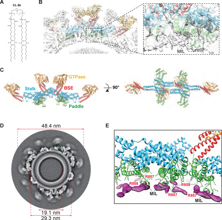
Figure 3. CryoEM reconstruction of human S-OPA1 bound to CL-Br-enriched membranes.
(A) Structure of tetrabrominated CL. (B) The side view for surface representation and corresponding ribbon diagram of the S-OPA1 tetramer 1 oriented into the cryoEM density map. Inset window shows the close-up view of the paddle domain (green) and conserved MIL region interacting with membranes. (C) Tetrameric ribbon model of the S-OPA1 bound to CL-Br membranes. The four structural domains are colored as follows: GTPase (orange), BSE (red), Stalk (blue), and Paddle (green). (D) A gray scale slice of the cryoEM 3D reconstruction of the membrane-bound S-OPA1 filament. The green rectangle indicates the position of the magnified view shown in panel E. (E) The difference map calculated from brominated and native protein-lipid reconstructions shows additional densities (magenta) located near the PDs (green) of S-OPA1.

