

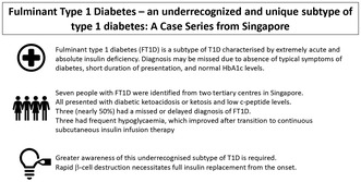
Abstract
Fulminant type 1 diabetes (FT1D) is a unique subtype of type 1 diabetes, characterized by acute absolute insulin deficiency, severe ketosis, and increased risk of hypoglycemia, glycemic variability and microvascular complications. Seven people with FT1D were identified from two tertiary centers in Singapore. Six were Chinese, the mean age was 35 years and all were lean (mean body mass index 20.3 kg/m2). All presented with diabetes ketosis or ketoacidosis and low C‐peptide. All but one had low glutamic acid decarboxylase antibodies. Nearly half had a missed/delayed diagnosis of FT1D. Three had frequent hypoglycemia, which improved after transition to continuous subcutaneous insulin infusion therapy. Individuals with FT1D experience unique diagnostic and management challenges associated with rapid absolute insulin deficiency. Greater awareness about this clinical entity is required.
Keywords: Continuous subcutaneous insulin infusion, Diabetic ketoacidosis, Fulminant type 1 diabetes
Greater awareness about fulminant type 1 diabetes, a unique and underrecognized subtype of type 1 diabetes, is required, as a delay in diagnosis can be fatal. These individuals experience challenges, such as increased glycemic variability, hypoglycemia and microvascular complications.
INTRODUCTION
Fulminant type 1 diabetes (FT1D) is a subtype of type 1 diabetes (T1D) that was recognized in 2000, largely described among East Asian populations 1 , 2 . It is characterized by rapid pancreatic β‐cell destruction and the absence of autoantibodies 3 . Unlike classical autoimmune type 1 diabetes (AT1D), individuals with FT1D have a much shorter duration of hyperglycemic symptoms, present with a greater severity of ketoacidosis and hyperglycemia, yet have misleadingly normal glycated hemoglobin (HbA1c) at presentation 4 .
We describe a case of FT1D that was missed at first presentation, followed by a series of FT1D from multi‐ethnic Singapore, highlighting the importance of characterizing the correct diabetes subtype, as this informs optimal management from the outset.
METHODS
People diagnosed with FT1D between 2014 and 2022 at endocrinology units in two tertiary centers were identified using the 2012 diagnostic criteria for FT1D: (1) diabetes ketosis or ketoacidosis <7 days after onset of hyperglycemic symptoms, (2) plasma glucose ≥16 mmol/L and HbA1c <8.7% at presentation, and (3) serum C‐peptide <0.3 μg/L (fasting) or <0.5 μg/L (post‐meal) 1 . Clinical characteristics were collected. Statistical analysis was carried out using IBM SPSS Statistics 21.0 (IBM Corp., Armonk, NY, USA). Descriptive statistics are expressed as the mean ± standard deviation (continuous variables), and frequency (percentage; categorical variables).
RESULTS
A 27‐year‐old man (case 1) with no medical history presented with 1 day of polyuria and polydipsia. He was lean (body mass index 19.7 kg/m2). Investigations showed hyperglycemia (32.2 mmol/L), yet HbA1c was not raised (5.5%; normal hemoglobin level). The Emergency Department administered a single dose of subcutaneous aspart insulin (15 units), and discharged him with advice to see a general practitioner in a week. As his HbA1c was normal, he was not discharged with glucose‐lowering medications or insulin.
Within 24 h, the patient returned with nausea, epigastric pain and lethargy, and was tachycardic and dehydrated. He now had diabetes ketosis (glucose 34.4 mmol/L, ketones 2.5 mmol/L), and was immediately started on intravenous hydration and insulin. Amylase and lipase levels were not raised. He was discharged on multiple daily insulin injections. One month later, C‐peptide was undetectable (<0.01 μg/L, glucose 6.7 mmol/L). Anti‐glutamic acid decarboxylase (GAD) levels were weakly positive (0.9 U/mL, reference 0–0.8 U/mL) and anti‐islet cell antibodies were negative. A diagnosis of FT1D was made, and the patient was continued on multiple daily insulin injections.
CASE SERIES
Over 8 years, seven people with FT1D were identified. The mean age was 35.0 ± 6.4 years, 6/7 (85.7%) were Chinese, and three (42.9%) were female. None were overweight (body mass index 20.3 ± 1.6 kg/m2). Symptom duration was very short (2.4 ± 1.2 days). Osmotic symptoms and abdominal pain were present in six (85.7%) people, whereas only one (16.7%) reported flu‐like symptoms. Two had infective triggers, of whom one had COVID‐19 infection.
Excluding case 1 who had been pretreated with insulin, the others presented with moderate to severe diabetes ketosis (mean glucose 40.9 ± 16.3 mmol/L, ketone level 5.3 ± 2.0 mmol/L, pH 7.18 ± 0.13). The mean HbA1c was not in the diabetes range (6.0 ± 0.5%). All but case 1 had negative GAD and ICA. C‐peptide levels were low (0.05 ± 0.08 μg/L). Four people had abdominal imaging: one had diffuse pancreatic enlargement, another had peri‐pancreatic fluid (Tables 1 and 2).
Table 1.
Baseline characteristics
| Characteristics | Results (Singapore cohort) | Results (Japanese cohort) 9 |
|---|---|---|
| Sex | ||
| Female (%) | 3 (42.9%) | – |
| Male (%) | 4 (57.1%) | |
| Age at diagnosis (years) | 35.0 ± 6.4 | 39.1 ± 15.7 |
| BMI (kg/m2) | 20.3 ± 1.6 | 20.7 ± 3.9 |
| Symptoms at presentation | ||
| Flu‐like symptoms (%) | 1 (14.3%) | 71.7% |
| Abdominal pain (%) | 6 (85.7%) | 72.5% |
| Osmotic symptoms (%) | 6 (85.7%) | 93.7% |
| Duration of symptoms (days) | 2.4 ± 1.2 | 4.4 ± 3.1 |
| Diabetic ketosis or ketoacidosis at presentation (%) | 7 (100%) | 100% |
| HbA1c at presentation (%) | 6.0 ± 0.5 | 6.4 ± 0.9 |
| Glucose level at presentation (mmol/L) | 40.9 ± 16.3 | 44.4 ± 20.0 |
| Ketone level at presentation (mmol/L) | 5.3 ± 2.0 | – |
| pH at presentation | 7.18 ± 0.13 | 7.13 ± 0.13 |
| Glutamic acid decarboxylase antibody‐positive (%) | 1 (14.3%) | 5.07% |
| Islet cell antibody‐positive (%) | 0 (0%) | – |
|
C‐peptide level (ug/L), All paired glucose >5.5 mmol/L |
0.05 ± 0.08 | 0.10 ± 0.10 |
| Amylase levels (U/L; reference range 38–149 U/L) | 221.6 ± 164.1 | – |
| Lipase levels (U/L; reference range: 8–55 U/L) | 290.8 ± 239.7 | – |
| Total daily dose of insulin/weight (u/kg) | 0.62 ± 0.14 | – |
Data are presented as mean ± standard deviation, or total number (%). BMI, body mass index; HbA1c, glycated hemoglobin.
Table 2.
Case details
| Case | Age (years) | Sex | Ethnicity | BMI (kg/m2) | Precipitant | HbA1c (%) | C‐peptide (ug/L) | Pancreatic imaging | Treatment | Total daily dose of insulin (units/kg) | Complications | Frequent hypoglycemia | |
|---|---|---|---|---|---|---|---|---|---|---|---|---|---|
| Macrovascular | Microvascular | ||||||||||||
| 1 | 27 | M | Malay | 19.7 | NA | 5.5 | 0.01 | NA | MDI | 0.604 | No | Yes (mild non‐proliferative retinopathy) | No |
| 2 | 40 | F | Chinese | 20.1 | NA | 7.1 | 0.2 | NA | MDI | 0.828 | No | No | No |
| 3 | 32 | M | Chinese | 21.7 | NA | 6.2 | NA | NA | MDI | 0.679 | No | No | No |
| 4 | 44 | F | Chinese | 19 | NA | 5.5 | 0.01 | Diffuse pancreatic enlargement with mild fat stranding | CSII | 0.556 | No | No | Yes |
| 5 | 39 | M | Chinese | 19.9 | Infection | 6.2 | 0.02 | Normal pancreas | CSII | 0.444 | No | No | Yes |
| 6 | 35 | F | Chinese | 26.7 | NA | 6 | 0.01 | Mild fluid around pancreatic tail | CSII | 0.434 | No | No | No |
| 7 | 28 | M | Chinese | 18.7 | COVID‐19 infection | 5.6 | 0.06 | Normal pancreas | CSII | 0.787 | No | No | Yes |
BMI, body mass index; CSII, continuous subcutaneous insulin infusion; F, female; HbA1c, glycated hemoglobin; M, male, MDI, multiple daily insulin injections; NA, not available.
All patients were discharged with multiple daily insulin injections; four were later transitioned to continuous subcutaneous insulin infusion therapy (CSII), of whom three were on advanced hybrid closed loop systems (Table 3). They had experienced frequent symptomatic hypoglycemia episodes, which improved after structured education and transition to CSII. During the mean follow‐up duration of 4.9 ± 2.9 years, one patient developed mild non‐proliferative retinopathy 5 years after FT1D onset.
Table 3.
Changes to glycemic parameters among people with fulminant type 1 diabetes initiated on advanced hybrid closed loop therapy
| Glycemic variables | Pre‐AHCL | Post‐AHCL |
|---|---|---|
| Case 4 | ||
| HbA1c | 5.5% | 5.6% |
| Time in range | 50% | 92% |
| Time below range | 37% | 5% |
| Coefficient of variation | 37.3% | 29.4% |
| Case 5 | ||
| HbA1c | 6.9% | 6.2% |
| Time in range | 56% | 84% |
| Time below range | 5% | 3% |
| Coefficient of variation | 46% | 36.1% |
| Case 7 | ||
| HbA1c | 6.6% | 6.6% |
| Time in range | 61% | 81% |
| Time below range | 15% | 1% |
| Coefficient of variation | – | 34.1% |
ACHL, advanced hybrid closed loop; HbA1c, glycated hemoglobin.
DISCUSSION
We describe the first case series of seven people with FT1D from Singapore. FT1D is not limited to East Asian populations, and presents unique diagnostic and management challenges.
Diabetes subtyping can be challenging among ethnic groups in Asia, where type 2 diabetes is more common, and develops at a younger age and lower body mass index than in people of white European ancestry 5 . Conversely, type 1 diabetes is rare (<1%), and the prevalence of positive GAD and ICA is low (maximum 41.5%) compared with white populations 6 . These overlapping phenotypes make it difficult to accurately subtype Asians with diabetes at presentation; the choice between insulin replacement versus sensitizing therapy becomes challenging.
FT1D is an even rarer and more recently identified subtype of type 2 diabetes. Data are scarce, and limited to case reports outside of East Asia. FT1D differs from AT1D in its rapid onset of β‐cell destruction, with our cohort presenting after a very short duration of osmotic symptoms, absence of weight loss and non‐diabetes range HbA1c levels. Individuals with FT1D tended to be older than adult AT1D patients 2 , and GAD and ICA were mostly negative. Given the lack of typical features associated with AT1D, the diagnosis was initially missed or delayed in three people. One was discharged without insulin, and two were initially diagnosed with acute pancreatitis, the latter being a common misdiagnosis in people with FT1D, as they often have elevated pancreatic enzymes (up to 98%) 1 and pancreatic swelling on imaging (36%) 7 .
Six of seven individuals were of Chinese ethnicity, consistent with the high prevalence of FT1D (7–20% of type 1 diabetes) previously described amongst East Asians 8 . This is postulated to be related to genetic susceptibility unique to the Asian population, due to class II human leukocyte antigen haplotypes (DRB1*04:05‐DQ1B*04:01 and DRB1*09:01‐DQB1*0303) 8 . Baseline characteristics and clinical features were similar between the present cohort and that of Imagawa et al. 9 , suggesting particular attention should be paid to individuals of East Asian descent presenting with features of FT1D, regardless of the country of diagnosis.
The failure to diagnose FT1D can be fatal. Unlike individuals with AT1D who experience a gradual decline in pancreatic β‐cell function, individuals with FT1D develop absolute insulin deficiency from the onset, resulting in greater severity of hyperglycemia and acidosis at presentation. Prompt recognition and full insulin replacement from the onset is critical 1 .
Even after diagnosis, long‐term management is complex. Individuals with FT1D experience high glycemic variability (GV), a higher frequency of severe hypoglycemia, higher total daily insulin doses and higher incidence of microvascular complications within the first 5 years of diagnosis compared with AT1D 10 . In the present study, nearly half of the patients experienced high GV and frequent symptomatic hypoglycemia, and underwent early transition to CSII (<1 year from diagnosis), with demonstrable improvements in GV, TIR and hypoglycemia rates. Although the role of CSII in FT1D is limited to case reports 11 , CSII clearly improves TIR and hypoglycemia rates in people with type 1 diabetes 12 ; people with FT1D would be anticipated to similarly benefit from CSII and advanced hybrid closed loop systems.
This first case series of FT1D from Singapore highlights the need to promote awareness of this underrecognized subtype of type 1 diabetes. Normal HbA1c levels or negative GAD should not negate the diagnosis. Rapid β‐cell destruction in these individuals necessitates immediate insulin therapy. Early use of CSII could help address the higher GV and hypoglycemia.
DISCLOSURE
The authors declare no conflict of interest.
Approval of the research protocol: SingHealth Centralized Institutional Review Board (CIRB Reference Number 2022/2671).
Informed Consent: Informed consent was obtained from all patients.
Approval date of Registry and the Registration No. of the study/trial: SingHealth Centralized Institutional Review Board (CIRB Reference Number 2022/2671, approved 14/11/2022).
Animal Studies: NA.
SOURCES OF FUNDING
None.
REFERENCES
- 1. Imagawa A, Hanafusa T, Awata T, et al. Report of the Committee of the Japan Diabetes Society on the research of fulminant and acute‐onset type 1 diabetes mellitus: New diagnostic criteria of fulminant type 1 diabetes mellitus (2012). J Diabetes Investig 2012; 3: 536–539. [DOI] [PMC free article] [PubMed] [Google Scholar]
- 2. Song SO, Yun J, Ko S, et al. Prevalence and clinical characteristics of fulminant type 1 diabetes mellitus in Korean adults: A multi‐institutional joint research. J Diabetes Investig 2022; 13: 47–53. [DOI] [PMC free article] [PubMed] [Google Scholar]
- 3. Imagawa A, Hanafusa T, Miyagawa J, et al. A novel subtype of type 1 diabetes mellitus characterized by a rapid onset and an absence of diabetes‐related antibodies. N Engl J Med 2000; 342: 301–307. [DOI] [PubMed] [Google Scholar]
- 4. Luo S, Ma X, Li X, et al. Fulminant type 1 diabetes: A comprehensive review of an autoimmune condition. Diabetes Metab Res Rev 2020; 36: e3317. [DOI] [PubMed] [Google Scholar]
- 5. Shai I, Jiang R, Manson JE, et al. Ethnicity, obesity, and risk of type 2 diabetes in women. Diabetes Care 2006; 29: 1585–1590. [DOI] [PubMed] [Google Scholar]
- 6. Lee YS, Ng WY, Thai AC, et al. Prevalence of ICA and GAD antibodies at initial presentation of type 1 diabetes mellitus in Singapore children. J Pediatr Endocrinol Metab 2001; 14: 767–772. [DOI] [PubMed] [Google Scholar]
- 7. Kahara T, Takamura T, Sakurai M, et al. Pancreatic exocrine and endocrine events occur concomitantly but independently during the course of fulminant type 1 diabetes. Diabetes Res Clin Pract 2006; 71: 241–246. [DOI] [PubMed] [Google Scholar]
- 8. Imagawa A, Hanafusa T. Fulminant type 1 diabetes—East and west. J Clin Endocrinol Metab 2023; 108: e1473–e1478. [DOI] [PubMed] [Google Scholar]
- 9. Imagawa A, Hanafusa T, Uchigata Y, et al. Fulminant type 1 diabetes: A Nationwide survey in Japan. Diabetes Care 2003; 26: 2345–2352. [DOI] [PubMed] [Google Scholar]
- 10. Murase Y, Imagawa A, Hanafusa T, et al. Fulminant type 1 diabetes as a high risk group for diabetic microangiopathy—A nationwide 5‐year‐study in Japan. Diabetologia 2007; 50: 531–537. [DOI] [PubMed] [Google Scholar]
- 11. Li CY, Li Y, You ZY, et al. Fulminant type 1 diabetes mellitus in pregnancy. Braz J Med Biol Res 2020; 53: e9633. [DOI] [PMC free article] [PubMed] [Google Scholar]
- 12. Matejko B, Juza A, Kieć‐Wilk B, et al. Transitioning of people with type 1 diabetes from multiple daily injections and self‐monitoring of blood glucose directly to MiniMed 780G advanced hybrid closed‐loop system: A two‐center, randomized, controlled study. Diabetes Care 2022; 45: 2628–2635. [DOI] [PMC free article] [PubMed] [Google Scholar]