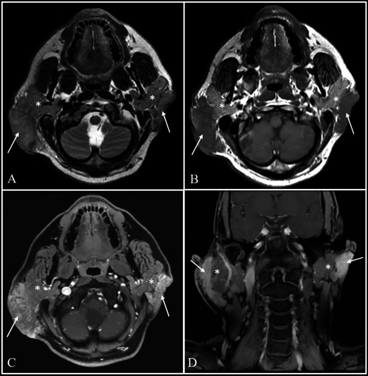
Figure 2. Evaluation with MRI.
Magnetic resonance imaging (MRI) with axial T2-weighted (A), axial T1-weighted (B), axial and coronal post-gadolinium T1-weighted images (C and D) showing T2 intermediate intensity lesions with heterogeneous post-contrast enhancement in bilateral infraauricular and retroauricular regions (arrows). The lesions are seen superficial to and separately from both the parotid glands, which is best appreciated on Pre-Gadolinium T1-weighted images (B). The parotid glands show normal signal intensity on both sides (asterisks).

