Abstract
We have developed a new strategy for immunization of wild rabbit populations against myxomatosis and rabbit hemorrhagic disease (RHD) that uses recombinant viruses based on a naturally attenuated field strain of myxoma virus (MV). The recombinant viruses expressed the RHDV major capsid protein (VP60) including a linear epitope tag from the transmissible gastroenteritis virus (TGEV) nucleoprotein. Following inoculation, the recombinant viruses induced specific antibody responses against MV, RHDV, and the TGEV tag. Immunization of wild rabbits by the subcutaneous and oral routes conferred protection against virulent RHDV and MV challenges. The recombinant viruses showed a limited horizontal transmission capacity, either by direct contact or in a flea-mediated process, promoting immunization of contact uninoculated animals.
Myxomatosis and rabbit hemorrhagic disease (RHD) are considered the major viral diseases affecting European rabbit (Oryctolagus cuniculus) populations. Myxoma virus (MV), the causative agent of myxomatosis, is a large virus with a double-stranded DNA genome of 163 kb which replicates in the cytoplasm of infected cells. MV belongs to the Leporipoxvirus genus of the Poxviridae family (39). The virus induces a benign disease in its natural host, Sylvilagus rabbits in the Americas. In European rabbits, however, MV causes myxomatosis, a systemic and usually fatal disease. The virus is spread by blood-feeding arthropod vectors such as mosquitoes or fleas. Epidemics occur annually or less frequently, depending on the emergence of large numbers of susceptible young rabbits and the availability of arthropod vectors (for recent reviews on myxomatosis, see references 19 and 26). MV was deliberately released as a biological control agent for the European rabbit initially in Australia (1950) and soon after in France (1952), whence it rapidly spread across the entire rabbit range in Europe, and has become endemic since then. Following initial release, devastating epizootics occurred in both continents, with a mortality rate of around 99.5%. The evolution of attenuated viral strains along with the development of host resistance led to a diminished incidence of the disease (18, 19). Nevertheless, recent studies carried out in Europe and Australia indicate that myxomatosis still regulates rabbit population numbers (57, 61). Control of myxomatosis among domestic rabbits is currently achieved by vaccination using heterologous vaccines based on Shope fibroma virus, a less virulent leporipoxvirus, or homologous vaccines based on cell culture-attenuated strains of MV (1, 47, 55).
RHD, an acute and highly contagious disease in wild and domestic rabbits (13, 44), was first reported by Liu et al. (30) in the People's Republic of China. The disease spread throughout Europe between 1987 and 1989 (37). Recently it was accidentally released in Australia (40). Infected rabbits usually die within 48 to 72 h of necrotizing hepatitis. The disease is responsible for high economic losses in rabbitries as well as high mortality rates in wild rabbit populations (31, 40, 59, 60). The etiological agent, RHD virus (RHDV), is a member of the Caliciviridae family (43, 48) which has been designated by the International Committee on Taxonomy of Viruses as the type species of the new genus Lagovirus (51). The virions (30 to 40 nm in diameter) have a 7.4-kb single-stranded positive-sense RNA genome and are nonenveloped and icosahedral. The RHDV capsids are made of a major protein component of 60 kDa (VP60) and a recently described minor polypeptide of 12.9 kDa (VP10) (62). Commercial vaccines against RHD are prepared from the livers of experimentally infected rabbits (2, 46), since in vitro systems are not available for efficient virus propagation. This approach involves the need to handle large amounts of highly infectious material. In recent years, the RHDV capsid protein gene has been successfully expressed in several heterologous systems. To date, VP60 protein has been produced in Escherichia coli (8); Saccharomyces cerevisiae (9), recombinant virus-based systems such as baculovirus (28, 32, 41, 56) and poxvirus (4, 5, 20), and plants (12). The recombinant VP60 obtained in all these systems has been shown to induce protection of rabbits against a lethal challenge with RHDV. In some cases the recombinant VP60 protein self-assembled to form virus-like particles which were highly immunogenic (9, 28, 41, 56). Both virus-like particles (50) and a recombinant vaccinia virus-VP60 virus (5) have been shown to confer protection against RHDV by the oral route.
While the above-mentioned results offer promising opportunities for the protection of domestic rabbits against myxomatosis and RHD, control of both diseases in wild rabbit populations remains an unsolved problem of great concern. In this regard, it should be noted that the European rabbit plays a key ecological role in Mediterranean ecosystems; in addition, rabbits are among the most important small game species in several European countries.
For large-scale wild rabbit immunization against myxomatosis and RHD, efficient vaccination by the oral route would be desirable. Furthermore, an appropriate immunization procedure for wild rabbit populations most likely would require some extent of horizontal vaccine transmission among individuals, in order to reach a fraction of immunized animals within the rabbit population sufficient to reduce the spread of both diseases. With this goal in mind, we have recently isolated the naturally attenuated MV strain 6918. This strain was selected from a survey of 20 MV field isolates currently circulating in Spain, which were analyzed for virulence and the ability to spread horizontally among rabbits by contact transmission (3a). MV strain 6918 presented characteristics suitable for use as a vaccine against myxomatosis in wild rabbit populations. It was virtually nonpathogenic and elicited high levels of MV-specific antibodies on inoculated rabbits, inducing protection against a virulent MV challenge. Furthermore, strain 6918 was capable of limited horizontal spreading by contact transmission, inducing protection against myxomatosis on uninoculated rabbits (3a).
To extend the use of such a transmissible vaccine to immunize wild rabbits against RHDV, we constructed recombinant MVs based on the 6918 strain that expressed RHDV VP60 protein. The ability of these recombinant viruses to promote transmissible protection against myxomatosis and RHD was analyzed both by direct contact and by flea-mediated transmission after immunization by the subcutaneous (s.c.) or oral route. To monitor the spread and efficacy of the recombinant virus vaccine in the field, a peptide tag was included within the recombinant VP60 protein to differentiate between naturally infected and immunized animals.
MATERIALS AND METHODS
Cells and viruses.
MV strain 6918 (3a) and recombinant MVs were propagated in RK-13 (rabbit kidney) cell line grown in Dulbecco's minimum essential medium (DMEM) supplemented with 5% fetal bovine serum, 2 mM l-glutamine, penicillin (100 U/m), and streptomycin (100 μg/ml). SIRC (rabbit cornea) cells were used for the preparation of MV recombinants as described below. Both rabbit cell lines were obtained from the American Type Culture Collection. MV Laussane strain, provided by Laboratorios Hipra, and the AST/89 strain of RHDV (48) were used to challenge rabbits in the immunization experiments.
Rabbits.
Common rabbits (brown colored) 2 months old, weighing around 1 kg, and free from anti-MV and anti-RHDV antibodies, were provided by a commercial breeder. These rabbits are routinely used for restocking in the field and hereafter will be referred to as wild rabbits.
Fleas.
The fleas used in this study, Xenopsylla cunicularis, are specific parasites of wild rabbits in the occidental Mediterranean region. A laboratory colony has been maintained in culture since 1993, using the method described by Cooke (14). Adults were obtained from a wild rabbit shot in Zaragoza, Spain. Adult fleas were allowed to feed on domestic rabbits, and the larvae were cultured in hermetic boxes with larval rearing medium made from dried bovine blood and powdered brewer's yeast mixed with sand. Cocoons were sifted from the sand and stored in plastic jars for adult emergence. Larvae and cocoons are maintained in an environmental chamber at 22°C and 70 to 80% relative humidity. Unfed adult fleas, 2 to 4 days old, were used in the experiments involving flea-mediated recombinant virus transmission.
Amplification and cloning of VP60 gene constructs.
The VP60 gene from RHDV AST/89 strain (nucleotides 5305 to 7125) was previously cloned in plasmid pMA91 (35), yielding expression vector pMAVP60 (9). In this construct, the VP60 gene is flanked by BglII restriction sites. In addition, two other constructs, designated VP60-T1 and VP60-T2, were made by adding a DNA sequence coding for a linear epitope from the transmissible gastroenteritis virus (TGEV) nucleoprotein, which is recognized by monoclonal antibody (MAb) DA3 (33). In VP60-T1, the tag was inserted within the 5′ region of the VP60 gene; in VP60-T2, the TGEV epitope sequence was placed at the 3′ end.
The VP60-T1 BglII cassette was constructed by first inserting a 360-bp BglII/SphI DNA fragment from pMAVP60 into plasmid pUCHSV-TK (15). The resulting construct was BamHI/HindIII digested and subsequently ligated to a 1,731-bp BamHI/HindIII fragment from plasmid pT35 (8), resulting in plasmid pTKVP60. A 39-bp synthetic DNA fragment, coding for the DA3 tag, was generated by annealing oligonucleotides 5′-GATCTAGAAAATTATACAGATGTGTTTGATGACACACAG-3′ and 5′-GATCCTGTGTGTCATCAAACACATCTGTATAATTTTCTA-3′ and then inserted into the unique BamHI site of pTKVP60. After correct orientation of the synthetic DNA was checked, the resulting plasmid was digested with HindIII, blunt ended, and ligated to commercial linkers to generate a BglII site at the VP60 gene 3′ end.
The VP60-T2 BglII cassette was constructed by first inserting a 42-bp synthetic DNA, coding for the DA3 tag, into the unique BamHI site of pAcYM1 vector (34), resulting in plasmid pAcM1-DA3. The synthetic DNA fragment was generated by annealing oligonucleotides 5′-GATCCATAGAAAATTATACAGATGTGTTTGATGACACACAGT-3′ and 5′-GATCACTGTGTGTCATCAAACACATCTGTATAATTTTCTATG-3′. In parallel, the VP60 gene was PCR amplified by using oligonucleotide primers 0-5 (9) and 3′-Bgl (5′-CCAGTCCGATGAGATCTGACATAGG-3′), which generated BglII restriction sites at both ends of the VP60 gene, eliminating its natural stop codon. The PCR product was digested with BglII and cloned into BamHI-digested pAcM1-DA3 to generate pAcM1-VP60-DA3. This construct contained the complete VP60 gene fused at its 3′ end to the DA3 epitope-coding sequence followed by a translation stop codon. A BstEII/HindIII fragment from plasmid pAcM1-VP60-DA3 was then ligated to plasmid pTKVP60, generating plasmid pTKVP60-DA3. Finally, this plasmid was digested with HindIII, blunt ended, and ligated to commercial linkers to generate a BglII site at the 3′ end of the VP60 DA3 construct.
Construction of recombinant transfer vectors.
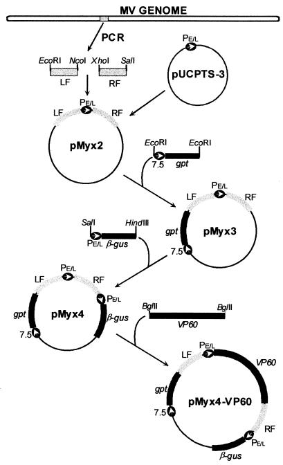
The procedure used to construct recombinant MV viruses was adapted from standard methods (16) previously described for vaccinia virus (VV). A transfer vector, pMyx4, with MV DNA flanking regions suitable to direct foreign gene insertion into MV genome by homologous recombination, was constructed as follows. Two MV DNA fragments from the intergenic site between the thymidine kinase (TK) gene (open reading frame [ORF] MJ2) and ORF MJ2a (22) were amplified by PCR using oligonucleotide primers 5′-CACCGAATTCTAGGACCCATGTTTGC-3′ (EcoRI site underlined) and 5′-TTTATTTTTCCATGGTTTTAAAAAAAATAACAT-3′ (NcoI site underlined) for the left flank (529 bp) and oligonucleotide primers 5′-TACAACTCGAGGTGTGGATCCTGTTATTTTTTT-3′ (XhoI site underlined BamHI site in boldface) and 5′-TATAAGACACGTCGACGTCACTGAA-3′ (SalI site underlined) for the right flank (424 bp). After PCR amplification and digestion with appropriate restriction enzymes, the flanking sequences were cloned into unique restriction sites in plasmid pUCPTS-3 (3), a derivative of plasmid pUC19 containing a VV synthetic early/late promoter, creating plasmid pMyx2 (Fig. 1).
FIG. 1.
Construction of pMyx4-VP60 transfer vector (see Materials and Methods for details). LF, left flank; RF, right flank; PE/L, synthetic early/late VV promoter; 7.5, VV 7.5 promoter; gpt, E. coli gpt gene; β-gus, E. coli β-gus gene. Plasmids pMyx4-VP60-T1 and pMyx4-VP60-T2 were obtained by insertion of a DNA fragment containing VP60-T1 or VP60-T2 respectively, in the pMyx4 vector.
A DNA construct containing the xanthine-guanine phosphoribosyltransferase gene (gpt) from E. coli under the control of VV 7.5 promoter was cloned into pMyx2. For this purpose, a DNA fragment containing the gpt cassette was PCR amplified from plasmid pGEM-gpt (6), using oligonucleotides 5′-CGGGCTCGAGCACTAATTCCAAACCCACCCGCTTT-3′ (XhoI site underlined) and 5′-ATTGGAGCTCGCCTGAAGTTAAAAAGAACAACGCC-3′ (SacI site underlined). The amplified DNA fragment was digested with XhoI and SacI and inserted into plasmid pGEM-7-Z(f-) (Promega). A sequence of 130 bp between the VV 7.5 promoter and the gpt gene was excised by digestion with BamHI and BglII and religation. The shortened gpt cassette was amplified by PCR using oligonucleotide primers 5′-GAGCACGAATTCAAACCCACCCG-3′ (EcoRI site underlined) and 5′-CGCCTGAATTCAAAAAGAACAACGC-3′ (EcoRI site underlined). The amplified DNA fragment was digested with EcoRI and ligated to the EcoRI site of plasmid pMyx2, generating pMyx3 (Fig. 1).
Plasmid pMyx3 was further modified by insertion of a DNA construct containing the β-glucuronidase gene (β-gus) from E. coli under the control of the VV synthetic early/late promoter. A XhoI/HindIII DNA fragment containing the β-gus cassette derived from plasmid pRB21-β-gus (7) was inserted into SalI/HindIII-digested plasmid pMyx3 to create pMyx4 (Fig. 1).
Finally, the BglII DNA fragments containing VP60, VP60-T1, and VP60-T2 derivatives of the RHDV major capsid gene were cloned into BamHI-digested pMyx4, yielding plasmids pMyx4-VP60, pMyx4-VP60-T1, and pMyx4-VP60-T2, respectively.
Isolation of recombinant viruses.
The recombinant MVs were isolated by the transient dominant selection (TDS) approach (17) by adaptation of previous procedures (23). Preconfluent monolayers of SIRC cells grown in T-25 flasks were infected with MV strain 6918 at a multiplicity of infection of about 0.2 PFU. The virus was allowed to adsorb for 1 h at 37°C, and cells were transfected with 5 μg of calcium phosphate-precipitated plasmid DNA. Eighteen hours after infection, the transfection medium was removed and replaced with selective medium: DMEM–2.5% fetal bovine serum supplemented with mycophenolic acid (25 μg/ml), xanthine (250 μg/ml), and hypoxanthine (15 μg/ml). The cells were then incubated at 37°C until complete cytopathic effect was reached (normally 5 to 6 days), and progeny viruses were passaged once in the presence of selective medium (mycophenolic acid, xanthine, hypoxanthine) to replicate in recombinant viruses expressing the gpt gene. Viruses resulting from a single recombinant event exhibiting a gpt+ β-gus+ phenotype (blue plaques) were isolated by plaque assay on monolayers of SIRC cells grown in selective culture medium containing 1% (wt/vol) low-melting-point agarose and 300 μg of 5-bromo-4-chloro-3-indolyl-β-d-glucuronide (X-Gluc) per ml. Two further passages in SIRC cells were performed without selection to allow amplification of viruses deleted for the gpt gene. The viral preparations were then subjected to plaque assay on monolayers of SIRC cells overlaid with nonselective medium containing 1% (wt/vol) low-melting-point agarose, 300 μg of X-Gluc per ml, and 1% neutral red, and white plaques (β-gus−) were selected. Finally, the double-recombinant viral clones that had incorporated the different versions of the VP60 gene were identified by PCR analysis using oligonucleotides MV1 (5′-CGCAAATATCCTGTCTATATTGC-3′), which hybridizes with the left flanking region of the insertion site in the MV genome, and VP60-1 (5′-GAAAACATCATTATAATAAAAGTTCG-3′), which hybridizes with the 5′ region of the VP60 gene. PCR-positive virus clones were plaque purified twice and then amplified by infection of RK-13 cells. The genomic structures of the recombinant viruses were further analyzed by PCR to confirm that inserts of the correct size had integrated in the expected sites. DNA from infected cells was used as the template for PCRs using specific oligonucleotide primers derived from the MV genomic sequence flanking the insertion site. The oligonucleotides used were MV1 and 5′-CGCTGTAGTATTTTTTTATCGTATT-3′ (MV2). The amplification of a 3.3-kb PCR product, instead of the 1.0-kb product obtained from wild-type MV, was indicative of correct insertion of the VP60 constructs. To analyze the genetic stability of the constructions, the three recombinant viruses were subjected to 15 serial passages in RK-13 cell monolayers, after which the genomic structure of the resulting progeny virus was analyzed by the same PCR protocol (using oligonucleotides MV1 and MV2).
Western-ECL immunoblotting.
Monolayers of RK-13 cells were infected with MV strain 6918 or recombinant viruses at a multiplicity of infection of approximately 5 PFU per cell. When complete cytopathic effect was reached, the monolayers were washed with cold phosphate-buffered saline (PBS) and treated with lysis buffer (50 mM Tris HCl [pH 8.0], 150 mM NaCl, 2 mM EDTA, 1% NP-40, 1 μg of leupeptin per ml, 1 mM phenylmethylsulfonyl fluoride) for 20 min on ice. Subsequently, the infected cell extracts were scraped and centrifuged at 14,000 × g for 5 min (4°C). The proteins present in the supernatants were resolved by sodium dodecyl sulfate–10% polyacrylamide gel electrophoresis and transferred onto nitrocellulose membranes by electroblotting. The membranes were then saturated overnight at 4°C with PBS–5% nonfat dry milk and incubated for 1 h at 37°C either with a rabbit hyperimmune antiserum against RHDV for detecting VP60 protein or with MAb DA3 (33). After several washes with PBS–0.05% Tween 20, the membranes were incubated for 1 h at 37°C with the appropriate secondary antibody: goat anti-rabbit immunoglobulin conjugated with horseradish peroxidase (Chemicon) or goat anti-mouse immunoglobulin conjugated with horseradish peroxidase (Bio-Rad). Membranes were extensively washed with PBS–0.05% Tween 20 and incubated for 1 min with a 1:1 mix of solution A (2.5 mM luminol [Sigma], 0.4 mM p-coumaric acid [Sigma], 100 mM Tris HCl [pH 8.5]) and solution B (0.018% H2O2, 100 mM Tris HCl [pH 8.5]) and exposed for 15 s to an autoradiography film.
Immunization of rabbits with recombinant viruses.
To analyze the immunogenic potential of the recombinant viruses, a group of 10 wild rabbits were inoculated by s.c. injection at the back with a 0.1-ml (104-PFU) dose of each virus in DMEM. The rabbits were observed for a period of 56 days, and clinical symptoms due to the virus inoculation were recorded. Serum samples extracted from the marginal ear vein of the rabbits at 0, 21, 35, and 56 days post immunization (dpi) were used to evaluate the serological responses against MV, RHDV, and DA3 epitope by using an enzyme-linked immunosorbent assay (ELISA). For this, separate ELISA plate wells (Polysorp; Nunc) were coated with specific antigens: 5 μg of semipurified MV from infected RK-13 cells (3a), 0.1 μg of purified RHDV from infected rabbit liver extracts (48), or 0.2 μg of purified TGEV virus from infected swine testis cells (25). Binding of specific antibodies present in serial dilutions of serum samples was visualized by incubation with protein G conjugated with horseradish peroxidase (Pierce) and subsequent addition of substrate solution [2,2′-azinobis(3-ethylbenzthiazolinesulfonic acid) (ABTS); Sigma]. After 10 min of substrate incubation, the reaction was stopped by addition of 1% sodium dodecyl sulfate and color development was recorded. Serum titers were defined as the inverse of the highest dilution giving an A405 value twofold over the background level (negative control rabbit sera).
Transmissible immunization of rabbits with recombinant 6918VP60-T2.
To analyze the ability of the recombinant 6918VP60-T2 to disseminate among rabbits by horizontal contact transmission and induce protection against myxomatosis and RHD in directly immunized rabbits, as well as in “contacted” uninoculated animals, two groups of six wild rabbits were injected once s.c. at the back with 104 PFU of 6918VP60-T2 virus (immunized groups A and B). Three days later, each inoculated rabbit group was placed in a cage together with another group of six rabbits (first-passage groups A and B, respectively) for 6 days. Subsequently, rabbits from the first-passage groups were separated from the inoculated animals and placed in another cage together with a new group of six rabbits (second-passage groups A and B, respectively) for 6 more days, after which the different groups of rabbits were placed in separate cages; 35 days after the initial immunization with recombinant 6918VP60-T2, a blood sample was taken from all rabbits. The same day, the rabbits from the three A groups together with four unvaccinated control rabbits were challenged intramuscularly with 100 50% lethal doses (LD50) of RHDV (AST/89 strain), whereas the animals from the three B groups and four unvaccinated control rabbits were challenged with 103 PFU of virulent MV (Laussane strain) by intradermal (i.d.) injection. Rabbits were monitored daily for symptoms of myxomatosis or RHD up to day 56, when a blood sample was extracted from all survivors. The serological responses against MV and RHDV were evaluated by ELISA as described above.
A similar approach was used to evaluate the flea-mediated transmission of recombinant 6918VP60-T2 virus and the concomitant induction of protection against myxomatosis and RHD. For this, special cages divided into two contiguous compartments separated by two metallic nets 25 cm apart were used. A group of six wild rabbits were placed in one of the compartments of each cage and were injected s.c. in the back with 104 PFU of 6918VP60-T2 virus (immunized groups A and B). Three days later, 150 fleas (25 per rabbit) were released among each group of inoculated rabbits, and another group of six rabbits was placed in the adjacent compartment of each cage (first-passage groups A and B, respectively). Under these conditions there was no direct contact between the inoculated and first-passage groups of rabbits, but the fleas readily spread through the whole cage. After 6 days, the inoculated rabbits were carefully combed to release the fleas present in their fur into the cage and were replaced by a new group of six rabbits (second-passage groups A and B, respectively) for another 6 days. The different groups of rabbits were then placed in separate cages and challenged either with virulent MV or RHDV as described above.
Induction of oral protection with recombinant 6918VP60-T2 was also analyzed. Two groups of two rabbits were immunized by oral administration of 107 PFU of 6918VP60-T2 virus (immunized groups A and B). Three days later, each immunized rabbit group was placed in the same cage with another 2 rabbits (first-passage groups A and B, respectively) for 6 days, after which the different groups of rabbits were separated; 35 days after the initial oral administration of recombinant 6918VP60-T2, a blood sample was extracted from all rabbits. The same day, the rabbits from the two A groups together with two unvaccinated control rabbits were challenged intramuscularly with 100 LD50 of RHDV (AST/89 strain), whereas the rabbits from the B groups and two unvaccinated control rabbits were challenged by i.d. injection with 103 PFU of virulent MV (Laussane strain). Rabbits were monitored daily for symptoms of myxomatosis or RHD up to day 56, when a blood sample was taken from each of the survivors and serological responses against MV and RHDV were evaluated by ELISA.
RESULTS
Construction of MV-VP60 recombinants.
A general-purpose MV recombination and expression vector was constructed as outlined in Fig. 1. First, recombination flanks were generated by PCR and cloned in plasmid pUCPTS-3, which contains a synthetic VV early/late promoter (3). Those flanks directed the insertion of foreign DNA between the MJ2 (TK gene) and MJ2a ORFs, an intergenic site which has been successfully used to clone heterologous genes into the MV genome (23, 24). In addition, gpt and β-gus cassettes were placed outside the recombination flanks to facilitate selection and identification of single- and double-recombination events. Plasmid pMyx4 has unique XhoI and BamHI restriction sites downstream of the promoter sequence, which can be used to clone the gene of interest.
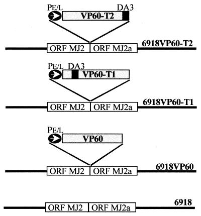
Three different constructs of the VP60 gene were cloned into the pMyx4 vector and subsequently inserted into the genome of MV strain 6918 by homologous recombination using the TDS approach (17). Figure 2 summarizes the VP60 gene constructs and the names assigned to the resulting MV-VP60 recombinants. The first gene construct consisted of the complete RHDV VP60 gene. Constructs VP60-T1 and VP60-T2 were gene fusions of the VP60 gene with a DNA fragment (39 and 42 nucleotides, respectively) coding for a linear epitope tag from the TGEV nucleoprotein, recognized by MAb DA3 (33). This peptide tag was inserted within the VP60 gene near the 5′ end (VP60-T1) or at the 3′ end (VP60-T2). The resulting MV-VP60 recombinants 6918VP60, 6918VP60-T1, and 6918VP60-T2 were named according to the inserted VP60 gene cassette.
FIG. 2.
Summary of MV-VP60 gene constructs. PE/L, synthetic early/late VV promoter; DA3; peptide tag from the TGEV nucleoprotein recognized by MAb DA3 (33).
The genomic structures of the three recombinant viruses were analyzed by PCR using oligonucleotide primers external to the insertion region. The amplification of a 3.3-kb PCR product, instead of the 1.0-kb product obtained from wild-type MV, confirmed that a DNA fragment of the correct length had been inserted into the intended location in the MV genome (not shown). After 15 serial passages of the recombinant viruses in RK-13 cell monolayers, the same 3.3-kb product was amplified by PCR with no detection of the wild-type MV 1.0-kb product (not shown), indicating that the different VP60 versions were stably integrated in the MV genome. The growth rates, virus titers, and plaque size phenotypes of the recombinant viruses in cell cultures were similar to those of the parental virus.
Expression of VP60 in recombinant MV-infected cells.
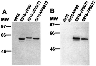
Western blot analysis of RK-13 cells infected with 6918 or recombinant MVs showed the presence of specific polypeptides with the expected sizes in cell extracts from cultures infected with recombinant MVs (Fig. 3).
FIG. 3.
Expression of VP60 in recombinant MV-infected RK-13 cells. Lysates of cells infected with the indicated viruses were analyzed by Western blot analysis using a rabbit hyperimmune antiserum against RHDV (A) or MAb DA3 (B). Positions of molecular weight (MW) markers are shown in kilodaltons.
A hyperimmune antiserum against RHDV recognized a protein with an apparent molecular mass of 60 kDa in 6918VP60-infected cell lysates, which corresponded to the monomeric form of the VP60 capsid protein, in agreement with published values (4, 8, 20, 28, 32, 41, 56). This polypeptide was not detected in lysates of cells infected by parental 6918 virus. The mobility of the detected polypeptides in the 6918VP60-T1 and 6918VP60-T2-infected cell extracts was slightly slower than that of 6918VP60-infected cell lysates, reflecting the presence of the peptide tag. Accordingly, these slowly moving VP60 bands found only in 6918VP60-T1 and 6918VP60-T2-infected cell extracts were also recognized by MAb DA3 (Fig. 3).
Immune responses induced by recombinant viruses.
After s.c. injection of domestic or wild rabbits with the recombinant MVs, only a small transient lesion at the inoculation site (usually around 0.5 cm) and, in some cases, localized discrete secondary nodules were observed. Presence of virus could be detected by PCR in a time range of 2 to 10 dpi. The virus was detected at the inoculation site, the draining lymph node, and samples of skin and in secondary nodules when these were present. No virus was detected in internal organs (blood, spleen, liver, and lungs). No febrile response or loss of body weight was detected in the inoculated rabbits, and the overall health of the rabbits was largely unaffected. The clinical symptoms appeared 5 to 7 dpi and completely resolved in all inoculated rabbits normally by 15 dpi. Thus, the symptomatology induced by the recombinant viruses in the inoculated rabbits was similar to that previously described for 6918 MV strain (3a), confirming the attenuated nature of these viruses.
To evaluate the immune responses elicited by the inoculated rabbits, serum samples obtained at different times postinoculation were monitored by ELISA for the presence of anti-MV, anti-RHDV, and anti-TGEV (anti-peptide tag) antibodies.
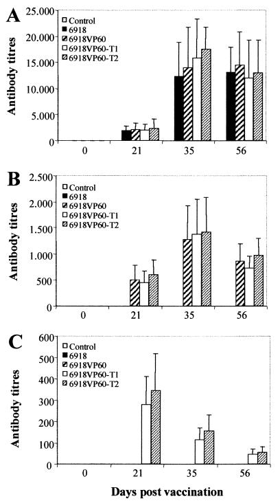
Rabbits inoculated with MV strain 6918 or the recombinant viruses progressively developed anti-MV antibody titers which were highest at 35 dpi. These levels were maintained up to 56 dpi (Fig. 4A). There was no gross difference in the antibody titers induced by the four viruses.
FIG. 4.
Serum antibody responses (ELISA) in rabbits immunized by s.c. injection with MV strain 6918 and MV-VP60 recombinants. Anti-MV (A), anti-RHDV (B), and anti-TGEV (C) antibody titers are reported as means plus standard errors of the means (n = 10). Serum titers were defined as the inverse of the highest dilution giving an A405 twofold over the background level (a negative control rabbit serum). The control group consisted of nonvaccinated animals.
Anti-VP60 antibodies were detected in rabbits inoculated with the recombinant viruses, while animals immunized with 6918 MV strain and the nonvaccinated rabbits remained seronegative (Fig. 4B). The time course of the antibody response against RHDV was similar to that obtained against MV.
As expected, antibodies recognizing TGEV in ELISA were detected only in rabbits inoculated with the recombinant viruses 6918VP60-T1 and 6918VP60-T2 (Fig. 4C). In this case, the highest antibody response was obtained 21 dpi. The antibody titers diminished significantly by day 35 and were still detectable after 56 days.
Transmissible protection induced by 6918VP60-T2.
Since both 6918VP60-T1 and 6918VP60-T2 MV recombinants showed grossly similar characteristics in terms of specific antibody response, one of them, 6918VP60-T2, was selected for further analysis.
To determine whether administration of 6918VP60-T2 could protect wild rabbits from virulent MV or RHDV infections, and if this protection could be transmitted by direct contact to uninoculated rabbits, a challenge experiment was conducted. Groups of six rabbits (immunized groups A and B) injected s.c. with 6918VP60-T2 were held in contact with uninoculated rabbits (first-passage groups A and B). Subsequently, the first-passage rabbits were placed in the same cage with a new group of uninoculated rabbits (second-passage groups A and B). The different groups of rabbits were then separated, and 35 days after the initial immunization with 6918VP60-T2, rabbits were challenged with RHDV or virulent MV. The results are shown in Table 1.
TABLE 1.
Protection against virulent RHDV and MV induced by contact transmission of 6918VP60-T2
| Group | Vaccine administration (6918VP60-T2) | Virulent challengea | Seropositive rabbitsb | Mean antibody titer of seropositive rabbit
|
Survival | |||
|---|---|---|---|---|---|---|---|---|
| Anti-RHDV
|
Anti-MV
|
|||||||
| 35 dpv | 56 dpv | 35 dpv | 56 dpv | |||||
| Control | ||||||||
| A | RHDV | 0/4 | NDc | NTd | ND | NT | 0/4 | |
| B | MV | 0/4 | ND | NT | ND | NT | 0/4 | |
| Immunized | ||||||||
| A | 104 PFU (s.c.) | RHDV | 6/6 | 940 | 4,375 | 8,750 | 6,250 | 6/6 |
| B | 104 PFU (s.c.) | MV | 6/6 | 1,250 | 900e | 9,375 | 20,000e | 5/6 |
| 1st passage | ||||||||
| A | Contact with immunized group A rabbits | RHDV | 3/6 | 200 | 625 | 175 | 100 | 3/6 |
| B | Contact with immunized group B rabbits | MV | 4/6 | 60 | 175 | 75 | 3,750 | 4/6 |
| 2nd passage | ||||||||
| A | Contact with 1st-passage group A rabbits | RHDV | 1/6 | 5 | 1,000 | 25 | 50 | 1/6 |
| B | Contact with 1st-passage group B rabbits | MV | 0/6 | ND | NT | ND | NT | 0/6 |
Rabbits were challenged intramuscularly with 100 LD50 of RHDV (strain AST/89) or by i.d. injection with 1,000 PFU of MV (Laussane strain). The challenge was administered 35 days postvaccination (dpv).
Number of rabbits seropositive to RHDV and MV (35 and 56 dpv)/total number of rabbits.
ND, not detected.
NT, not tested, (all rabbits had died).
Antibody titers 56 dpv corresponded to five seropositive rabbits, as one rabbit had died before analysis.
Direct immunization with 6918VP60-T2 induced high anti-RHDV and anti-MV antibody titers. Furthermore, all rabbits directly immunized with 6918VP60-T2 survived the RHDV lethal challenge, and five of six resisted the virulent MV challenge. In contrast, none of the control unvaccinated rabbits survived the lethal challenge with RHDV or virulent MV. Analysis of the serological responses after the challenge revealed a high increase in the specific postchallenge anti-RHDV or anti-MV antibody titers. Detectable levels of antibodies against the TGEV peptide tag were observed in all the rabbits at 35 and 56 days postvaccination (not shown).
Around 50% (7 of 12) of the first-passage rabbits were seropositive for both RHDV and MV by day 35 postvaccination (32 days after initial contact with immunized rabbits) as well as for the TGEV peptide tag (not shown). These rabbits survived the challenge with RHDV (three of six) or virulent MV (four of six). By contrast, the rabbits that were seronegative for RHDV, MV, and TGEV peptide tag did not resist virulent challenges. This result indicated that the recombinant 6918VP60-T2 was able to pass from inoculated to uninoculated rabbits by contact transmission and that this was sufficient to induce a protective immune response in contact transmission rabbits, although the antibody titers in these rabbits were lower than those exhibited by directly immunized animals. The proportion of seropositive rabbits in the second-passage groups was greatly reduced. Only 1 of 12 rabbits had detectable anti-RHDV and anti-MV antibody titers by day 35 postvaccination (26 days after initial contact with first-passage rabbits) and survived the challenge with RHDV. None of the six rabbits challenged with virulent MV survived.
Once the ability of recombinant 6918VP60-T2 to spread among rabbits by contact transmission and induce transmissible protection against both viruses had been established, it was of interest to determine whether the recombinant vaccine could be propagated among rabbits by flea-mediated transmission, as this is the most important means of MV diffusion in nature. For this, a challenge experiment involving flea-mediated transmission of recombinant 6918VP60-T2 was conducted as described in Materials and Methods.
As shown in Table 2, the results obtained closely resembled those of the contact transmission experiment described before. The directly immunized rabbits elicited high anti-RHDV and anti-MV antibody titers and were seropositive for the TGEV peptide tag. All of these rabbits survived the challenge with RHDV or virulent MV. Fifty percent (6 of 12) of the first-passage rabbits were seropositive for MV, RHDV, and TGEV peptide tag and resisted the challenge with RHDV (3 of 6) or MV (3 of 6). Only 1 of 12 second-passage rabbits had detectable anti-RHDV and anti-MV antibody titers by day 35 postvaccination. This rabbit survived the challenge with MV. None of the six rabbits challenged with RHDV survived. The protection transmission observed in this experiment was shown to be dependent on the presence of fleas, as in a parallel experiment carried out in the absence of fleas, no seroconversion was observed in passage rabbits and none of them resisted the lethal challenge with virulent MV or RHDV (data not shown).
TABLE 2.
Protection against virulent RHDV and MV induced by flea-mediated transmission of 6918VP60-T2
| Group | Vaccine administration (6918VP60-T2) | Virulent challengea | Seropositive rabbitsb | Mean antibody titer of seropositive rabbit
|
Survival | |||
|---|---|---|---|---|---|---|---|---|
| Anti-RHDV
|
Anti-MV
|
|||||||
| 35 dpv | 56 dpv | 35 dpv | 56 dpv | |||||
| Control | ||||||||
| A | RHDV | 0/4 | NDc | NTd | ND | NT | 0/4 | |
| B | MV | 0/4 | ND | NT | ND | NT | 0/4 | |
| Immunized | ||||||||
| A | 104 PFU (s.c.) | RHDV | 6/6 | 2,300 | 4,000 | 5,625 | 3,375 | 6/6 |
| B | 104 PFU (s.c.) | MV | 6/6 | 2,000 | 1,938 | 6,875 | 17,500 | 6/6 |
| 1st-passage | ||||||||
| A | Contact with immunized group A rabbits | RHDV | 3/6 | 200 | 1,625 | 225 | 250 | 3/6 |
| B | Contact with immunized group B rabbits | MV | 3/6 | 250 | 375 | 350 | 1,750 | 3/6 |
| 2nd passage | ||||||||
| A | Contact with 1st-passage group A rabbits | RHDV | 0/6 | ND | NT | ND | NT | 0/6 |
| B | Contact with 1st-passage group B rabbits | MV | 1/6 | ND | 5 | 50 | 2,500 | 1/6 |
Rabbits were challenged intramuscularly with 100 LD50 of RHDV (strain AST/89) or by i.d. injection with 1,000 PFU of MV (Laussane strain). The challenge was administered 35 days postvaccination (dpv).
Number of rabbits seropositive to RHDV and MV (35 and 56 dpv)/total number of rabbits.
ND, not detected.
NT, not tested (all rabbits had died).
Finally, the feasibility of immunizing rabbits with recombinant 6918VP60-T2 by the oral route was addressed (Table 3). Following oral administration of a high dose (107 PFU) of recombinant 6918VP60-T2, the immunized rabbits developed high anti-RHDV and anti-MV antibody titers and survived challenges with either RHDV or virulent MV. In addition, protection induced by oral administration of 6918VP60-T2 could be transmitted to unvaccinated rabbits by contact transmission, as the passage rabbits were seropositive to RHDV and MV and survived virulent challenges.
TABLE 3.
Oral vaccination with 6918VP60-T2 and protection by contact transmission against virulent RHDV and MV
| Group | Vaccine administration (6918VP60-T2) | Virulent challengea | Seropositive rabbitsb | Mean antibody titer of seropositive rabbit
|
Survival | |||
|---|---|---|---|---|---|---|---|---|
| Anti-RHDV
|
Anti-MV
|
|||||||
| 35 dpv | 56 dpv | 35 dpv | 56 dpv | |||||
| Control | ||||||||
| A | RHDV | 0/2 | NDc | NTd | ND | NT | 0/2 | |
| B | MV | 0/2 | ND | NT | ND | NT | 0/2 | |
| Immunized | ||||||||
| A | 107 PFU (oral) | RHDV | 2/2 | 3,000 | 5,575 | 6,125 | 5,350 | 2/2 |
| B | 107 PFU (oral) | MV | 2/2 | 2,300 | 1,870 | 5,500 | 19,625 | 2/2 |
| Passage | ||||||||
| A | Contact with immunized group A rabbits | RHDV | 2/2 | 135 | 1,340 | 250 | 225 | 2/2 |
| B | Contact with immunized group B rabbits | MV | 2/2 | 175 | 150 | 135 | 2,325 | 2/2 |
Rabbits were challenged intramuscularly with 100 LD50 of RHDV (strain AST/89) or by i.d. injection with 1,000 PFU of MV (Laussane strain). The challenge was administered 35 days postvaccination (dpv).
Number of rabbits seropositive to RHDV and MV (35 and 56 dpv)/total number of rabbits.
ND, not detected.
NT, not tested (all rabbits had died).
DISCUSSION
A number of vaccines are available to protect rabbits against myxomatosis and RHD. Immunization against myxomatosis relies on the use of cell culture-attenuated strains of MV (1, 47, 55), whereas vaccination against RHD is achieved with inactivated RHDV (2, 47). These vaccines have proven effective in the control of both diseases among domestic rabbits, but they are not suited to be used for wild rabbit vaccination.
Immunization of wildlife is difficult to achieve because such animals are free ranging, thus precluding the use of vaccines that require individual administration by conventional veterinary practices. In these cases, the oral route is considered a feasible way of vaccine administration. For example, oral vaccination is being used to control enzootic sylvatic rabies in Europe and North America by means of a recombinant VV-rabies vaccine delivered by baiting (10). Another possibility could be use of viral vectors capable of spreading within an animal population. This is a potentially useful approach for delivering antigens to wild animals, especially when the distribution, size, and turnover rate of a population preclude capture or baiting techniques as the only means for antigen delivery. The European rabbit (O. cuniculus) is an example of such a population. With this in mind, we have explored the possibility of wild rabbit vaccination against both myxomatosis and RHD by using an MV-VP60 recombinant capable of spreading through rabbit populations by horizontal transmission. Hopefully, administration of the recombinant vaccine to a small number of rabbits will eventually lead to the immunization of a fraction of animals within the population which is sufficient to reduce the spread of both diseases.
Several facts led us to anticipate that the proposed transmissible vaccine could be useful for the control of myxomatosis and RHD among rabbit populations. First, recombinant poxvirus systems have been successfully developed as vectors for delivering a wide range of vaccine antigens to humans and animals. Viruses used include VV (38), avipoxviruses (20), raccoon poxvirus (21), capripoxvirus (53), swinepox virus (58), and myxoma virus (4, 27). Second, the expression of RHDV VP60 capsid protein in several heterologous systems has been shown to induce protective immunity (4, 5, 8, 9, 20, 28). Remarkably, no indications of toxicity or side effects associated to the expression of VP60 have been reported. In addition, molecular epidemiology studies have revealed a low genomic variation (less than 10% nucleotide divergence) within isolates collected from different geographic areas and over a period of several years (29, 42). This result suggests that to date, a single RHDV serotype exists. Since this is also the case of MV, vaccination with a recombinant MV-VP60 is expected to provide effective protection against all currently circulating strains of MV and RHDV. Third, MV has some features that makes it a good choice for the development of transmissible recombinant vaccines for rabbits, with respect to efficacy and safety. Suitable insertion sites for heterologous genes have been described (23, 24), and the ability of MV-based recombinants to induce a strong immune response, including mucosal immunity, has been established (27). Doses as low as 5 PFU of a recombinant MV induced high titers of specific antibodies in inoculated rabbits (27), suggesting that the viral doses delivered by arthropod vectors in nature, which are in the range of 1 to 100 PFU (18), can be sufficient to elicit adequate antibody responses against recombinant antigens. In addition, field trials carried out in Australia have addressed issues related to the introduction, spread, and monitoring of recombinant MVs released into wild rabbit populations in areas where MV is endemic (52). Concerning safety issues, MV exhibits a very narrow host range, as it infects only rabbits (both Sylvilagus and Oryctolagus spp.). The virus has been widely distributed throughout Europe, Australia, and the Americas for nearly 50 years with no evidence of infection of other species. Thus, the host-restricted nature of MV minimizes the risk of recombinant vaccine spreading to nontarget species in nature. Moreover, given the current widespread geographic distribution of MV, which is similar to the distribution of RHDV, the field use of a recombinant MV-RHDV vaccine would not involve the introduction of a virus that does not already exist in a particular area.
A critical step in the development of the transmissible vaccine was the choice of an appropriate parental MV strain for construction of the MV-VP60 recombinants, i.e., an MV strain with virtually no pathogenic potential and capable of horizontal transmission among rabbits. Results obtained so far by several authors indicate that we are unlikely to obtain such an MV strain by cell culture passage attenuation of virulent field strains, as these attenuated viruses usually lack the ability to spread through horizontal transmission (47, 54, 55). Presumably, these attenuated MV variants have lost gene functions that are not essential for virus replication in cell cultures but are necessary for virus dissemination in vivo. This seems to be the case of MV strain SG33, a nontransmissible cell culture-attenuated strain used in France to immunize rabbits against myxomatosis (55). SG33 has a deletion of approximately 13 kb (49), which includes the Serp2 gene, which has been shown to be important in the pathobiology of MV (36). As an alternative approach, we decided to select a suitable viral strain from among currently circulating field strains. The rationale was that if a suitable nonpathogenic strain could be isolated from the field, it should be capable of horizontal spread among rabbits, since the virus was circulating in nature. Indeed, an attenuated MV field strain, 6918, with remarkable biological characteristics was isolated (3a). Strain 6918 causes a nonpathogenic infection comparable to that of attenuated MV strains derived from cell culture passages yet retains some extent of horizontal transmission potential. These features of strain 6918 make it an interesting MV variant with which to study the mechanisms involved in MV dissemination and pathobiology. Research aimed at its molecular characterization is in progress.
Since preservation of the biological properties of the original MV strain was of major importance, we considered the effect of insertion of foreign DNA into the MV genome. The conventional procedure for the isolation of recombinant poxviruses based on TK gene disruption results in a severe attenuation of the virus (11). Provided that strain 6918 was already a nonvirulent MV strain, further attenuation was not desirable since this could result in a loss of vaccine efficacy and affect the virus transmissibility. Thus, we decided not to disrupt any viral gene. The different VP60 constructs were inserted in the intergenic site between the ORFs MJ2 (TK gene) and MJ2a, as recombinant MVs with a foreign gene inserted at this site had been previously shown to retain overall wild-type biological properties (24).
The TDS two-step selection process (17) was used for the isolation of the recombinant viruses. This procedure enabled the construction of three MV-VP60 recombinants without any marker genes inserted in the final recombinant viral genomes. This is a very desirable feature for recombinant viruses intended for field release, since the environmental risks associated to selectable marker genes (i.e., antibiotic resistance genes or genes encoding metabolic enzymes such as β-galactosidase) is a problem of major concern.
Recently, two MV-VP60 recombinants, obtained by coinsertion of the RHDV VP60 gene and the E. coli lacZ (β-galactosidase) marker gene into the genome of MV, have been reported to simultaneously protect domestic rabbits against myxomatosis and RHD (4). However, these recombinant viruses are not well suited for vaccination of wild rabbit populations since they are based on the nontransmissible MV strain SG33, which was further attenuated by deletion of the TK gene (22) or the MV virulence factors myxoma growth factor and M11L (45). In addition, there was no indication of effective protective immunization by the oral route (4).
The MV-VP60 recombinants described in this work expressed the different VP60 gene constructs to high levels in infected cells (Fig. 3). Upon s.c. injection of wild rabbits, the recombinant viruses induced specific antibody responses against MV (Fig. 4A) and RHDV (Fig. 4B). In addition, the peptide tag present in 6918VP60-T1 and 6918VP60-T2 elicited a specific antibody response (Fig. 4C). The time course of the antibody response against the peptide tag differed from that against MV and RHDV. This difference may be due to the difference in nature of the antigens. In the case of the peptide tag (11 amino acids), only one (or few) epitopes are exposed to the immune system of rabbits upon inoculation with the recombinant virus, whereas the kinetics of the antibody responses against MV and RHDV result from the concurrent presentation of multiple epitopes (several viral proteins and the whole VP60, respectively).
Direct immunization of rabbits with recombinant 6918VP60-T2 either by a single s.c. injection or by oral administration induced complete protection against a lethal challenge with RHDV. Furthermore, protection could be transmitted to unvaccinated rabbits by contact transmission (Table 1) or by flea-mediated transmission (Table 2). On the whole, 50% of the rabbits in a first passage and around 10% in a second passage were protected from RHDV challenge. Indeed, all rabbits with detectable anti-VP60 antibody titers resisted a lethal RHDV challenge, in agreement with previous results (8, 28, 48), which indicated that protective immunity against RHD is efficient as soon as antibodies against VP60 can be detected in animal sera. Similar results were obtained when rabbits were challenged with virulent MV, with nearly complete protection of directly immunized rabbits, 50% protection of the first-passage rabbits, and around 10% protection in second-passage rabbits.
The fact that the antibody titers against MV and RHDV begin to drop by 56 dpi (Fig. 3) raises questions about the efficiency of 6918VP60-T2 in long-term protection. Ongoing experiments indicate that antibody titers against MV and RHDV continue to drop over time but are still detectable at least 8 months after inoculation (data not shown), suggesting that inoculated rabbits may be protected over this period of time. On the other hand, it was shown that infection of immunized rabbits with virulent MV or RHDV induced a high increase in antibody titers. This result indicates that the immunity evoked by 6918VP60-T2 is readily reinforced by exposure to virulent virus. In areas where myxomatosis or RHD is endemic, vaccinated rabbits will be readily reexposed to the viruses. Therefore, a high level of immunity is likely to be maintained in vaccinated rabbits over a prolonged period of time. It should be borne in mind that the virulent challenges were carried out with a high dose (1,000 PFU) of the Laussane strain (virulence grade I) or RHDV (100 LD50). Thus, the challenge conditions used in the experiments reported in this work were by far more severe than those occurring in the field.
Interestingly, oral administration of a high dose of 6918VP60-T2 was able to induce transmissible protection against a virulent challenge with MV and RHDV (Table 3). This result opens the possibility of combining both characteristics of the 6918VP60-T2 vaccine, i.e., oral administration and horizontal transmissibility among rabbits, in field immunization of wild rabbits to control myxomatosis and RHD.
On the basis of the results presented in this paper, along with experimental data addressing further safety and efficacy issues (to be published elsewhere), the recombinant 6918VP60-T2 has been subjected to the mandatory risk assessment process relative to the release of genetically modified organisms. Authorization of a limited field trial is currently being considered by the Spanish National Committee of Biosafety to assess the efficacy and safety of the vaccine under controlled field conditions, with respect to its use in a large-scale program for the control of myxomatosis and RHD among wild rabbit populations.
ACKNOWLEDGMENT
This work was supported by a grant from the Fundación para el Estudio y Defensa de la Naturaleza y la Caza.
REFERENCES
- 1.Argüello J L. Contribución a la profilaxis de la mixomatosis del conejo mediante el uso de una cepa homóloga. Med Vet. 1986;3:91–103. [Google Scholar]
- 2.Argüello J L. Viral haemorrhagic disease of rabbits: vaccination and immune response. Rev Sci Tech Off Int Epiz. 1991;10:471–480. [PubMed] [Google Scholar]
- 3.Bárcena J, Blasco R. Recombinant swinepox virus expressing β-galactosidase: investigation of viral host range and gene expression levels in cell culture. Virology. 1998;243:396–405. doi: 10.1006/viro.1998.9053. [DOI] [PubMed] [Google Scholar]
- 3a.Baŕcena, J., A. Pagès-Manté, R. March, M. Morales, M. A. Ramírez, J. M. Sánchez-Vizcaíno, and J. M. Torres. Isolation of an attenuated myxoma virus field strain that can confer protection against myxomatosis on contacts of vaccinates. Arch. Virol., in press. [DOI] [PubMed]
- 4.Bertagnoli S, Gelfi J, Gall G, Boilletot E, Vautherot J F, Rasschaert D, Laurent S, Petit F, Boucraut-Baralon C, Milon A. Protection against myxomatosis and rabbit viral hemorrhagic disease with recombinant myxoma viruses expressing rabbit hemorrhagic disease virus capsid protein. J Virol. 1996;70:5061–5066. doi: 10.1128/jvi.70.8.5061-5066.1996. [DOI] [PMC free article] [PubMed] [Google Scholar]
- 5.Bertagnoli S, Gelfi J, Petit F, Vautherot J F, Rasschaert D, Laurent S, Gall G, Boilletot E, Chantal J, Boucraut-Baralon C. Protection of rabbits against rabbit viral haemorrhagic disease with a vaccinia-RHDV recombinant virus. Vaccine. 1996;14:506–510. doi: 10.1016/0264-410x(95)00232-p. [DOI] [PubMed] [Google Scholar]
- 6.Blasco R, Cole N B, Moss B. Sequence analysis, expression, and deletion of a vaccinia virus gene encoding a homolog of profilin, a eukaryotic actin-binding protein. J Virol. 1991;65:4598–4608. doi: 10.1128/jvi.65.9.4598-4608.1991. [DOI] [PMC free article] [PubMed] [Google Scholar]
- 7.Blasco R, Moss B. Selection of recombinant vaccinia viruses on the basis of plaque formation. Gene. 1995;158:157–162. doi: 10.1016/0378-1119(95)00149-z. [DOI] [PubMed] [Google Scholar]
- 8.Boga J A, Casais R, Marín M S, Martín-Alonso J M, Cármenes R S, Prieto M, Parra F. Molecular cloning, sequencing and expression in Escherichia coli of the capsid protein gene from rabbit haemorrhagic disease virus (Spanish isolate AST/89) J Gen Virol. 1994;75:2409–2413. doi: 10.1099/0022-1317-75-9-2409. [DOI] [PubMed] [Google Scholar]
- 9.Boga J A, Martín-Alonso J M, Casais R, Parra F. A single dose immunization with rabbit haemorrhagic disease virus major capsid protein produced in Saccharomyces cerevisiae induces protection. J Gen Virol. 1997;78:2315–2318. doi: 10.1099/0022-1317-78-9-2315. [DOI] [PubMed] [Google Scholar]
- 10.Brochier B, Aubert M F, Pastoret P P, Masson E, Schon J, Lombard M, Chappuis G, Languet B, Desmettre P. Field use of a vaccinia-rabies recombinant vaccine for the control of sylvatic rabies in Europe and North America. Rev Sci Tech Off Int Epizoot. 1996;15:947–970. doi: 10.20506/rst.15.3.965. [DOI] [PubMed] [Google Scholar]
- 11.Buller R M L, Smith G L, Cremer K, Notkins A L, Moss B. Decreased virulence of recombinant vaccinia virus expression vectors is associated with a thymidine kinase-negative phenotype. Nature (London) 1985;317:813–815. doi: 10.1038/317813a0. [DOI] [PubMed] [Google Scholar]
- 12.Castañón S, Marín M S, Martín-Alonso J M, Boga J A, Casais R, Humara J M, Ordás R J, Parra F. Immunization with potato plants expressing VP60 protein protects against rabbit hemorrhagic disease virus. J Virol. 1999;73:4452–4455. doi: 10.1128/jvi.73.5.4452-4455.1999. [DOI] [PMC free article] [PubMed] [Google Scholar]
- 13.Chasey D. Rabbit haemorrhagic disease: the new scourge of Oryctolagus cuniculus. Lab Anim. 1997;31:33–44. doi: 10.1258/002367797780600279. [DOI] [PubMed] [Google Scholar]
- 14.Cooke B D. Notes on the comparative reproductive biology and the laboratory breeding of the rabbit flea Xenopsylla cunicularis Smit (Siphonaptera: Pulicidae) Aust J Zool. 1990;38:527–534. [Google Scholar]
- 15.de la Peña P, Zasloff M. Enhancement of mRNA nuclear transport by promoter elements. Cell. 1987;50:613–619. doi: 10.1016/0092-8674(87)90034-1. [DOI] [PubMed] [Google Scholar]
- 16.Earl P L, Moss B. Generation of recombinant vaccinia viruses. In: Ausubel F M, Brent R, Kingston R E, Moore D D, Seidman J G, Smith J A, Struhl K, editors. Current protocols in molecular biology. New York, N.Y: Greene Publishing Associates and Wiley Interscience; 1991. pp. 16.17.1–16.17.16. [Google Scholar]
- 17.Falkner F G, Moss B. Transient dominant selection of recombinant vaccinia viruses. J Virol. 1990;64:3108–3111. doi: 10.1128/jvi.64.6.3108-3111.1990. [DOI] [PMC free article] [PubMed] [Google Scholar]
- 18.Fenner F, Ratcliffe F N. Myxomatosis. Cambridge, England: Cambridge University Press; 1965. [Google Scholar]
- 19.Fenner F, Ross J. Myxomatosis. In: Thompson H V, King C M, editors. The European rabbit. The history and biology of a successful colonizer. Oxford, England: Oxford University Press; 1994. pp. 205–240. [Google Scholar]
- 20.Fischer L, Le Gros F X, Mason P W, Paoletti E. A recombinant canarypox virus protects rabbits against a lethal rabbit hemorrhagic disease virus (RHDV) challenge. Vaccine. 1997;15:90–96. doi: 10.1016/s0264-410x(96)00102-8. [DOI] [PubMed] [Google Scholar]
- 21.Hu L, Esposito J J, Scott F W. Raccoon poxvirus feline panleukopenia virus VP2 recombinant protects cats against FPV challenge. Virology. 1996;218:248–252. doi: 10.1006/viro.1996.0186. [DOI] [PubMed] [Google Scholar]
- 22.Jackson R J, Bults H G. The myxoma virus thymidine kinase gene: sequence and transcriptional mapping. J Gen Virol. 1992;73:323–328. doi: 10.1099/0022-1317-73-2-323. [DOI] [PubMed] [Google Scholar]
- 23.Jackson R J, Bults H G. A myxoma virus intergenic transient dominant selection vector. J Gen Virol. 1992;73:3241–3245. doi: 10.1099/0022-1317-73-12-3241. [DOI] [PubMed] [Google Scholar]
- 24.Jackson R J, Hall D F, Kerr P J. Construction of recombinant myxoma viruses expressing foreign genes from different intergenic sites without associated attenuation. J Gen Virol. 1996;77:1569–1575. doi: 10.1099/0022-1317-77-7-1569. [DOI] [PubMed] [Google Scholar]
- 25.Jiménez G, Correa I, Melgosa M P, Bullido M J, Enjuanes L. Critical epitopes in transmissible gastroenteritis virus neutralization. J Virol. 1986;64:6270–6273. doi: 10.1128/jvi.60.1.131-139.1986. [DOI] [PMC free article] [PubMed] [Google Scholar]
- 26.Kerr P J, Best S M. Myxoma virus in rabbits. Rev Sci Tech Off Int Epizoot. 1998;17:256–268. doi: 10.20506/rst.17.1.1081. [DOI] [PubMed] [Google Scholar]
- 27.Kerr P J, Jackson R J. Myxoma virus as a vaccine vector for rabbits: antibody levels to influenza virus haemagglutinin presented by a recombinant myxoma virus. Vaccine. 1995;13:1722–1726. doi: 10.1016/0264-410x(95)00113-f. [DOI] [PubMed] [Google Scholar]
- 28.Laurent S, Vautherot J F, Madelaine M F, Le Gall G, Rasschaert D. Recombinant rabbit hemorrhagic disease virus capsid protein expressed in baculovirus self-assembles into viruslike particles and induces protection. J Virol. 1994;68:6794–6798. doi: 10.1128/jvi.68.10.6794-6798.1994. [DOI] [PMC free article] [PubMed] [Google Scholar]
- 29.Le Gall G, Arnauld C, Boilletot E, Morisse J P, Rasschaert D. Molecular epidemiology of rabbit haemorrhagic disease virus outbreaks in France during 1988–1995. J Gen Virol. 1998;79:11–16. doi: 10.1099/0022-1317-79-1-11. [DOI] [PubMed] [Google Scholar]
- 30.Liu S J, Xue H P, Pu B Q, Qian S H. A new viral disease in rabbits. Anim Husb Vet Med. 1984;16:253–255. [Google Scholar]
- 31.Marchandeau S, Chantal J, Portejoie Y, Barraud S, Chaval Y. Impact of viral hemorrhagic disease on a wild population of European rabbits in France. J Wildl Dis. 1998;34:429–435. doi: 10.7589/0090-3558-34.3.429. [DOI] [PubMed] [Google Scholar]
- 32.Marín M S, Martín-Alonso J M, Perez-Ordoyo L I, Boga J A, Argüello-Villares J L, Casais R, Venugopal K, Jiang W, Gould E A, Parra F. Immunogenic properties of rabbit haemorrhagic disease virus structural protein VP60 expressed by a recombinant baculovirus: an efficient vaccine. Virus Res. 1995;39:119–128. doi: 10.1016/0168-1702(95)00074-7. [DOI] [PubMed] [Google Scholar]
- 33.Martín-Alonso J M, Balbin M, Garwes D J, Enjuanes L, Gascón S, Parra F. Antigenic structure of transmissible gastroenteritis virus nucleoprotein. Virology. 1992;188:168–174. doi: 10.1016/0042-6822(92)90746-C. [DOI] [PMC free article] [PubMed] [Google Scholar]
- 34.Matsuura Y, Posee R D, Overton H A, Bishop D H L. Baculovirus expression vectors: the requirements for high level expression of proteins, including glycoproteins. J Gen Virol. 1987;68:1233–1250. doi: 10.1099/0022-1317-68-5-1233. [DOI] [PubMed] [Google Scholar]
- 35.Mellor J, Dobson M J, Roberts N A, Tuite M F, Emtage J S, White S, Lowe P A, Patel T, Kingsman A J, Kingsman S M. Efficient synthesis of enzymatically active calf chymosin in Saccharomyces cerevisiae. Gene. 1983;24:1–14. doi: 10.1016/0378-1119(83)90126-9. [DOI] [PubMed] [Google Scholar]
- 36.Messud-Petit F, Gelfi J, Delverdier M, Amardeilh M F, Py R, Sutter G, Bertagnoli S. Serp2, an inhibitor of the interleukin-1β-converting enzyme, is critical in the pathobiology of myxoma virus. J Virol. 1998;72:7830–7839. doi: 10.1128/jvi.72.10.7830-7839.1998. [DOI] [PMC free article] [PubMed] [Google Scholar]
- 37.Morise J P, Le Gall G, Boilleot E. Hepatitis of viral origin in leporidae: introduction and aetiological hypotheses. Rev Sci Tech Off Int Epizoot. 1991;10:283–295. [PubMed] [Google Scholar]
- 38.Moss B. Genetically engineered poxviruses for recombinant gene expression, vaccination, and safety. Proc Natl Acad Sci USA. 1996;93:11341–11348. doi: 10.1073/pnas.93.21.11341. [DOI] [PMC free article] [PubMed] [Google Scholar]
- 39.Murphy F A, Fauquet C M, Bishop D H L, Ghabrial S A, Jarvis A W, Martelli G P, Mayo M A, Summers M D, editors. Virus taxonomy: classification and nomenclature of viruses. Sixth report of the International Committee for the Taxonomy of Viruses. Archives of Virology, Suppl. 10. Vienna, Austria: Springer Verlag; 1995. [Google Scholar]
- 40.Mutze G, Cooke B, Alexander P. The initial impact of rabbit hemorrhagic disease on European rabbit populations in South Australia. J Wildl Dis. 1998;34:221–227. doi: 10.7589/0090-3558-34.2.221. [DOI] [PubMed] [Google Scholar]
- 41.Nagesha H S, Wang L F, Hyatt A D, Morrissy C J, Lenghaus C, Westbury H A. Self-assembly, antigenicity, and immunogenicity of the rabbit haemorhagic disease virus (Czechoslovakian strain V-351) capsid protein expressed in baculovirus. Arch Virol. 1995;140:1095–1108. doi: 10.1007/BF01315418. [DOI] [PubMed] [Google Scholar]
- 42.Nowotny N, Ros-Bascunana C, Ballagi-Pordany A, Gavier-Widen D, Uhlén M, Belak S. Phylogenetic analysis of rabbit haemorrhagic disease and European brown hare syndrome viruses by comparison of sequences from the capsid protein gene. Arch Virol. 1997;142:657–673. doi: 10.1007/s007050050109. [DOI] [PubMed] [Google Scholar]
- 43.Ohlinger V F, Haas B, Meyers G, Weiland F, Thiel H J. Identification and characterization of the virus causing rabbit hemorrhagic disease. J Virol. 1990;64:3331–3336. doi: 10.1128/jvi.64.7.3331-3336.1990. [DOI] [PMC free article] [PubMed] [Google Scholar]
- 44.Ohlinger V F, Thiel H J. Rabbit hemorrhagic disease (RHD): characterization of the causative calicivirus. Vet Res. 1993;24:103–116. [PubMed] [Google Scholar]
- 45.Opgenorth A, Graham K A, Nation N, Strayer D, McFadden G. Deletion analysis of two tandemly arranged virulence genes in myxoma virus: M11L and myxoma growth factor. J Virol. 1992;66:4720–4731. doi: 10.1128/jvi.66.8.4720-4731.1992. [DOI] [PMC free article] [PubMed] [Google Scholar]
- 46.Pagès A, Costa L. Control de la enfermedad hemorrágica vírica del conejo (RHDV) mediante vacunación. Med Vet. 1990;7:93–96. [Google Scholar]
- 47.Pagès A, Espuña E. Estudios sobre la aplicación de vacunas homólogas de mixomatosis en conejos silvestres. Med Vet. 1988;5:365–374. [Google Scholar]
- 48.Parra F, Prieto M. Purification and characterization of a calicivirus as the causative agent of a lethal hemorrhagic disease in rabbits. J Virol. 1990;64:4013–4015. doi: 10.1128/jvi.64.8.4013-4015.1990. [DOI] [PMC free article] [PubMed] [Google Scholar]
- 49.Petit F, Boucraut-Baralon C, Py R, Bertagnoli S. Analysis of myxoma virus genome using pulsed-field gel electrophoresis. Vet Microbiol. 1996;50:27–32. doi: 10.1016/0378-1135(96)00014-4. [DOI] [PubMed] [Google Scholar]
- 50.Plana-Duran J, Bastons M, Rodriguez M J, Climent I, Cortés E, Vela C, Casal I. Oral immunization of rabbits with VP60 particles confers protection against rabbit hemorrhagic disease. Arch Virol. 1996;141:1423–1436. doi: 10.1007/BF01718245. [DOI] [PubMed] [Google Scholar]
- 51.Pringle C R. Virus taxonomy—San Diego 1998. Arch Virol. 1998;143:1449–1459. doi: 10.1007/s007050050389. [DOI] [PubMed] [Google Scholar]
- 52.Robinson A J, Jackson R, Kerr P, Merchant J, Parer I, Pech R. Progress towards using recombinant myxoma virus as a vector for fertility control in rabbits. Reprod Fertil Dev. 1997;9:77–83. doi: 10.1071/r96067. [DOI] [PubMed] [Google Scholar]
- 53.Romero C H, Barrett T, Chamberlain R W, Kitching R P, Fleming M, Black D N. Recombinant capripoxvirus expressing the hemagglutinin protein gene of rinderpest virus: protection of cattle against rinderpest and lumpy skin disease viruses. Virology. 1994;204:425–429. doi: 10.1006/viro.1994.1548. [DOI] [PubMed] [Google Scholar]
- 54.Saito J K, McKercher D G, Castrucci G. Attenuation of the myxoma virus and use of the living attenuated virus as an immunizing agent for myxomatosis. J Infect Dis. 1964;114:417–427. doi: 10.1093/infdis/114.5.417. [DOI] [PubMed] [Google Scholar]
- 55.Saurat P, Gilbert Y, Ganière J P. Etude d'une souche de virus myxomateux modifie. Rev Med Vet. 1978;129:415–451. [Google Scholar]
- 56.Sibilia M, Boniotti M B, Angoscini P, Capucci L, Rossi C. Two independent pathways of expression lead to self-assembly of the rabbit hemorrhagic disease virus capsid protein. J Virol. 1995;69:5812–5815. doi: 10.1128/jvi.69.9.5812-5815.1995. [DOI] [PMC free article] [PubMed] [Google Scholar]
- 57.Trout R C, Ross J, Tittensor A M, Fox A P. The effect on a British wild rabbit population (Oryctolagus cuniculus) of manipulating myxomatosis. J Appl Ecol. 1993;29:679–686. [Google Scholar]
- 58.Van der Leek M L, Feller J A, Sorensen G, Isaacson W, Adams C L, Borde D J, Pfeiffer N, Tran T, Moyer R W, Gibbs E P J. Evaluation of swinepox virus as a vaccine vector in pigs using an Aujeszky's disease (pseudorabies) virus gene insert coding for glycoproteins gp50 and gp63. Vet Rec. 1994;134:13–18. doi: 10.1136/vr.134.1.13. [DOI] [PubMed] [Google Scholar]
- 59.Villafuerte R, Calvete C, Blanco J C, Lucientes J. Incidence of viral hemorrhagic disease in wild rabbit populations in Spain. Mammalia. 1995;59:651–659. [Google Scholar]
- 60.Villafuerte R, Calvete C, Górtazar Z, Moreno S. First epizootic of rabbit hemorrhagic disease in free-living populations of Oryctolagus cuniculus at Doñana National Park, S. W. Spain. J Wildl Dis. 1994;30:176–179. doi: 10.7589/0090-3558-30.2.176. [DOI] [PubMed] [Google Scholar]
- 61.Williams K, Parer I, Coman B, Burley J, Braysher M. Managing vertebrate pests: rabbits. Bureau of Resource Sciences/CSIRO Division of Wildlife and Ecology. Canberra, Australia: Australian Government Publishing Service; 1995. [Google Scholar]
- 62.Wirblich C, Thiel H J, Meyers G. Genetic map of the calicivirus rabbit hemorrhagic disease virus as deduced from in vitro translation studies. J Virol. 1996;70:7974–7983. doi: 10.1128/jvi.70.11.7974-7983.1996. [DOI] [PMC free article] [PubMed] [Google Scholar]


