



Abstract
Objective
Early articular cartilage lesion (CL) is a vital sign in the onset of posttraumatic knee osteoarthritis (PTOA) in patients with anterior cruciate ligament deficiency (ACLD). Researchers have suggested that altered kinematics could accelerate CLs and, therefore, lead to the onset of PTOA. However, little is known about whether specific knee kinematics exist that lead to early CL in chronic ACLD knees. Level walking is the most frequent and relevant in vivo activity, which greatly impacts knee health. We hypothesized that the knee kinematics during level walking in chronic ACLD knees with early tibiofemoral CL would significantly differ from those of chronic ACLD knees without early tibiofemoral CL.
Methods
Thirty patients with a chronic ACLD history, including 18 subjects with CLs and 12 subjects without CLs, and 35 healthy control subjects were recruited for the study from July 2020 to August 2022. The knee kinematic data during level walking were collected using a three‐dimensional motion analysis system. The kinematic differences between groups were compared using statistical parametric mapping with one dimension for One‐Way ANOVA. The cartilage statuses of the ACLD knees were assessed via MRI examination. The CLs distribution of subjects was evaluated using a modified Noyes scale and analyzed by chi‐square tests.
Results
ACLD knees with CLs had significantly greater posterior tibial translation (7.7–8.0mm, 12%–18% gait cycle GC, p = 0.014) compared to ACLD knees without CLs during level walking. ACLD knees with CLs had greater posterior tibial translation (4.6–5.5mm, 0%–23% GC, p < 0.001; 5.8–8.0mm, 86%–100% GC, p < 0.001) than healthy controls during level walking. In the group of ACLD knees with CLs, CL is mainly located in the back of the tibia plateau and front of load bearing area of the medial femoral condyle (p < 0.05).
Conclusion
Chronic anterior cruciate ligament deficient knees with cartilage lesions have increased posterior tibial translation compared to anterior cruciate ligament deficient knees without cartilage lesions and healthy subjects. The posterior tibial translation may play an important role in knee cartilage degeneration in ACLD knees. The increased posterior tibial translation and cartilage lesion characteristics may improve our understanding of the role of knee kinematics in cartilage degeneration and could be a helpful potential reference for anterior cruciate ligament deficient therapy, such as physical training to improve abnormal kinematic behavior.
Keywords: Anterior Cruciate Ligament Deficiency, Cartilage Lesion, Knee Kinematics, Level Walking, Post‐Traumatic Knee Osteoarthritis
ACLD knees with cartilage lesions have increased posterior tibial translation during level walking than those without cartilage lesions and healthy subjects. The increased posterior tibial translation may play an important role in knee cartilage lesions of ACLD patients.

Introduction
Post‐traumatic knee osteoarthritis (PTOA) is the main long‐term outcome of ACL deficiency (ACLD). 1 , 2 The development of PTOA in ACLD knees will eventually lead to knee disability and decreased life quality, and this is becoming a critical issue for patients. 3 It is important to clarify the mechanism of PTOA development.
Of the multi‐factors of PTOA development, 4 mechanical factors have been suggested to play a vital role in the progression of PTOA. 5 , 6 Early cartilage lesion (CL) is a sensitive key sign of the onset of PTOA. 3 , 7 , 8 , 9 Research, including that of Chaudhari et al., 5 has posited that specific aberrant kinematics and altered load distribution are key contributors to the development of CLs and subsequent early‐onset osteoarthritis post‐ACL injury. This theory is gaining traction among researchers; for instance, an animal study by Frank et al. demonstrated a direct correlation between irregular knee kinematics and the early stages of osteoarthritis in sheep with ACLD with medial collateral ligament (MCL) injuries. 10 Further investigations, such as those by Zaid et al., 11 have linked abnormal tibial movements with the initial formation of CLs in ACLR patients under specific conditions. Similarly, findings by Li et al. 6 and Zampeli et al. 12 have underscored the association between peculiar kinematic behaviors and the emergence of new CLs post‐ACLR, highlighting the critical impact of knee dynamics on cartilage health.
Early articular CLs may initially reveal contact area characteristics and provide a unique window into and evidence with which to explore the possible kinematic‐induced mechanism of cartilage degeneration and the onset of PTOA after an ACL injury. 5 , 13 Level walking is the most frequent and relevant in vivo activity during daily life, and it could be crucial to the development of osteoarthritis in ACLD knees because of its cumulative effect and load‐bearing property. 5 Previous researchers focused on the influence of in vivo kinematics during level walking on articular cartilage strain changes in their investigations and identified a significant relationship between them. 14 , 15 However, limited data has explored whether specific in vivo kinematics during level walking are associated with the early development of CLs in patients with ACLD knees.
The purpose of the study was to investigate (i) whether specific in vivo knee kinematics during level walking were associated with early development of CLs in patients with ACLD knees; (ii) how the CL distributed in those ACLD patients with early CLs. It was hypothesized that knee kinematics of ACLD patients with early tibiofemoral CLs would be significantly different from those of ACLD patients without early tibiofemoral CLs, and there would be significant area differences in CL distribution in those ACLD patients with early CLs. Exploring the hypothesis could enhance our understanding of the mechanism of how knee osteoarthritis is induced by ACLD, and the result could be used as a reference for delaying knee osteoarthritis in patients who suffer from ACL injuries. 7
Methods
Subjects
The study was approved by the ethics committee of Guangdong Provincial People's Hospital (No. 2019‐226H‐1). Informed content was obtained from all subjects. Patients with ACLD knees were recruited via hospital advertisement. The present study enrolled patients with chronic ACLD between 1 and 3 years which was in the early CL stage of ACLD knees. 16 , 17 The inclusion criteria for patients were as follows: (1): complete, unilateral, isolated ACLD confirmed through MRI imaging; (2) follow‐up between 12 and 36 months from the time of ACL injury; (3) no severe CL was identified by MRI imaging at the time of ACL injury; (4) no surgery or severe lower limbs injury or deformity before/after ACL injury; (5) no presence of musculoskeletal or neurological diseases that affected lower limbs; (6) no multi‐ligament injuries (collateral ligament injury/posterior cruciate ligament injury with ACL injury), and (7) no radiographic sign of PTOA. Patients were excluded if they were: (1) suffering from secondary structural injuries including meniscus tear and ligament injuries during follow‐up meetings; (2) unable to walk daily or walk with severe knee discomfort; (3) suffering from severe symptoms or pain after ACL injuries, and (4) underwent special rehabilitation training program.
A total of 74 ACLD patients were initially enrolled in the study (Figure 1). However, during the follow‐ups, 44 patients developed secondary meniscus tears that were confirmed by MRI examination (20 subjects had medial meniscus tear, 18 subjects had lateral meniscus tear, and six subjects had both medial and lateral meniscus tear). Meniscus injuries are a progressive factor in knee CLs. 18 Hence, the remaining 30 patients were finally enrolled in the experiment. After the assessment of the patients' CLs, the 30 patients were divided into ACLD with CLs (18 subjects) and without CL (12 subjects) groups. Thirty‐five healthy subjects with no history of injuries or symptoms in the lower limbs were recruited as healthy controls.
FIGURE 1.
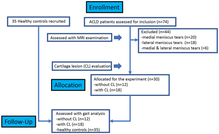
Flow diagram of the study.
MRI examination and cartilage assessment
To determine if and how the subjects were affected by tibiofemoral articular CLs, MRI examinations were conducted using a GE Signa HDxt 3.0 T magnetic resonance instrument (GE Medical System, Milwaukee, USA). A standard protocol was employed to examine the three planes of the patients' knees, covering the ACL, articular cartilage, meniscus, and other joint structures. The MRI imaging protocol included T2 axial fat saturation fast‐spin echo (TE (time of echo) 80 ms, TR (time of repetition) 4680 ms), coronal proton density (PD) fat saturation (TE 29 ms, TR 2740 ms), sagittal PD fat saturation (TE 29 ms, TR 2740 ms), and sagittal T1 fast‐spin echo (TE 15 ms, TR 420m s) sequences. Articular cartilage assessment utilized coronal and sagittal PD fat saturation sequences. The tibiofemoral articular CLs in patients were evaluated based on the modified Noyes scale. 19 Articular cartilage status was classified as follows: Level 0 for normal cartilage, Level I for signal change without surface defect, Level II for partial‐thickness defect less than 50%, Level III for partial‐thickness defect greater than 50%, and Level IV for full‐thickness defect.
Tibiofemoral cartilage was categorized into distinct subareas utilizing the Whole‐Organ Magnetic Resonance Imaging Score system (WORMS) with minor adjustments (Figure 2). 6 The tibial plateau was segregated into medial and lateral plateaus. These plateaus were further segmented into anterior, middle, and posterior subareas, demarcated by the anterior and posterior borders of the bare cartilage regions (Figure 2c). Corresponding to the tibial plateau, the femoral condyles (loading area, F1–F3, Figure 2a) were then divided in alignment with the subareas of the tibial plateau (Figure 2b). Upon diagnosing a patient with a CL, an assessment of tibiofemoral subareas for CLs was conducted. In order to evaluate the reliability of the MRI assessment, two musculoskeletal radiologists conducted the CL assessments. A Kappa test was conducted to assess the inter‐rater classification agreement between the two radiologists. The differences in the cartilage assessments were discussed and determined by the two radiologists.
Figure 2.
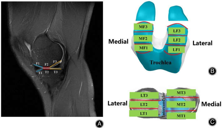
Diagram of the subareas of tibiofemoral cartilage. To assess cartilage lesion distribution, the femoral condyles and tibia plateau were divided into several subareas. Chart A showed the sagittal plane of the subareas. Charts B and C were the diagrams of the subareas of the femoral condyles. Combining the abbreviations used for the subareas of the femoral condyles and tibia plateau, the first letter represented the compartments (M for the medial compartment, L for the lateral compartments). The second letter represented the condyle or plateau (F for femoral condyle and T for tibia plateau). The final number represented the area marked on Chart A (1 for anterior, 2 for middle, 3 for posterior).
Clinical outcome scores
International Knee Documentation Committee (IKDC) subjective scores and Lysholm scores were obtained to describe the subjects.
Three‐dimensional knee kinematics during level walking
Knee kinematics were captured using a three‐dimensional (3D) motion capture system (Opti_Knee, Innomotion Inc., Shanghai, China). 20 The setup included two marker sets, a surgical navigation stereo infrared tracking device (Polaris Spectra; Northern Digital Inc., Canada), a high‐speed optical camera, a handheld digitizer, a bidirectional treadmill, and a computer workstation. The tracking device's accuracy is reported to be 0.3 mm root mean square (RMS), 21 and the gait system boasts a repeatability accuracy of less than 1.3° in rotation and 0.9 mm in translation. 22 Participants had markers attached mid‐thigh and calf with straps. Prior to collecting data, bony landmarks were identified using the handheld digitizer to create personalized 3D coordinate systems for the tibia and femur in a standing neutral position. Data collection involved participants walking barefoot on the treadmill. To ensure comfort and adaptability, participants practiced walking barefoot on the treadmill for 5 min before the actual data collection began. They then walked for 15 s at a self‐selected speed, during which knee kinematic data for about 15 gait cycles was gathered.
The knee kinematic data included measurements of the tibia's movement relative to the femur. This encompassed the knee flexion/extension angle (degrees, °), adduction/abduction angle (degrees, °), internal/external rotation angle (degrees, °), anterior/posterior translation (millimeters, mm), distal/proximal translation (millimeters, mm), and medial/lateral translation (millimeters, mm). The average kinematics during the gait cycle (GC) for each participant was calculated and segmented into two phases: the stance phase, accounting for 62% of the GC, and the swing phase, making up the remaining 38% of the GC. 23 Specifically, the stance phase was subdivided into initial contact (0%–2%), loading response (3%–12%), mid‐stance phase (13%–31%), terminal stance (32%–50%), and pre‐swing phase (51%–62%), and the swing phase was further divided into initial swing (63%–75%), mid‐swing (76%–87%), and terminal swing phase (88%–100%). 24 , 25
Statistical analysis
For demographic data (age, gender, height, weight, and BMI) and clinical outcome score comparisons, chi‐squared tests, one‐way ANOVA tests, and independent t‐tests were conducted to compare the differences between groups, respectively (SPSS version 24.0, IBM Corp., Armonk, NY, USA). 26 The primary outcome of the present study is the comparison of knee kinematics between the ACLD knees with CL and without CL groups. The secondary outcomes were the kinematic comparisons between the control group and the ACLD knees with CL and without CL groups. The statistical parametric mapping with one‐dimensional (SPM1D) One‐way ANOVA and post‐hoc independent t‐tests were used to evaluate the difference of kinematics above. The significant statistical level was set at 0.05. The SPM1D package available for MATLAB (m.0.4.8, http://www.spm1d.org) was used. The SPM1D used Random Field Theory expectations regarding smooth, one‐dimensional Gaussian Fields to examine statistical inferences for a set of one‐dimensional measurements. 27
Categorical variables, such as the distribution of CLs, were compared using chi‐squared tests, or Fisher's exact tests with Bonferroni correction as needed, with a statistical significance threshold set at 0.05. The inter‐rater agreement between the two radiologists was evaluated using MedCalc Statistical Software version 15.8 (MedCalc Software bvba, Ostend, Belgium). The agreement, measured by the Kappa value, was classified as poor (<0.20), fair (0.21–0.40), moderate (0.41–0.60), good (0.61–0.80), or excellent (0.81–1.00). 28
Results
Demographic characteristics, clinical scores
A total of 30 chronic ACLD patients met the inclusion and exclusion criteria, and the cartilage status was evaluated via MRI examination. According to the modified Noyes scale, 19 18 patients suffered from significant tibiofemoral CL (Noyes scale ≥ I) while 12 patients did not (normal articular cartilage). Details of demographic characteristics and clinical outcomes were shown in Table 1. There were no significant demographic differences among healthy controls, and ACLD patients with CL and without CL (p > 0.05). There was no significant difference in injury time between these two groups (p = 0.324). No significant differences in Lysholm and IKDC subjective scores were found between ACLD with and without CL groups (p > 0.05).
TABLE 1.
The demographic characteristics and clinical scores of subjects.
| Parameters | Average ± Standard Deviation | p‐value | ||
|---|---|---|---|---|
| Controls | ACLD with CL | ACLD without CL | ||
| Age (years) | 28.3 ± 3.1 | 30.5 ± 9.6 | 27.3 ± 5.6 | 0.289 |
| Gender (male: female) | 25:10 | 16:2 | 10:2 | 0.365 |
| Height (cm) | 169.7 ± 7.0 | 171.9 ± 9.7 | 171.2 ± 5.0 | 0.566 |
| Weight (kg) | 68.4 ± 6.8 | 70.4 ± 11.9 | 66.5 ± 10.1 | 0.509 |
| Body mass index (kg/m2) | 23.8 ± 2.5 | 24.2 ± 2.8 | 22.6 ± 2.7 | 0.275 |
| ACLD with CL | ACLD without CL | |||
|---|---|---|---|---|
| Time from injury (months) | 12.7 ± 1.4 | 13.6 ± 1.5 | 0.115 | |
| Lysholm | 89.4 ± 3.6 | 88.6 ± 1.4 | 0.212 | |
| IKDC Subjective Score | 83.8 ± 3.7 | 85.6 ± 3.8 | 0.602 | |
Motion analysis during level walking
The knee kinematic characteristics of patients with ACLD without and with CLs were shown in Figures 3, 4, 5, 6. ACLD knees with CL had significantly greater posterior tibial translation (7.7–8.0mm, 12%–18% gait cycle (GC), p = 0.014, Figure 3) compared to ACLD knees without CL. ACLD knees with CL exhibited increased posterior tibial translation (4.5–5.5mm, 0%–23% GC, p < 0.001 & 5.8–8.0mm, 86%–100%, p = 0.002, Figure 3) compared to the healthy controls. The range of motion (ROM) of flexion/extension of ACLD with CL was 10.1° lower than that of the healthy controls (p = 0.010, Figure 6). The ROM of flexion/extension of ACLD without CL was 9.9° lower than that of the healthy controls (p = 0.036, Figure 6). There were no significant differences in knee kinematics of the coronal plane (adduction/abduction, medial/lateral tibial translation) and transverse plane (internal/external tibial rotation, proximal/distal tibial translation).
FIGURE 3.
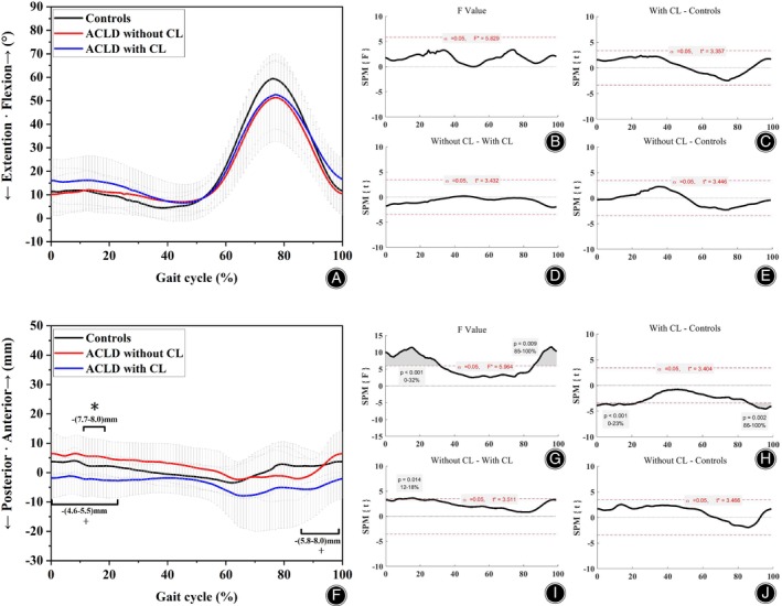
Chart of sagittal knee kinematics during level walking. Charts A&F showed the alterations of sagittal knee kinematics. Charts B–E & G–J showed the statistical values of SPM analysis. * indicates that significant alterations (p < 0.05) were found between ACLD knees with CL and ACLD knees without CL. + indicated that significant alterations (p < 0.05) were found between ACLD knees with CL and those of healthy controls. The numbers below/above the symbols represented the magnitude range of kinematics differences between the groups.
FIGURE 4.
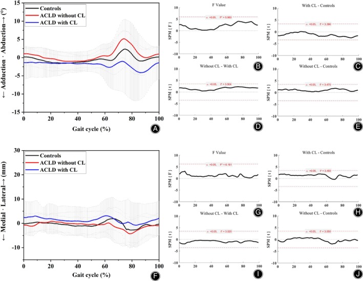
Chart of coronal knee kinematics during level walking. Charts A&F showed the alterations of coronal knee kinematics. Charts B–E & G–J showed the statistical values of SPM analysis. The results showed that there were no significant differences in knee kinematics between groups in the coronal plane.
FIGURE 5.
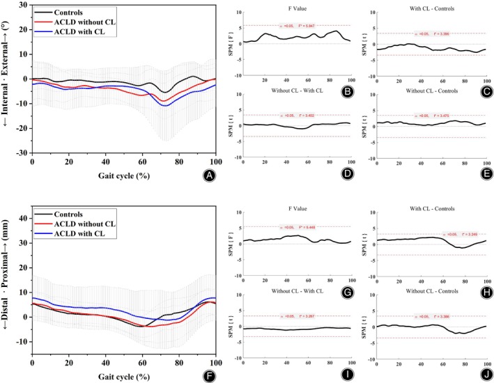
Chart of transverse knee kinematics during level walking. Charts A&F showed the alterations of transverse knee kinematics. Charts B‐E & G‐J showed the statistical values of SPM analysis. The results showed that there were no significant differences of knee kinematics between groups in transverse plane.
FIGURE 6.
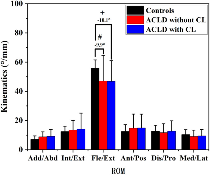
Chart of range of motion of knee kinematics during level walking. # indicated that significant alterations (p < 0.05) were found between ACLD knees without CL and those of healthy controls. + indicated that significant alterations (p < 0.05) were found between ACLD knees with CL and those of healthy controls.
CL distribution
The CL distributions in subareas were shown in Table 2. The inter‐rater agreement for the Noyes score for the subareas was excellent with a Kappa value of 0.93, indicating good reliability of MRI assessment. In the medial femoral condyle, MF1 area had more severe CLs than the other areas (MF2 & MF3, p = 0.001). In the medial tibial plateau, MT3 area had more severe CLs than the other areas (MT1 & MT2, p = 0.001). In the lateral tibial plateau, LT3 area had more severe CL than the other areas (LT1 & LT2, p = 0.019). There was no significant difference of CL distribution in lateral femoral condyle (p = 0.327).
TABLE 2.
The CL distribution in subareas of the tibiofemoral compartments in ACLD knees with CL.
| Cartilage lesions (%) | χ2 | p‐value | ||||||
|---|---|---|---|---|---|---|---|---|
| 0 | I | II | III | IV | ||||
| Medial compartment | ||||||||
| Medial femoral condyle | MF1 | 11.1 | 50.0 | 33.3 | 5.6 | ‐ | 18.922 | 0.001 |
| MF2 | 11.1 | 72.2 | 16.7 | ‐ | ‐ | |||
| MF3 | 61.1 | 38.9 | ‐ | ‐ | ‐ | |||
| Medial tibial plateau | MT1 | 22.2 | 72.2 | 5.6 | ‐ | ‐ | 19.170 | 0.001 |
| MT2 | 27.8 | 72.2 | ‐ | ‐ | ‐ | |||
| MT3 | 5.6 | 38.9 | 50.0 | ‐ | 5.6 | |||
| Lateral compartment | ||||||||
| Lateral femoral condyle | LF1 | 22.2 | 55.6 | 22.2 | ‐ | ‐ | 6.584 | 0.327 |
| LF2 | 16.7 | 44.4 | 33.3 | 5.6 | ‐ | |||
| LF3 | 44.4 | 44.4 | 11.1 | ‐ | ‐ | |||
| Lateral tibial plateau | LT1 | 33.3 | 61.1 | 5.6 | ‐ | ‐ | 13.297 | 0.019 |
| LT2 | 27.8 | 66.7 | 5.6 | ‐ | ‐ | |||
| LT3 | 11.1 | 38.9 | 22.2 | 27.8 | ‐ | |||
Note: Bold indicates significant values.
Discussion
The results confirm our hypothesis. The main findings of this study are as follows: (1) ACLD patients with CL had more posterior tibial translation than ACLD patients without CL and healthy controls during level walking, and these results may reveal the possible role of posterior tibial translation during walking in accelerating cartilage degeneration and the onset of PTOA; (2) There were significant differences of CL distribution between tibiofemoral loading regions in the group of ACLD patients with CL: (1) CLs were found mostly located in posterior tibial plateaus and (2) there were more severe CLs located in anterior loading region of medial femoral condyles. How knee kinematics play a role in cartilage degeneration remains a difficult but critical concern in delaying cartilage degeneration in patients with ACLD knees. Previous investigators proposed the theory of “loading shift leading to articular cartilage degeneration in both originated loaded and new loaded areas”. 5 , 14 Our results further showed the evidence that increased posterior tibial translation during level walking may be correlated with cartilage degenerations in chronic ACLD patients. In the group of ACLD patients with CL, we found more severe CLs located in the back of tibial plateau and anterior loading region of lateral femoral regions. Consistent with the theories' assumption, this result further showed that some areas of tibiofemoral cartilage (posterior tibial plateau and anterior loading region of lateral femoral region) would mostly degenerate in the beginning onset of post‐traumatic osteoarthritis of ACLD patients.
Increased Posterior Tibial Translation in ACLD Knees
The abnormal kinematics in patients with ACLD is an inherent problem, even after successful ACLR. 29 , 30 , 31 ACLD can result in articular cartilage degeneration and eventually the happening of PTOA. However, the mechanism of cartilage degeneration has not been fully clarified or proved. The degeneration of cartilage in ACLD knees was considered to be a multifactorial mechanism. 32 As the main alteration that occurs after an ACL injury, abnormal knee kinematics was thought to be a critical factor in the progression of CLs. Promising theories that specific kinematics inducing PTOA was proposed and discussed. 5 According to the theory of Chaudhari et al., 5 specific abnormal kinematics lead to abnormal contact and loading‐bearing in the cartilage areas that are not commonly loaded in healthy knee and this could lead to the degeneration of cartilage and osteoarthritis over time. However, the evidence remains insufficient. In this study, we tested and supported the theory proposed by Chaudhari et al. 5 by evaluating knee‐walking kinematic differences between ACLD with CL and without CL. CL development after ACLD is a key sign of the onset of PTOA. 3 , 5 , 7 Knee kinematic differences between ACLD patients with and without early CL could initially reveal whether kinematic factors accelerate CL development. Increased posterior tibial translation during level walking was found in ACLD patients with CL (Figure 3). This may indicate the important role that posterior tibial translation plays in the cartilage degeneration of ACLD knees. ACLR is known as one of the main therapeutic methods for ACLD. 33 Similar results of increased posterior tibial translation were reported in ACLR patients. Zaid et al. found abnormal (posterior) tibial position to be significantly correlated with cartilage degeneration in patients with ACLR. 11 Ikuta et al. suggested that increased posterior tibial translation could be a factor in accelerating medial knee osteoarthritis progression. 34 Nevertheless, some other studies suggested that increased posterior tibial translation was a sign of a “stiffening strategy” and caused increased compressive joint forces, which could lead to PTOA. 35 These studies indicated that increased posterior tibial translation could be an important parameter indicating cartilage degeneration in ACL‐deficient patients.
CL Distribution of ACLD Knees
The CL distribution (Table 2) showed us the early onset characteristics of PTOA in ACLD patients. Our results showed that the CLs tended to happen in the anterior and back parts of the tibial plateau and anterior regions of medial femoral condyles in ACLD patients (Table 2). This information provided us further understanding of the characteristics of cartilage degeneration in ACLD patients. As is discussed, increased posterior tibial translation could be correlated with contact area shifting and increased compressive joint forces. So, the possible explanation of the CL distribution could be that: the cartilage of the anterior and posterior tibial plateau (under the meniscus) suffered early degeneration because of new increased compressive forces, and the cartilage of the middle tibia plateau mildly degenerated because of their used powerful capability of bearing load although they also suffered increased compressive forces; the anterior region of medial condyle degenerated severely because of increased contact force between condyle and steep edge of medial plateau due to forward movement of femur. However, how posterior tibial translation actually leads to such CL characteristics remains unclear. Further study should be designed to explore their relationship.
The Strengths and Limitations of this Study
Our findings provide an extensive view of the possible kinematic mechanism of cartilage degeneration and emphasize the important role of abnormal knee kinematics in the onset of PTOA in ACLD knees. This could be helpful for clinicians to develop new solutions for the delay of cartilage degeneration and PTOA progression for those who suffer an ACL injury, such as muscle training to modify the kinematics, surgery techniques, or new kinematic‐based braces. The main limitation of this study is the small sample size of 30 ACLD subjects and 35 healthy controls. This was partially because only a small portion of ACLD patients delayed their therapy, and because over half of the ACLD patients (44 patients) suffered from secondary meniscus injuries and were excluded. This was also because the comprehensive and careful experimental procedure included an MRI examination, 3D gait analysis, and clinical assessment. However, the present study contains important emerging evidence about the relationship between specific kinematic characteristics during level walking and the progressive pathogenesis of early CLs in ACLD patients. Another limitation of this study was the use of skin markers in gait analysis, which can cause errors due to skin movement. The surgical navigation stereo infrared tracking device was reported to have an accuracy level of 0.3 mm root mean square (RMS) 21 , and the gait system was reported to have a repeatability of less than 1.3° in rotation and 0.9 mm in translation. 22 We took precautions to minimize the skin movement error by having a well‐trained orthopaedic clinician with 3 years of experiences place all the markers and acquire the kinematic data during level walking. Kinematics and CL were measured around the early stage of chronic ACLD (1–3 years) without PTOA. This could get rid of the influence of osteophytes and other factors of PTOA allowing us to study this relationship. Besides, it has been reported that CL couldn't affect kinematics and loading distribution. 36 The allocation of ACLD with and without CL could allow us to study the contribution of kinematics on CL causally. In addition, in this study, we didn't investigate this other risk‐factors, like tibial slope, AP, and rotational instability of the tibia. Future study should comprehensively consider these factors.
Conclusion
The main finding of the study is that significantly increased posterior tibial translation during gait cycles was identified in anterior cruciate ligament‐deficient patients with cartilage lesions compared to those of anterior cruciate ligament‐deficient patients without cartilage lesions and healthy controls. The increased posterior tibial translation may play an important role in knee cartilage lesions in anterior cruciate ligament‐deficient knees. The findings may improve our understanding of the role of kinematics in cartilage degeneration and could be a useful reference for anterior cruciate ligament‐deficient therapy.
Author Contributions
XZ: conceptualization, methodology, statistical analysis, writing. FL: writing, methodology. WH: Investigation. LK: Investigation. JZ: Investigation. Da Guo: methodology. Yu Zhang: methodology. DL: conceptualization, funding acquisition.
Ethics Statement
The study was approved by the independent ethics committee (No. 2019‐226H‐1) of Guangdong Provincial People's Hospital. Informed consent was obtained from each participant.
Acknowledgments
The study was supported by the Project of Administration of Traditional Chinese Medicine of Guangdong Province (No.Z155080000004), Chinese Medicine Leading Talents Project (Qihuang Scholar, No. [2022]6 of Chinese Department of TCM Education), Lin Dingkun's Guangdong Province Famous Traditional Chinese Medicine Inheritance Studio Construction Project (No. 0103030912), Lingnan Orthopedic School Inheritance Studio Construction Project (No. E43607), Science and Technology Planning Project of Guangdong Province (No. 2023B1212060063), Shi Yangshan Academic Experience Inheritance Studio Construction Project (No.E43715), and Guangdong Provincial Hospital of Chinese Medicine Scientific and Technological Research Special Project (YN2020MS05).
Xiaolong Zeng and Fangzheng Lin are co‐first authors.
Disclosure: The authors declare that they have no conflict of interest.
Contributor Information
Xiaolong Zeng, Email: xiaolzeng@126.com.
Yu Zhang, Email: luck_2001@126.com.
Dingkun Lin, Email: lindingkuntcm@126.com.
References
- 1. Ajuied A, Wong F, Smith C, Norris M, Earnshaw P, Back D, et al. Anterior cruciate ligament injury and radiologic progression of knee osteoarthritis: a systematic review and meta‐analysis. Am J Sports Med. 2014;42(9):2242–2252. https://www.ncbi.nlm.nih.gov/pubmed/24214929 [DOI] [PubMed] [Google Scholar]
- 2. Poulsen E, Goncalves GH, Bricca A, Roos EM, Thorlund JB, Juhl CB. Knee osteoarthritis risk is increased 4‐6 fold after knee injury – a systematic review and meta‐analysis. Br J Sports Med. 2019;53(23):1454–1463. https://www.ncbi.nlm.nih.gov/pubmed/31072840 [DOI] [PubMed] [Google Scholar]
- 3. Bodkin SG, Werner BC, Slater LV, Hart JM. Post‐traumatic osteoarthritis diagnosed within 5 years following ACL reconstruction. Knee Surg Sports Traumatol Arthrosc. 2020;28(3):790–796. https://www.ncbi.nlm.nih.gov/pubmed/30887068 [DOI] [PubMed] [Google Scholar]
- 4. Glyn‐Jones S, Palmer AJ, Agricola R, Price AJ, Vincent TL, Weinans H, et al. Osteoarthritis. Lancet. 2015;386(9991):376–387. https://www.ncbi.nlm.nih.gov/pubmed/25748615 [DOI] [PubMed] [Google Scholar]
- 5. Chaudhari AM, Briant PL, Bevill SL, Koo S, Andriacchi TP. Knee kinematics, cartilage morphology, and osteoarthritis after ACL injury. Med Sci Sports Exerc. 2008;40(2):215–222. https://www.ncbi.nlm.nih.gov/pubmed/18202582 [DOI] [PubMed] [Google Scholar]
- 6. Li G, Li JS, Torriani M, Hosseini A. Short‐term contact kinematic changes and longer‐term biochemical changes in the cartilage after ACL reconstruction: a pilot study. Ann Biomed Eng. 2018;46(11):1797–1805. https://www.ncbi.nlm.nih.gov/pubmed/29946971 [DOI] [PMC free article] [PubMed] [Google Scholar]
- 7. Svantesson E, Hamrin Senorski E, Webster KE, Karlsson J, Diermeier T, Rothrauff BB, et al. Clinical outcomes after anterior cruciate ligament injury: panther symposium ACL injury clinical outcomes consensus group. Knee Surg Sports Traumatol Arthrosc. 2020;28(8):2415–2434. https://www.ncbi.nlm.nih.gov/pubmed/32767052 [DOI] [PMC free article] [PubMed] [Google Scholar]
- 8. Biz C, Maso G, Gambato M, Belluzzi E, Pozzuoli A, Favero M, et al. Challenging surgical treatment of displaced articular Tibial plateau fractures: do early knee radiographic features have a predictive value of the mid‐term clinical functional outcomes? Orthop Surg. 2019;11(6):1149–1162. [DOI] [PMC free article] [PubMed] [Google Scholar]
- 9. Belluzzi E, Olivotto E, Toso G, Cigolotti A, Pozzuoli A, Biz C, et al. Conditioned media from human osteoarthritic synovium induces inflammation in a synoviocyte cell line. Connect Tissue Res. 2019;60(2):136–145. [DOI] [PubMed] [Google Scholar]
- 10. Frank CB, Beveridge JE, Huebner KD, Heard BJ, Tapper JE, O'Brien EJ, et al. Complete ACL/MCL deficiency induces variable degrees of instability in sheep with specific kinematic abnormalities correlating with degrees of early osteoarthritis. J Orthop Res. 2012;30(3):384–392. https://www.ncbi.nlm.nih.gov/pubmed/21919045 [DOI] [PubMed] [Google Scholar]
- 11. Zaid M, Lansdown D, Su F, Pedoia V, Tufts L, Rizzo S, et al. Abnormal tibial position is correlated to early degenerative changes one year following ACL reconstruction. J Orthop Res. 2015;33(7):1079–1086. https://www.ncbi.nlm.nih.gov/pubmed/25721417 [DOI] [PMC free article] [PubMed] [Google Scholar]
- 12. Zampeli F, Pappas E, Velonakis G, Roumpelakis IM, Poulou LS, Papagiannis GI, et al. Development of new cartilage lesions after ACL reconstruction is associated with abnormal knee rotation. Knee Surg Sports Traumatol Arthrosc. 2022;30(3):842–851. https://www.ncbi.nlm.nih.gov/pubmed/33528592 [DOI] [PubMed] [Google Scholar]
- 13. Stergiou N, Ristanis S, Moraiti C, Georgoulis AD. Tibial rotation in anterior cruciate ligament (ACL)‐deficient and ACL‐reconstructed knees: a theoretical proposition for the development of osteoarthritis. Sports Med. 2007;37(7):601–613. https://www.ncbi.nlm.nih.gov/pubmed/17595155 [DOI] [PubMed] [Google Scholar]
- 14. Andriacchi TP, Koo S, Scanlan SF. Gait mechanics influence healthy cartilage morphology and osteoarthritis of the knee. J Bone Joint Surg Am. 2009;91(Suppl 1):95–101. https://www.ncbi.nlm.nih.gov/pubmed/19182033 [DOI] [PMC free article] [PubMed] [Google Scholar]
- 15. Crook BS, Collins AT, Lad NK, Spritzer CE, Wittstein JR, DeFrate LE. Effect of walking on in vivo tibiofemoral cartilage strain in ACL‐deficient versus intact knees. J Biomech. 2021;116:110210 https://www.ncbi.nlm.nih.gov/pubmed/33444927 [DOI] [PMC free article] [PubMed] [Google Scholar]
- 16. Haughom B, Schairer W, Souza RB, Carpenter D, Ma CB, Li X. Abnormal tibiofemoral kinematics following ACL reconstruction are associated with early cartilage matrix degeneration measured by MRI T1rho. Knee. 2012;19(4):482–487. https://www.ncbi.nlm.nih.gov/pubmed/21807522 [DOI] [PMC free article] [PubMed] [Google Scholar]
- 17. Li AK, Ochoa JK, Pedoia V, Amano K, Souza RB, Li X, et al. Altered tibiofemoral position following ACL reconstruction is associated with cartilage matrix changes: a voxel‐based relaxometry analysis. J Orthop Res. 2020;38(11):2454–2463. https://www.ncbi.nlm.nih.gov/pubmed/32369216 [DOI] [PubMed] [Google Scholar]
- 18. Arno S, Hadley S, Campbell KA, Bell CP, Hall M, Beltran LS, et al. The effect of arthroscopic partial medial meniscectomy on tibiofemoral stability. Am J Sports Med. 2013;41(1):73–79. https://www.ncbi.nlm.nih.gov/pubmed/23149019 [DOI] [PubMed] [Google Scholar]
- 19. Gold GE, Chen CA, Koo S, Hargreaves BA, Bangerter NK. Recent advances in MRI of articular cartilage. AJR Am J Roentgenol. 2009;193(3):628–638. https://www.ncbi.nlm.nih.gov/pubmed/19696274 [DOI] [PMC free article] [PubMed] [Google Scholar]
- 20. Zhang Y, Yao Z, Wang S, Huang W, Ma L, Huang H, et al. Motion analysis of Chinese normal knees during gait based on a novel portable system. Gait Posture. 2015;41(3):763–768. https://www.ncbi.nlm.nih.gov/pubmed/25743776 [DOI] [PubMed] [Google Scholar]
- 21. Elfring R, de la Fuente M, Radermacher K. Assessment of optical localizer accuracy for computer aided surgery systems. Comput Aided Surg. 2010;15(1–3):1–12. https://www.ncbi.nlm.nih.gov/pubmed/20233129 [DOI] [PubMed] [Google Scholar]
- 22. Zhang Y, Huang W, Yao Z, Ma L, Lin Z, Wang S, et al. Anterior cruciate ligament injuries Alter the kinematics of knees with or without meniscal deficiency. Am J Sports Med. 2016;44(12):3132–3139. https://www.ncbi.nlm.nih.gov/pubmed/27511793 [DOI] [PubMed] [Google Scholar]
- 23. Levine (Veterinary physical therapist) D, Whittle M, Whittle MGA. Whittle's gait analysis. In: Levine D, Richards J, Whittle MW, editors. 5th ed. Edinburgh; New York, Scotland: Churchill Livingstone/Elsevier; 2012. [Google Scholar]
- 24. Perry J, Burnfield JM. Gait analysis: normal and pathological function. 2nd ed. Thorofare, NJ: SLACK; 2010. [Google Scholar]
- 25. Levine D, Richards J, Whittle M, Whittle M. Whittle's gait analysis. 5th ed. New York: Churchill Livingstone/Elsevier, Edinburgh; 2012. [Google Scholar]
- 26. Chang WX, Xu N, Kumagai T, Shiraishi T, Kikuyama T, Omizo H, et al. The impact of Normal range of serum phosphorus on the incidence of end‐stage renal disease by a propensity score analysis. PloS One. 2016;11(4):e0154469 https://www.ncbi.nlm.nih.gov/pubmed/27123981 [DOI] [PMC free article] [PubMed] [Google Scholar]
- 27. Pataky TC. One‐dimensional statistical parametric mapping in python. Comput Methods Biomech Biomed Engin. 2012;15(3):295–301. [DOI] [PubMed] [Google Scholar]
- 28. Altman DG. Practical statistics for medical research, Chapman & Hall/CRC. Boca Raton: Fla; 1999. [Google Scholar]
- 29. Slater LV, Hart JM, Kelly AR, Kuenze CM. Progressive changes in walking kinematics and kinetics after anterior cruciate ligament injury and reconstruction: a review and meta‐analysis. J Athl Train. 2017;52(9):847–860. https://www.ncbi.nlm.nih.gov/pubmed/28985125 [DOI] [PMC free article] [PubMed] [Google Scholar]
- 30. Shabani B, Bytyqi D, Lustig S, Cheze L, Bytyqi C, Neyret P. Gait changes of the ACL‐deficient knee 3D kinematic assessment. Knee Surg Sports Traumatol Arthrosc. 2015;23(11):3259–3265. https://www.ncbi.nlm.nih.gov/pubmed/25026934 [DOI] [PubMed] [Google Scholar]
- 31. Zampeli F, Terzidis I, Espregueira‐Mendes J, Georgoulis JD, Bernard M, Pappas E, et al. Restoring tibiofemoral alignment during ACL reconstruction results in better knee biomechanics. Knee Surg Sports Traumatol Arthrosc. 2018;26(5):1367–1374. https://www.ncbi.nlm.nih.gov/pubmed/29067474 [DOI] [PubMed] [Google Scholar]
- 32. Diermeier T, Rothrauff BB, Engebretsen L, Lynch AD, Svantesson E, Hamrin Senorski E, et al. Treatment after anterior cruciate ligament injury: panther symposium ACL treatment consensus group. J ISAKOS. 2021;6(3):129–137. https://www.ncbi.nlm.nih.gov/pubmed/34006576 [DOI] [PubMed] [Google Scholar]
- 33. Biz C, Cigolotti A, Zonta F, Belluzzi E, Ruggieri P. ACL reconstruction using a bone patellar tendon bone (BPTB) allograft or a hamstring tendon autograft (GST): a single‐center comparative study. Acta Biomed. 2019;90(12‐s):109–117. [DOI] [PMC free article] [PubMed] [Google Scholar]
- 34. Ikuta F, Yoneta K, Miyaji T, Kidera K, Yonekura A, Osaki M, et al. Knee kinematics of severe medial knee osteoarthritis showed tibial posterior translation and external rotation: a cross‐sectional study. Aging Clin Exp Res. 2020;32(9):1767–1775. https://www.ncbi.nlm.nih.gov/pubmed/31598915 [DOI] [PubMed] [Google Scholar]
- 35. Chmielewski TL, Ramsey DK, Snyder‐Mackler L. Evidence for differential control of tibial position in perturbed unilateral stance after acute ACL rupture. J Orthop Res. 2005;23(1):54–60. https://www.ncbi.nlm.nih.gov/pubmed/15607875 [DOI] [PubMed] [Google Scholar]
- 36. Van Rossom S, Khatib N, Holt C, Van Assche D, Jonkers I. Subjects with medial and lateral tibiofemoral articular cartilage defects do not alter compartmental loading during walking. Clin Biomech (Bristol, Avon). 2018;60:149–156. https://www.ncbi.nlm.nih.gov/pubmed/30366244 [DOI] [PubMed] [Google Scholar]