Abstract
We recently developed a system for the generation of infectious bovine respiratory syncytial virus (BRSV) from cDNA. Here, we report the recovery of fully viable chimeric recombinant BRSVs (rBRSVs) that carry human respiratory syncytial virus (HRSV) glycoproteins in place of their BRSV counterparts, thus combining the replication machinery of BRSV with the major antigenic determinants of HRSV. A cDNA encoding the BRSV antigenome was modified so that the complete G and F genes, including the gene start and gene end signals, were replaced by their HRSV A2 counterparts. Alternatively, the BRSV F gene alone was replaced by that of HRSV Long. Each antigenomic cDNA directed the successful recovery of recombinant virus, yielding rBRSV/A2 and rBRSV/LongF, respectively. The HRSV G and F proteins or the HRSV F in combination with BRSV G were expressed efficiently in cells infected with the appropriate chimeric virus and were efficiently incorporated into recombinant virions. Whereas BRSV and HRSV grew more efficiently in bovine and human cells, respectively, the chimeric rBRSV/A2 exhibited intermediate growth characteristics in a human cell line and grew better than either parent in a bovine line. The cytopathology induced by the chimera more closely resembled that of BRSV. BRSV was confirmed to be highly restricted for replication in the respiratory tract of chimpanzees, a host that is highly permissive for HRSV. Interestingly, the rBRSV/A2 chimeric virus was somewhat more competent than BRSV for replication in chimpanzees but remained highly restricted compared to HRSV. This showed that the substitution of the G and F glycoproteins alone was not sufficient to induce efficient replication in chimpanzees. Thus, the F and G proteins contribute to the host range restriction of BRSV but are not the major determinants of this phenotype. Although rBRSV/A2 expresses the major neutralization and protective antigens of HRSV, chimpanzees infected with this chimeric virus were not significantly protected against subsequent challenge with wild-type HRSV. This suggests that the growth restriction of rBRSV/A2 was too great to provide adequate antigen expression and that the capacity of this chimeric vaccine candidate for replication in primates will need to be increased by the importation of additional HRSV genes.
Bovine respiratory syncytial virus (BRSV) and Human respiratory syncytial virus (HRSV) are closely related members of the Pneumovirus genus within the subfamily Pneumovirinae, family Paramyxoviridae, order Mononegavirales (6, 36). BRSV is a major cause of respiratory tract disease in cattle (32, 44). HRSV is the most important causative agent of viral pediatric respiratory disease worldwide (6). A licensed vaccine against HRSV is not available, though several promising candidates for attenuated live vaccines recently have been developed (references 17, 46, 47, and 48, and references therein).
The genomes of HRSV and of BRSV are single-stranded, negative-sense RNAs of 15,222 nucleotides (nt) (HRSV A2) and 15,140 nt (BRSV ATue51908) (1, 4). Their genome organizations are identical, comprising 10 genes (encoding 10 mRNAs containing 11 translational open reading frames [ORFs]) in the order 3′-NS1-NS2-N-P-M-SH-G-F-M2-1/M2-2-L-5′. Like all members of Mononegavirales, the respiratory syncytial virus (RSV) genomic RNA is tightly encapsidated by the nucleoprotein (N) and is associated with the phosphoprotein (P) and the viral RNA-dependent RNA polymerase (L). This encapsidated RNA is the template for replication and transcription (13, 49). The viral polymerase enters the genome in the 3′ leader region and synthesizes the 10 monocistronic mRNAs by linear sequential start-stop transcription. This is directed by the respective gene start signals, which are located on the upstream end of each gene and are highly conserved between BRSV and HRSV, and the gene end/polyadenylation signals, which are located at the downstream end of each gene and are semiconserved (21, 22, 50). RNA replication yields a full-length, positive-sense intermediate, the antigenome, which serves as template for the synthesis of full-length genomic RNA. The N, P, and L proteins are necessary and sufficient to direct RNA replication, whereas efficient synthesis of full-length mRNAs and efficient sequential transcription also require the M2-1 transcription antitermination factor (5, 9, 13, 14, 49).
In addition, RSV encodes four envelope-associated structural proteins. One of these is the nonglycosylated matrix (M) protein, which is essential for virion formation (42) and is thought to mediate interaction between the nucleocapsid and the envelope. The other three are transmembrane surface glycoproteins: the attachment glycoprotein G (25), the fusion glycoprotein F (45), and the small hydrophobic protein SH (2). For both BRSV and HRSV, the G and F proteins are the major neutralization and protective antigens (7, 31, 41). The G protein is highly variable between the HRSV subgroups (16) and between HRSV and BRSV (24), with a conserved central structure (16) that might be involved in receptor binding. Between the BRSV strain ATue51908 and the HRSV A2 used in this work, the overall amino acid identity of the G protein is 28%. The F protein mediates viral penetration and syncytium formation (45). F is synthesized as an F0 precursor which is cleaved endoproteolytically into the F1 and F2 subunits that remain linked by disulfide bonds. BRSV ATue51908 and HRSV A2 F proteins are 82% identical.
Live vaccinia virus has the distinction of being both the first vaccine and the most successful, having led to the eradication of smallpox as a human disease. The original vaccinia virus introduced by Jenner in 1798 was of bovine origin. The use of an animal virus as a live vaccine against its human counterpart is called the “Jennerian” approach. This strategy requires that the human virus of interest has an animal counterpart which is attenuated in the nonnatural human host, yet grows sufficiently well and is sufficiently related antigenically that it can induce effective protection against the human virus. As a current example, bovine parainfluenzavirus (BPIV) type 3 is under clinical evaluation as vaccine against human parainfluenzavirus (HPIV) type 3 and appears to have appropriate levels of attenuation and immunogenicity (19). The recently licensed quadrivalent rotavirus vaccine exemplifies both the Jennerian approach and a “modified Jennerian” approach (33). This vaccine is based on a rhesus rotavirus that is attenuated in humans and has sufficient antigenic relatedness to the human rotavirus serotype 3 that it can confer resistance in humans to this human rotavirus serotype. Effective immunoprophylaxis against human rotavirus disease also requires resistance to the three other major serotypes of human rotavirus. Therefore, the rhesus rotavirus was used to construct, by the natural mechanism of gene reassortment during mixed infection in cell culture, three single-gene reassortant viruses. In each, the rhesus rotavirus gene encoding VP7, a major neutralization antigen, was replaced by its counterpart from human rotavirus serotype 1, 2, or 4. The resulting three chimeric reassortant viruses were combined with the rhesus rotavirus parent to formulate the quadrivalent vaccine. Chimeric animal/human viruses represent the modified Jennerian strategy.
BRSV and HRSV are related antigenically (11, 32), and BRSV has been considered as a possible Jennerian vaccine against HRSV (34, 35). In previous work (34, 35), BRSV was evaluated in several experimental animal models, most notably in chimpanzee, which is the animal that is the most permissive for HRSV replication and disease. When BRSV was administered intranasally to two animals, replication was detected in only one animal, and only at a very low level, and the animals were not significantly resistant to subsequent HRSV challenge (35) (R. M. Chanock, personal communication).
Recently, we reported the cDNA cloning and sequencing of the entire BRSV genome and the establishment of a reverse genetics system allowing recovery of infectious recombinant BRSV (rBRSV) (1). In the present work, we reevaluated the Jennerian approach by assaying the replication of rBRSV in chimpanzees and its protective efficacy against HRSV. Furthermore, we made use of this system to generate chimeric rBRSV in which the glycoprotein F gene alone, or F and G genes together, were replaced by their HRSV counterpart(s), thus combining the replication features of BRSV and the antigenic properties of HRSV. This represents the first step in developing a chimeric BRSV/HRSV virus as a vaccine against HRSV, and this modified Jennerian vaccine candidate also was evaluated in chimpanzees. The construction of bovine/human chimeric viruses also addresses the fundamental issue of the contribution of the G and F glycoproteins in determining the host range of RSV.
MATERIALS AND METHODS
Construction of cDNAs encoding chimeric BRSV/HRSV antigenomes.
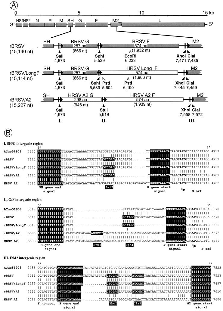
We previously constructed a cDNA encoding BRSV antigenomic RNA (1). This was modified by site-directed mutagenesis (20) in two ways: first, a unique synthetic NotI site in the NS1 noncoding region was removed to regenerate the ATue51908 wild-type sequence (GenBank accession no. AF 092942). Second, synthetic restriction sites were introduced into the SH/G (SalI, ATue51908 nt 4673), G/F (SphI, ATue51908 nt 5539), and F/M2 (XhoI, ATue51908 nt 7471 and ClaI, ATue51908 nt 7485) intergenic regions (Fig. 1B). The resulting plasmid was termed pBRSV18. This cDNA served as parent for the construction of BRSV/HRSV chimeras.
FIG. 1.
Construction of chimeric rBRSV in which the F gene alone was replaced by its counterpart from HRSV Long to create rBRSV/LongF or in which the F and G genes were both replaced by their counterparts from HRSV A2 to create rBRSV/A2. (A) Diagram of the rBRSV genome, illustrating the replacement of the F and G genes with those of HRSV. The location of the ORFs are shown as shaded (BRSV) or open (HRSV) rectangles. The G and F genes are shown as enlargements, in which gene end/polyadenylation signals are represented by bars, gene start signals are shown as filled triangles, and the locations of synthetic restriction sites are indicated by filled arrowheads with the position in the complete antigenome sequence shown underneath. Naturally occurring PstI and EcoRI restriction sites used for differentiation of PCR products obtained from recombinant viruses are indicated by open arrowheads. The lengths of the fragments framed by the marker restriction sites are indicated in parentheses. The genome length of each recombinant virus is indicated on the left in parentheses. The roman numerals (I, II, and III) refer to intergenic regions which are expanded in panel B. (B) Alignment of the SH/G (I), G/F (II), and F/M2 (III) intergenic regions of biologically derived BRSV strain ATue51908 (top line), its recombinant version rBRSV (second line), the chimeric virus rBRSV/LongF (third line), the recombinant chimeric virus rBRSV/A2 (fourth line), and rHRSV strain A2 (bottom line). The sequences are shown as DNA-positive strands. Gaps are indicated by dots, gene start and gene end signals are shaded, and translation start codons are in boldface. Restriction sites are indicated, with recognition sequences shaded. Numbering refers to the antigenomic positions (for BRSV, ATue51908, GenBank accession no. AF092942; for HRSV strain A2, GenBank accession no. M74568, further modified as described in reference 4).
The G and F genes of the previously reported recombinant version of HRSV A2 of antigenic subgroup A (GenBank accession no. M74568, further modified as described in reference 4) were amplified as a single cDNA (A2 nt 4674 to 7551) with oligonucleotide primers which introduced a SalI site immediately upstream of the G gene and an XhoI site immediately downstream of the F gene (Fig. 1B). The PCR product was cloned, and its sequence was confirmed. Subsequently, the SalI and XhoI restriction sites were used to transfer an approximately 2,885-bp fragment spanning the HRSV G and F genes, including gene start- and gene end/polyadenylation signals, into pBRSV18, replacing the respective BRSV genes. The resulting plasmid was termed pBRSV/A2-10.
A second antigenomic plasmid was constructed which contains the F gene of HRSV strain Long (26) in place of the BRSV F gene. Total RNA was made from HEp-2 cells infected with HRSV Long. Subsequently, reverse transcription (RT)-PCR was performed using primers FLa (5′-AGGAATTCGCATGCGGGGCAAATAACAATGGAGTTGCCAATC-3′), containing nt 1 to 28 of the HRSV Long F gene sequence preceded by an EcoRI/SphI adapter (underlined), and FLRa (5′-AGGAATTCTCGAGTTTTTATATAACTATAAACTAGGAATCTAC-3′), containing nt 1904 to 1875 of the Long F gene followed by an EcoRI/XhoI adapter (underlined). This yielded a PCR product of 1,931 bp. The cloned cDNA was sequenced, and a 1,906-bp SphI/XhoI fragment containing the complete Long F gene was excised and transferred into pBRSV18 in place of the BRSV F gene, resulting in plasmid pBRSV/LongF-12. Figure 1 represents a schematic overview of the genome organization of the recombinant viruses (rBRSV, rBRSV/A2, and rBRSV/LongF) recovered from pBRSV18, pBRSV/A2-10, and pBRSV/LongF-12, respectively.
Recovery of recombinant RSVs.
Recombinant RSVs were recovered from cDNA essentially as described before (1). Dishes (diameter, 32 mm) of subconfluent BSR T7/5 cells stably expressing phage T7 RNA polymerase were transfected with 5 μg of the respective antigenomic plasmid and a set of four support plasmids (2.5 μg of pN, 2.5 μg of pP, 0.5 μg of pL, and 0.5 μg of pM2) from which the BRSV N, P, L, and M2-1 proteins are expressed. All cDNA constructs were under control of a T7 promoter (1). Transfections were carried out by using Superfect (Qiagen). Two hours after transfection, the supernatant was removed, and cells were washed and maintained in minimum essential medium supplemented with 3% fetal calf serum (FCS). Three days after transfection, the cells were split in a 1 to 3 ratio. Cells and supernatant were harvested 7 days after transfection.
Viruses and cell culture.
Recombinant BRSV and BRSV/HRSV chimeric viruses were propagated in monolayer cultures of MDBK cells. Cells were infected at a multiplicity of infection (MOI) of 0.1 and were maintained at 37°C in minimum essential medium supplemented with 3% FCS. When the cytopathic effect (CPE) was pronounced, the medium was adjusted to 100 mM MgSO4 and to 50 mM HEPES (pH 7.5) (10), and cells and medium were collected and subjected to three rounds of freezing and thawing, and the clarified medium supernatants were stored at −70°C and titered by limiting dilution method on BSR T7/5 cells (1). HRSV Long (kindly provided by G. Herrler, Hannover, Germany) was propagated on HEp-2 cells by the same general procedure. HRSV Long is very similar to strain A2 with regard to sequence analysis (16, 26), with 98% amino acid sequence identity for the F protein, and also is closely related based on reactivity with most monoclonal antibodies (MAbs) (12). The Long strain was used as the wild-type HRSV control for the in vitro studies.
RT-PCR.
Total RNA of MDBK cells infected individually with each RSV was prepared (RNeasy, Qiagen) when extensive CPE was observed. To verify the identity of the recombinant viruses, regions containing the marker restriction sites shown in Fig. 1B were amplified by RT-PCR (1).
BRSV ATue51908, rBRSV, and rBRSV/A2 were reverse transcribed in a series of parallel reactions using positive-sense primers which were complementary to (i) the BRSV M gene (ATue51908 nt 3612 to 3635), (ii) the BRSV G gene (ATue51908 nt 5372 to 5392) or the HRSV G gene (HRSV A2 nt 5442 to 5463), or (iii) the BRSV F gene (ATue51908 nt 7218 to 7240) or HRSV F gene (A2 nt 7309 to 7329). An aliquot of the first-strand cDNA was used for PCR, and the following reactions were performed: the SH/G region was amplified with primer and first-strand cDNA from (i) above together with a BRSV G-specific primer (ATue51908 nt 4886 to 4862, negative sense) or an HRSV G-specific primer (A2 4878 to 4856, negative sense) (Fig. 2A), the G/F region was amplified with the appropriate primer and first-strand cDNA from (ii) above together with a BRSV F-specific primer (ATue51908 nt 5964 to 5941, negative sense) or an HRSV F-specific primer (A2 nt 6055 to 6033, negative sense) (Fig. 2B), or the F/M2 region was amplified with the appropriate first-strand cDNA and primer from (iii) above and a BRSV M2-specific primer (ATue51908 nt 7852 to 7832, negative sense) (Fig. 2C).
FIG. 2.
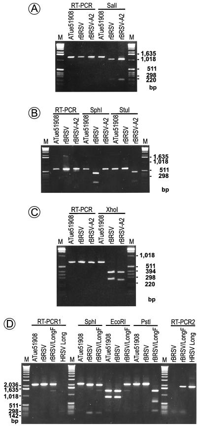
Demonstration of marker restriction sites in the genomes of biologically derived BRSV ATue51908, rBRSV, the chimeras rBRSV/A2 and rBRSV/LongF, and HRSV Long. RT-PCR was performed on infected-cell RNA, and the PCR products were subjected to restriction analysis and separated on a 2% (panels A and B), 3% (panel C), or 0.8% (panel D) agarose gel. M, 1-kb marker DNA ladder (Life Technologies) with the size of some marker fragments indicated. (A) Analysis of RT-PCR products encompassing the SH/G intergenic region and demonstration of the presence of a SalI site in rBRSV and rBRSV/A2 and its absence in biologically derived BRSV ATue51908. The RT-PCR products were consistent with the predicted size of approximately 1,274 bp, and the SalI digestion products were consistent with the predicted sizes of approximately 1,062 and 212 bp. (B) Analysis of RT-PCR products encompassing the G/F intergenic region and demonstration of the presence of an SphI site in rBRSV and a StuI site in rBRSV/A2. The PCR products were approximately 592 bp (ATue51908 and rBRSV) or 614 bp (rBRSV/A2). SphI digested only rBRSV, resulting in fragments of 425 and 167 bp. StuI digested only rBRSV/A2, resulting in fragments of 443 and 171 bp. (C) Analysis of RT-PCR products encompassing the F/M2 intergenic region, and demonstration of an XhoI site in rBRSV and rBRSV/A2 and its absence in BRSV ATue51908. The RT-PCR product was 637 (rBRSV/A2) or 648 bp (ATue51908 and rBRSV). XhoI digestion resulted in fragments of 381 (rBRSV and rBRSV/A2), 267 (rBRSV), or 256 bp (rBRSV/A2). (The last two bands each contain 14 additional nonviral nucleotides contributed by the oligonucleotide primers.) (D) Analysis of RT-PCR products confirming the identity of rBRSV/LongF. One series of reactions (RT-PCR1) was carried out using a BRSV-specific primer pair hybridizing upstream and downstream of the F gene. This yielded products of 2,187 bp (BRSV strain ATue51908 and rBRSV) or 2,161 bp (rBRSV/LongF), whereas the BRSV-specific primers did not produce a product from HRSV Long. SphI cleaved the products representing rBRSV and rBRSV/A2, yielding bands of 2,028 and 159 bp (rBRSV) or 2,002 and 159 bp (BRSV/LongF). BRSV strain ATue51908 was not cleaved. EcoRI cleaved BRSV ATue51908 and rBRSV at a naturally occurring site in the BRSV F gene to yield two fragments of 1,334 and 853 bp, whereas the fragment originating from rBRSV/LongF was not cleaved by EcoRI. PstI cleaved rBRSV/LongF at two naturally occurring sites in the Long F gene to yield fragments of 1,351, 586, and 224 bp, whereas rBRSV F gene in BRSV ATue51908 and rBRSV remained uncleaved. A second series of reactions (RT-PCR2) was performed using a primer pair that hybridized at either end of the HRSV Long F gene, yielding a PCR product of 1,904 bp for rBRSV/LongF and the HRSV Long parental virus. As expected, a product was not obtained from rBRSV.
BRSV ATue51908, rBRSV, rBRSV/LongF, and HRSV Long were compared in two different RT-PCRs. The first was performed with a primer pair hybridizing with the BRSV backbone upstream and downstream of the F gene, with the positive-sense upstream primer defined by nt 5380 to 5397 (G gene) and the negative-sense downstream primer defined by nt 7567 to 7550 (M2 gene). The second RT-PCR was performed with a primer pair hybridizing to either end of the HRSV Long F gene (primer FL, 5′-GGGGCAAATAACAATGGAGTTGCCAATC-3′, nt 1 to 28 of the HRSV Long F gene sequence, and primer FLR, 5′-TTTTTATATAACTATAAACTAGGAATCTAC-3′, HRSV Long F gene nt 1904 to 1875).
The RT-PCR products were subjected to restriction digestion and were analyzed on agarose gels. In addition, the origin and identity of the sequences of each region which was amplified by RT-PCR, and in particular each junction between BRSV and HRSV, was confirmed by partial nucleotide sequencing of the RT-PCR products with an automated sequencer (LI-COR, MWG-Biotech).
Indirect immunofluorescence assay.
HEp-2 cells were infected with an MOI of 0.1 with rBRSV, rBRSV/A2, rBRSV/LongF, or HRSV Long, were incubated for 36 h, were fixed with 80% acetone, and were incubated at 37°C for 30 min with MAb G66 directed to BRSV G (1:1,000 dilution), kindly provided by G. Taylor (11, 23), or MAb 021/1G directed against HRSV G (1:40 dilution), MAb 2F reacting with both HRSV and BRSV F (1:40 dilution), or MAb 44F, specific to HRSV F (1:40 dilution), kindly provided by J. A. Melero (12, 27). Cells were stained with a fluorescein-isothiocyanate-conjugated goat anti-mouse immunoglobulin G (Dianova, Hamburg, Germany) and were counterstained with 0.01% Evans Blue.
Growth analysis.
Subconfluent MDBK cells or HEp-2 cells were infected with rBRSV, rBRSV/A2, rBRSV/LongF, or HRSV strain Long at an MOI of 0.1. After 90 min of adsorption, the inoculum was removed and the cells were washed twice with medium containing 3% FCS and were incubated at 37°C. At various times after infection, the medium was adjusted to 100 mM MgSO4 and 50 mM HEPES (pH 7.5) (10), and the cells and medium were quickly frozen and thawed three times. The medium was clarified by centrifugation and was stored at −70°C and analyzed later in duplicate by plaque assay.
Electron microscopy.
For immunolabeling experiments, MDBK cells were infected with rBRSV, rBRSV/A2, rBRSV/LongF, or HRSV Long at an MOI of 0.1. At 72 h postinfection, when CPE was pronounced, the material was scraped off the plate and was collected by low-speed centrifugation, and the cell pellet was resuspended in a small amount of phosphate-buffered saline (PBS). Glow-discharged formvar-coated 300-mesh copper grids, stabilized with carbon, were floated on drops of cell suspension. Nonspecific adsorption of antibodies was blocked by treating the grids with 1% cold water fish gelatin (Sigma, Deisenhofen, Germany) in PBS containing 1% bovine serum albumin, fraction V (PBS-BSA). Subsequently, the grids were floated for 45 min on drops of PBS-BSA which each contained one of the four MAbs described above. After several washings in PBS-BSA, bound antibodies were detected with 10-nm-colloidal-gold-labeled goat anti-mouse immunoglobulin Fab (GAF10; BioCell Int., Cardiff, United Kingdom) and negatively stained with phosphotungstic acid, pH 6.0. The grids were examined using a transmission electron microscope (EM 400T; Philips).
Evaluation of replication and protective efficacy in chimpanzees.
Young chimpanzees were confirmed to be seronegative for RSV by enzyme-linked immunosorbent assay against HRSV F glycoprotein. Animals were inoculated by both the intranasal and intratracheal routes with a dose of 107 PFU per ml per site of rBRSV or rBRSV/A2 (see Table 2). Each virus was administered to two chimpanzees. Following inoculation, nasopharyngeal swab samples were taken daily on days 1 through 10 and 12, and tracheal lavage samples were taken on days 2, 5, 6, 8, and 12. Specimens were frozen, and RSV titers were measured later by plaque assay on HEp-2 cells. The amount of rhinorrhea, a measure of upper respiratory tract illness, was estimated daily and was assigned a score of 0 to 4 (0 = none, 1 = trace, 2 = mild, 3 = moderate, 4 = severe). The results were compared (see Table 2) to historic controls of animals which had been inoculated in the same way with 104 PFU of HRSV A2 wild-type virus per site (47) or inoculated with 105 PFU of the live attenuated cpts248/404 strain A2 vaccine candidate per site (46).
TABLE 2.
Replication of recombinant BRSV and chimeric rBRSV/A2 in the upper and lower respiratory tract of chimpanzees
| Chimpanzee no. | Virusa | Nasopharyngeal swab titer (log10 PFU/ml)
|
Tracheal lavage titer (log10 PFU/ml)
|
Peak rhinorrhea scoreb | ||||||||||||||||
|---|---|---|---|---|---|---|---|---|---|---|---|---|---|---|---|---|---|---|---|---|
| Day postinoculation
|
Peak | Day postinoculation
|
Peak | |||||||||||||||||
| 1 | 2 | 3 | 4 | 5 | 6 | 7 | 8 | 9 | 10 | 12 | 2 | 5 | 6 | 8 | 12 | |||||
| 95A016 | rBRSV | <0.7 | <0.7 | <0.7 | <0.7 | <0.7 | <0.7 | <0.7 | <0.7 | <0.7 | <0.7 | <0.7 | <0.7 | <0.7 | <0.7 | <0.7 | <0.7 | <0.7 | <0.7 | 0 |
| 95A018 | rBRSV | <0.7 | <0.7 | <0.7 | <0.7 | <0.7 | <0.7 | <0.7 | <0.7 | <0.7 | <0.7 | <0.7 | <0.7 | <0.7 | <0.7 | <0.7 | <0.7 | <0.7 | <0.7 | 0 |
| Mean | <0.7 | <0.7 | 0.0 | |||||||||||||||||
| 95A007 | rBRSV/A2 | <0.7 | <0.7 | 0.7 | 0.7 | 1.0 | <0.7 | <0.7 | <0.7 | <0.7 | <0.7 | <0.7 | 1.0 | <0.7 | 0.7 | 1.2 | <0.7 | <0.7 | 1.2 | 0 |
| 95A008 | rBRSV/A2 | <0.7 | <0.7 | 0.7 | <0.7 | 1.4 | 0.7 | <0.7 | <0.7 | 0.7 | 1.2 | <0.7 | 1.4 | <0.7 | 0.7 | 2.0 | <0.7 | <0.7 | 2.0 | 0 |
| Mean | 1.2 | 1.6 | 0.0 | |||||||||||||||||
| rA2cp248/404c | Mean | <0.7 | <0.7 | 1.1 | 2.2 | 1.5 | 2.0 | 1.4 | 1.4 | 1.0 | 0.7 | <0.7 | 2.5e | <0.7 | 1.0 | 1.0 | <0.7 | <0.7 | 1.4e | 0.8 |
| rA2d | Mean | <0.7 | 1.7 | 2.6 | 4.5 | 4.4 | 4.4 | 4.4 | 4.4 | 1.6 | <0.7 | <0.7 | 4.9e | 2.0 | 4.2 | 3.3 | 2.2 | <0.7 | 4.7e | 2.5 |
Each virus was administered at a dose of 7.0 log10 PFU/ml in a 1.0-ml inoculum intranasally and intratracheally. Nasopharyngeal swab samples were collected daily for 12 days and tracheal lavage samples were collected on days 2, 5, 6, 8, and 12.
The amount of rhinorrhea was estimated daily and assigned a score (0 to 4) that indicated extent and severity. Scores indicate severe (4), moderate (3), mild (2), trace (1), or no (0) rhinorrhea.
Virus rA2cp248/404 was evaluated in four chimpanzees at a dose of 105 PFU/ml.
Virus rA2 (rA2sites) was evaluated in four chimpanzees at a dose of 104 PFU/ml.
Mean peak titers are calculated by using the peak virus titer achieved in each animal. Because peak virus titers were achieved on different days for each animal, the mean peak titer exceeds that of the mean titer on any given day.
RESULTS
Construction of cDNAs encoding BRSV/HRSV chimeric antigenomes.
We previously described a system for generating rBRSV entirely from cDNA (1). The plasmid encoding the full-length BRSV antigenomic RNA (1) was modified to remove a synthetic NotI marker and thereby restore the wild-type sequence in the NS1 noncoding region. The antigenomic cDNA was further modified by insertion of synthetic SalI, SphI, XhoI, and ClaI restriction sites into the SH/G, G/F, and F/M2 intergenic regions (Fig. 1), resulting in plasmid pBRSV18. The length of the antigenome was unchanged. rBRSV recovered from this plasmid was indistinguishable from wild-type BRSV ATue51908 by in vitro growth characteristics (data not shown).
Subsequently, the BRSV G and F genes in pBRSV18 were replaced by the G and F genes of HRSV strain A2 by using the SalI and XhoI restriction sites in the SH/G and F/M2 intergenic regions, respectively (Fig. 1), as described in Materials and Methods. The resulting plasmid pBRSV/A2-10 encodes a chimeric BRSV antigenome with the G and F glycoprotein genes, including the gene start and gene end signals, derived from HRSV A2. The chimeric antigenome is 15,227 nt in length, 87 nt longer than the parental BRSV antigenome, and 5 nt longer than HRSV A2.
We chose to substitute entire genes because the transcription gene start and gene end signals are identical or almost identical between HRSV and BRSV. Specifically, between the A2 and ATue51908 viruses, the F gene start signals are identical while the G gene start signals differ only by a single nt at the 10th position (Fig. 1B), a position which is variable even within strain A2 (22). Between these two viruses, the G gene end signals are identical except at the sixth position, while the F gene end signals are identical except that the signal of strain A2 ends in four rather than five A residues (Fig. 1B). Both of these differences occur at positions which tend to be variable within and between viruses (22). With regard to intergenic regions, the SH/G region in the chimera is 28 nt compared to 38 and 44 nt for the BRSV and HRSV parents, respectively. The G/F intergenic region in the chimera was unchanged from that of its strain A2 parent, and at 52 nt, it is somewhat longer than its 27-nt BRSV counterpart. The F/M2 intergenic region in the chimera is 43 nt, compared to 55 and 46 nt for BRSV and HRSV, respectively (Fig. 1B). Based on studies with model minigenomes, the length and sequence content of the naturally occurring intergenic regions have essentially no influence on gene transcription (21).
It also was of interest to determine whether viable chimeric BRSV could be recovered in which the F gene alone was substituted. This was of interest because the G protein is highly divergent between BRSV and HRSV (28% identity), and it was possible that G and F might function only if paired with their respective homologous partner. Therefore, a second antigenomic plasmid, pBRSV/LongF-12, was constructed which encodes a BRSV antigenomic RNA in which the BRSV F gene has been replaced with its counterpart from HRSV Long, which is very closely related to strain A2 (16, 26). This chimeric antigenomic RNA contains 15,114 nt. With regard to transcription signals, the F gene start and gene end signals are identical between the Long and ATue519908 viruses, except that the number of A residues in the Long gene end signal has not been determined. The G/F and F/M2 intergenic regions of the BRSV/LongF chimera were 11 and 43 nt, respectively, compared to 27 and 55 nt for BRSV (Fig. 1B). The intergenic regions of the Long strain were not available for comparison.
Recovery and identification of chimeric rBRSV bearing HRSV glycoprotein genes.
Plasmids pBRSV/A2-10 and pBRSV/LongF-12, encoding chimeric BRSV/HRSV antigenomic RNAs, were transfected separately into BSR T7/5 cells, which stably express T7 RNA polymerase. A set of expression plasmids for the BRSV N, P, L, and M2-1 proteins was cotransfected. In parallel, transfections were done with pBRSV18 to generate parental rBRSV as a control. Five days after transfection, CPE consisting of rounding cells typical for RSV could be observed in all transfected dishes. The recovery rates of rBRSV, rBRSV/A2, and rBRSV/LongF were comparably high. Depending on the experiment, typical yields were approximately 30 to 300 foci per 32-mm-diameter dish as determined from duplicate monolayers which were overlaid with methylcellulose 2 h after transfection, were fixed 96 h posttransfection, and were analyzed by indirect immunofluorescence staining using a MAb directed to RSV F. Virus stocks were produced by two passages of the supernatants from transfections on MDBK cells.
The identities and structures of the recombinant chimeric viruses were analyzed by RT-PCR of total infected-cell RNA followed by restriction digestion and nucleotide sequencing of all BRSV/HRSV junction points (Fig. 2). This showed that the chimeric viruses and their recombinant and biological parents contained the expected marker restriction sites and that the junction sequences were correct.
Expression of the HRSV glycoproteins in cells infected with rBRSV/A2.
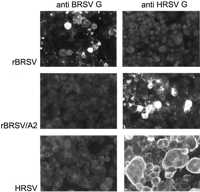
HEp-2 cells were infected with rBRSV, rBRSV/A2, or HRSV Long at an MOI of 0.1. Thirty-six hours postinfection, cells were fixed and stained with MAbs specific for the BRSV and HRSV G glycoproteins (Table 1 and Fig. 3) (see Materials and Methods). As expected, the BRSV G-specific MAb G66 reacted with rBRSV but not with rBRSV/A2 or HRSV. In contrast, the HRSV G-specific MAb 021/1G reacted with rBRSV/A2 and HRSV, but not with rBRSV. This confirmed that rBRSV/A2 directed the abundant expression of HRSV-specific G protein in infected cells and did not express BRSV-specific G protein.
TABLE 1.
Reactivity of MAbs with virus-infected cells in indirect immunofluorescence assay and immunoelectron microscopy
| MAb | Specificity | rBRSV | rBRSV/A2 | rBRSV/LongF | HRSV Long |
|---|---|---|---|---|---|
| G66 | BRSV G | + | − | + | − |
| 021/1G | HRSV G | − | + | − | + |
| 2F | RSV F | + | + | + | + |
| 44F | HRSV F | − | + | + | + |
FIG. 3.
Indirect immunofluorescence of HEp-2 cells infected with rBRSV (top panels), rBRSV/A2 (middle panels), or HRSV Long (bottom panels). Cells were infected at an MOI of 0.1 and were processed 36 h later for indirect immunofluorescence. The panels illustrate cells that were reacted with (left) MAb G66, specific to BRSV G protein, or (right) MAb O21/1G, specific to the HRSV G protein. The CPE produced by rBRSV and rBRSV/A2 in HEp-2 cells is comparable and not very pronounced. In contrast, HRSV produces an extensive CPE with large syncytia.
Additional HEp-2 cells were infected with rBRSV, HRSV Long, rBRSV/A2, or rBRSV/LongF; were fixed; and were stained with F-specific MAbs (Table 1) (see Materials and Methods). Staining with MAb 2F, which reacts with both HRSV and BRSV, showed that all of the viruses expressed high levels of F protein (not shown). Staining with MAb 44F, specific to HRSV, confirmed that, as expected, HRSV Long, rBRSV/A2, and rBRSV/LongF each expressed HRSV F protein, while rBRSV did not (not shown). Taken together, these results indicated that the recombinant chimeric viruses efficiently expressed the expected heterologous or homologous glycoproteins in cell culture.
Indirect immunofluorescence with G-specific MAbs (Fig. 3) showed that the chimeric rBRSV/A2 virus induced CPE in HEp-2 cells which was more similar to that of rBRSV than to that of HRSV. Specifically, in cultures infected with HRSV at an MOI of 0.1, nearly 100% of the cells showed positive immunofluorescence 36 h later, compared to 20% of cells infected with rBRSV and 20 to 30% of cells infected with rBRSV/A2. Thus, HRSV spreads more extensively in HEp-2 cells, and this property was not conferred on BRSV by transfer of the G and F glycoproteins alone. In addition, HRSV induced extensive CPE, including the formation of large syncytia, whereas rBRSV and rBRSV/A2 caused a reduced level of CPE and the formation of smaller syncytia. Thus, the capability to induce a relatively greater amount of CPE in HEp-2 cells also was not conferred on BRSV by the transfer of the HRSV G and F glycoproteins alone.
Growth of rBRSV/A2 in cell culture.
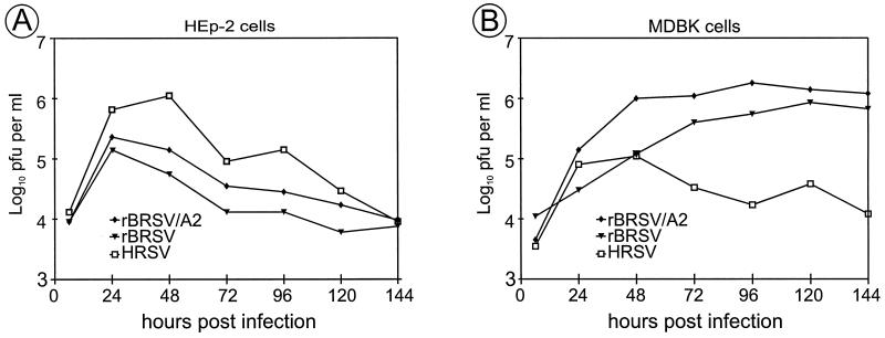
Multicycle growth of rBRSV, rBRSV/A2, and HRSV Long was compared in MDBK and HEp-2 cells which were infected at an MOI of 0.1. At various times postinfection, duplicate wells were harvested, and the virus in clarified medium supernatants was titrated (Fig. 4). In HEp-2 cells, HRSV produced approximately 10-fold more virus than did rBRSV (Fig. 4A), whereas the situation was reversed in MDBK cells (Fig. 4B). In MDBK cells, HRSV reached a maximum titer after 2 days, whereas rBRSV continued to grow and reached a maximum after 4 to 5 days. These observations identify a partial restriction of growth for BRSV and HRSV in human and bovine cells, respectively. The replication of the chimeric rBRSV/A2 virus in HEp-2 cells was intermediate between that of HRSV and BRSV, consistent with its chimeric nature. Thus, transfer of the human glycoproteins improved the growth of the chimera in human cells, but the effect was not complete, and the peak titer observed for the chimera more closely resembled that of its BRSV parent than that of HRSV. In bovine cells, the growth of the chimera more closely resembled that of BRSV although, interestingly, the chimeric virus grew somewhat better than its BRSV parent. The ability to replicate efficiently in this multicycle growth experiment indicated that the HRSV G and F proteins were functional in the rBRSV background and that the chimeric virus is fully competent for growth in two cell lines, one of human origin and one of bovine origin. The growth characteristics of rBRSV/LongF resembled those of rBRSV (data not shown), indicating that this chimera was fully competent for multicycle growth in both cell lines even though the G and F glycoproteins were of BRSV and HRSV, respectively.
FIG. 4.
Comparison of the multicycle growth of rBRSV, rBRSV/A2, and HRSV Long in human HEp-2 cells (A) and bovine MDBK cells (B). Duplicate cell monolayers in 24-well dishes were infected with the indicated virus at an MOI of 0.1. Monolayers were harvested at the indicated times, were stored at −70°C, and were titrated later in duplicate. Each value is the mean titer of material from two wells of infected cells.
Electron microscopy of the rBRSV/A2 and rBRSV/LongF chimeric viruses.
Virions of rBRSV/A2, rBRSV/LongF, rBRSV, and HRSV Long grown in MDBK cells were examined by immunoelectron microscopy using antibodies against the F and G proteins (Table 1), followed by labeling with gold-conjugated goat anti-mouse immunoglobulin and negative staining. The detected virions were filamentous with a diameter of 100 to 200 nm with highly variable lengths up to several micrometers. Some virions seemed to be branched. No spherical virion particles were detected (not shown).
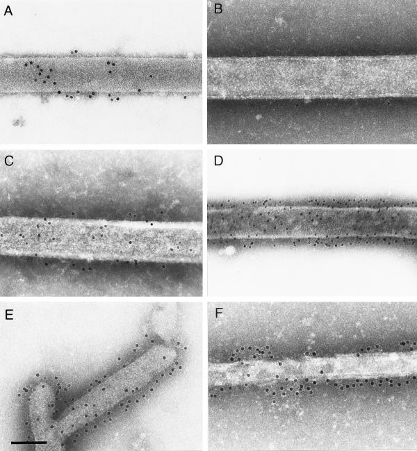
With MAb 44F, specific to HRSV F (Fig. 5B, D, and F), dense immunogold labeling of the rBRSV/A2 virion surface was observed (Fig. 5D), comparable to the labeling intensity of the HRSV virion surface (Fig. 5F), whereas no specific labeling of rBRSV virions was detected (Fig. 5B). Antibody 2F, with reacts with both BRSV and HRSV F protein, was used as control to demonstrate the intact antigenic structure of all virus preparations, including the BRSV preparation (5A, C, and E).
FIG. 5.
Immunogold labeling using MAbs reacting with both the HRSV F and BRSV F protein (MAb 2F; A, C, E) or exclusively with the HRSV F protein (MAb 44F; B, D, F). Labeling was performed on preparations of rBRSV (A, B), rBRSV/A2 (C, D), and HRSV Long (E, F). Bar, 150 nm.
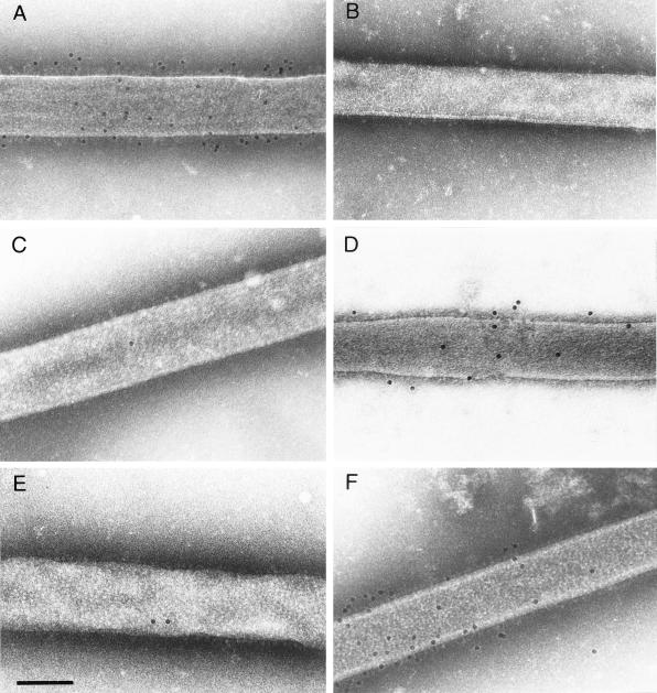
MAb 021/1G, specific to the HRSV G protein, reacted with rBRSV/A2 (Fig. 6D), as well as with HRSV particles (Fig. 6F), and failed to react with the rBRSV virion surface (Fig. 6B). MAb G66, specific to the BRSV G protein, reacted with the virion surface of rBRSV (Fig. 6A), but not with that of rBRSV/A2 (Fig. 6C) or HRSV (Fig. 6E). These results demonstrated that virions of the chimeric rBRSV/A2 virus contain abundant, appropriate levels of the HRSV-specific G and F glycoproteins.
FIG. 6.
Immunogold labeling using MAbs reacting with the BRSV G protein (MAb G66; A, C, E) or with the HRSV G protein (MAb 021/1G; B, D, F). Labeling was performed on preparations of rBRSV (A, B), rBRSV/A2 (C, D), and HRSV Long (E, F). Bar, 150 nm.
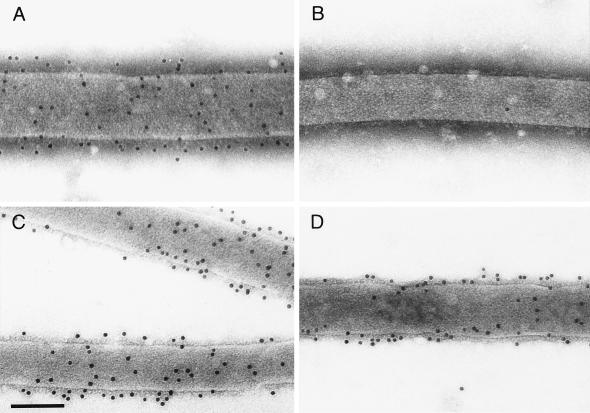
When virions of rBRSV/LongF were examined, dense surface staining was observed with MAb G66, which is specific to the BRSV G protein (Fig. 7A), and with antibody 44F, which is specific for HRSV F (Fig. 7D). This showed that both glycoproteins were efficiently incorporated into virions, even though the F protein was derived from HRSV and G was derived from BRSV.
FIG. 7.
Immunogold labeling of the surface proteins of rBRSV/LongF. Labeling was performed with the following MAbs: MAb G66 specific to the BRSV G protein (A), MAb 021/1G specific to the HRSV G protein (B), MAb 2F specific to the F proteins of both HRSV and BRSV (C), MAb 44F specific to HRSV F protein (D). Bar, 150 nm.
Evaluation of the replication and protective efficacy of the rBRSV/A2 chimeric virus in chimpanzees.
rBRSV and rBRSV/A2 were each administered to two chimpanzees by both the intranasal and intratracheal routes at the high dose of 107 PFU per ml per site (Table 2). Virus replication in the upper and lower respiratory tracts was monitored by nasopharyngeal swab and tracheal lavage, respectively. The results were compared (Table 2) to historic controls of animals which had been inoculated in the same way with 104 PFU of HRSV A2 wild-type virus per site (47) or 105 PFU of the live attenuated cpts248/404 strain A2 vaccine candidate per site (46).
Wild-type HRSV is highly permissive in seronegative chimpanzees and replicated to peak titers of more than 104.5 PFU per ml of nasopharyngeal swab or tracheal lavage (Table 2). The peak rhinorrhea score was 2.5. The live-attenuated vaccine candidate cpts248/404 replicated to peak titers of 102.5 and 101.4 PFU per ml of swab/lavage in the upper and lower respiratory tracts, respectively, and had a peak rhinorrhea score of 0.8. In contrast, there was no detectable replication of rBRSV in either the upper or lower respiratory tracts and no evidence of disease. Thus, even when administered at 100 to 1,000 times the dose of HRSV, rBRSV was highly restricted for replication in chimpanzees. This restriction is not absolute: although virus shedding was not detected here, in a previous experiment, one of two chimpanzees inoculated with BRSV shed a low level of virus (R. M. Chanock, personal communication).
Infection with the rBRSV/A2 chimera resulted in low levels of virus shedding over several days in both the upper and lower respiratory tract. That the shedding was not detected until day 3 or 5 indicates that it was not carryover from the inoculation, as does the length of time over which virus was recovered. The titers were much lower than those observed for wild-type HRSV and were moderately lower than those observed for the highly attenuated cpts248/404 vaccine candidate, even though the chimera had been administered at a dose that was 1,000-fold higher than that of the wild-type virus and 100-fold higher than that of the vaccine candidate. This indicates that the chimeric virus is very highly attenuated in chimpanzees despite the presence of the HRSV G and F glycoprotein genes. The serum antibody response was analyzed by neutralization assay against the homologous HRSV A2 and against the chimeric rBRSV/A2 virus. The neutralization titers were only slightly increased above background and were four- to eightfold lower than that observed with the highly attenuated cpts248/404 vaccine candidate (8) (data not shown). These very low levels of antibodies would be consistent with the interpretation that the replication of rBRSV and rBRSV/A2 were too low to be sufficiently immunogenic.
The animals which received rBRSV or rBRSV/A2 were challenged on day 30 with 104 PFU per site of wild-type HRSV administered intranasally and intratracheally (Table 3). Nasopharyngeal swabs and tracheal lavages were taken at 3, 5, 7, and 10 days following challenge, and virus titers were determined by plaque assay. Prior immunization with either virus did not provide a significant reduction in challenge virus replication in the upper or lower respiratory tract, although disease symptoms were reduced slightly.
TABLE 3.
Replication of wild-type RSV A2 challenge virus in chimpanzees previously inoculated with either rBRSV or rBRSV/A2a
| Chimpanzee no. | Virus | Nasopharyngeal swab titer (log10 PFU/ml)
|
Tracheal lavage titer (log10 PFU/ml)
|
Peak rhinorrhea scoreb | |||||||||
|---|---|---|---|---|---|---|---|---|---|---|---|---|---|
| Day postchallenge
|
Peak | Day postchallenge
|
Peak | ||||||||||
| 0 | 3 | 5 | 7 | 10 | 3 | 5 | 7 | 10 | |||||
| 95A016 | rBRSV | <0.7 | 3.7 | 4.1 | 1.0 | <0.7 | 4.1 | 5.2 | 6.0 | <0.7 | <0.7 | 6.0 | 1 |
| 95A018 | rBRSV | <0.7 | 3.8 | 3.0 | <0.7 | <0.7 | 3.8 | 2.0 | 4.5 | <0.7 | <0.7 | 4.5 | 1 |
| Mean | 4.0 | 5.3 | 1.0 | ||||||||||
| 95A007 | rBRSV/A2 | <0.7 | 4.6 | 4.3 | 1.6 | <0.7 | 4.6 | 5.7 | 6.3 | 1.7 | <0.7 | 6.3 | 2 |
| 95A008 | rBRSV/A2 | <0.7 | 2.2 | 5.4 | 1.7 | <0.7 | 5.4 | 3.2 | 6.3 | 2.8 | <0.7 | 6.3 | 2 |
| Mean | 5.0 | 6.3 | 2.0 | ||||||||||
| Controlsc | cpts248/404 | <0.7 | <0.7 | 2.0 | 1.0 | <0.7 | 2.0 | <0.7 | <0.7 | <0.7 | <0.7 | <0.7 | 0 |
Each chimpanzee was challenged on day 30 with wild-type RSV A2 administered intranasally and intratracheally at a dose of 4.0 log10 PFU/ml in a 1.0-ml inoculum. Nasopharyngeal swab samples and tracheal lavage samples were then collected on the indicated days. Virus titers are shown as log10 PFU/ml of sample.
See Table 2, footnote b.
Historic control animals (n = 2) from the study of Crowe et al. (8).
DISCUSSION
Previously, we described reverse genetics systems for generating rHRSV (4) or rBRSV (1) from cDNA. In the work described here, these systems were used to generate rBRSVs in which the F gene alone, or the F and G genes together, were replaced by their HRSV counterparts. The heterologous F glycoprotein alone, or F and G glycoproteins together, were efficiently incorporated into the virion envelope based on immunoelectron microscopy and appeared to be fully functional based on efficient multicycle growth of the chimeras in vitro. The chimeric virus rBRSV/A2, which was studied in greater detail because it bears the greater number of the HRSV surface proteins, more closely resembled BRSV than HRSV with respect to host range in cell culture and in chimpanzees. These data indicated that the G and F proteins contribute to the host range restriction of BRSV but, surprisingly, are not its major determinants. This study also evaluated Jennerian and modified Jennerian strategies for employing BRSV for the development of a live-attenuated vaccine against HRSV.
One potential obstacle to replacing the F and G proteins of BRSV with those of HRSV was that the proteins might be incompatible. By analogy to other mononegaviruses, it seemed likely that interaction between the internal domain of one or more glycoproteins and internal proteins such as M would be important in virion assembly and infectivity (3, 28, 29, 38). However, recent observations indicated that the G and SH proteins were unlikely to play critical roles, at least in the appropriate cell line. For example, RSV B1 multiply passaged at low temperature was recently shown to have sustained the spontaneous deletion of the SH and G genes, and this increased rather than decreased virus growth at low temperature in cell culture (18). Studies with rHRSV and rBRSV confirmed that the SH and G genes can be deleted without loss of growth fitness in cell culture (2) (M. Teng and P. L. Collins, unpublished data and U. J. Buchholz, unpublished data). In a minigenome system, the production of transmissible particles was strongly dependent on M and F, was less dependent on G, and was independent of SH (42). Taken together, these studies indicate that F and M are critical for virion formation and infectivity in vitro, whereas SH and G are dispensible under the appropriate in vitro conditions. The F proteins of these BRSV and HRSV strains share only 82% amino acid identity, with the cytoplasmic and transmembrane domains being 67 and 79% identical, respectively. This degree of divergence apparently had no effect on the ability of the chimeric virus to grow in vitro. This is not completely surprising, since we previously reported that the hemagglutinin-neuraminidase (HN) and F proteins of HPIV3 could be replaced by their HPIV1 counterparts without significant loss of growth efficiency even though the percentage of amino acid identity between serotypes was very low: HN transmembrane domain identity, 30%; F transmembrane identity, 22%; HN cytoplasmic tail identity, 9%; F cytoplasmic tail identity, 11% (40). It is also noteworthy that the HRSV F and BRSV G proteins could operate together in a single virus, rBRSV/LongF, without a loss in efficiency of incorporation into virions or of replication in cell culture. This was somewhat surprising, because the G proteins of HRSV and BRSV are very divergent, exhibiting 28% identity overall. The ability of nonmatched G and F glycoproteins to function in the same envelope might mean they do not interact closely or, alternatively, that this interaction is not highly sequence specific. This is in contrast to the situation with members of the Paramyxovirinae such as Sendai virus and other viruses of the genus Parainfluenzavirus, in which the processing and function of the HN and F proteins appear to be closely linked with respect to fusion (39, 43).
The chimpanzee is the experimental animal model that most closely resembles humans with regard to permissiveness for HRSV replication and disease. When a high dose of rBRSV was administered to two animals in this study, there was no evidence of virus shedding or disease. In a previous study, one of two chimpanzees inoculated with BRSV shed a low level of virus (35). In contrast, inoculation with a 1,000-fold-lower dose of HRSV resulted in high levels of virus shedding and the induction of disease. Thus, chimpanzees provide an unambiguous and highly authentic assay of the difference in host range between BRSV and HRSV. When a high dose of rBRSV/A2 was administered to two animals, virus shedding was detected in both animals but was very low. The observation that the transfer of the HRSV G and F proteins to BRSV improved its growth somewhat in chimpanzees indicates that these two glycoproteins contribute to the host range phenotype. However, the observation that the chimera remains highly restricted compared to HRSV indicates that one or more other viral proteins also make important contributions to the host range phenotype. It also is possible that cis-acting RNA sequences are involved, but this seems less likely because the transcription signals are highly conserved between HRSV and BRSV as noted above and because the leader region of HRSV can be substituted into BRSV with little effect (1).
These findings also have implications with regard to strategies for improving the permissiveness of convenient small experimental animals such as mice for HRSV infection and disease. One strategy would be to identify the human cellular receptor(s) for HRSV and express it in a transgenic mouse, such as has been done for measles virus (30, 37). However, since the permissiveness of BRSV for replication in chimpanzees was not greatly improved by the insertion of G and F glycoproteins of HRSV origin, it seems unlikely that expression in a mouse of the human molecules which interact with G and perhaps F would have a major effect on improving permissiveness for HRSV.
Reverse genetics is being used to engineer rHRSV to develop candidate live-attenuated vaccines (references 2, 15, 17, 46, 47, and 48, and references therein). In particular, the HRSV reverse genetics system was used to characterize and combine sets of attenuating mutations present in HRSV strains attenuated by classical passage and mutagenesis methods (references 17, 47, and 48, and references therein). Because of the strategy by which they were selected, most of these mutations confer the temperature-sensitive phenotype in addition to the attenuation phenotype. One limitation of the temperature-sensitive phenotype is that the virus can be overly restricted for replication in the lower respiratory tract while being underattenuated in the upper respiratory tract. This is because there is a temperature gradient within the respiratory tract, with temperature being higher (more restrictive) in the lower respiratory tract and lower (less restrictive) in the upper respiratory tract. The ability of an attenuated virus to replicate in the upper respiratory tract can result in complications, including congestion, rhinitis, fever, and otitis media. Thus, attenuation achieved primarily by temperature-sensitive mutations may not be ideal. One solution has been to supplement the temperature-sensitive mutations with deletion mutations, such as deletion of the entire SH or NS2 gene (2, 46). Such mutations are not temperature sensitive and, furthermore, would be genetically more stable than point mutations.
Here, we have pursued an alternative approach to develop a live-attenuated HRSV vaccine, namely to make use of the host range differences between HRSV and BRSV. It would be anticipated that a growth restriction in a nonnatural host would involve multiple genes and many sequence differences, and this is consistent with the present findings that the G and F glycoproteins contribute only part of this phenotype. Not surprisingly, experience with other viruses indicates that a host range restriction is a very stable phenotype (19, 33). Also, host range restrictions typically are not associated with temperature sensitivity. We reexamined the Jennerian approach, in which BRSV (in this case a recombinant version) was used directly as a live-attenuated vaccine against HRSV. This had been investigated previously in rodents, in monkeys, and in two chimpanzees with the B/097 strain (35). Whereas previous studies in rodents or monkeys suggested that BRSV might be a satisfactory Jennerian vaccine, the present results with strain ATue51908 confirm that BRSV itself has very low efficacy in chimpanzees as a vaccine against HRSV. This is likely due in large part to its very poor replication as described above.
We also sought to use reverse genetics to improve BRSV as a live-attenuated vaccine against HRSV, pursuing a modified Jennerian strategy. The first step, described here, involved substituting the G and F glycoproteins of BRSV with those of HRSV. Since G and F are the major protective antigens of RSV, their substitution would address the issue of antigenic divergence and also had the potential for improving the efficiency of replication of BRSV in primates. As noted above, the replication of BRSV in chimpanzees was only marginally improved. Since rBRSV/A2 remains overattenuated, the next step will be to improve its capacity for replication in primates by substituting additional HRSV genes. Specifically, the difference between rBRSV/A2, which is highly restricted in chimpanzees, and HRSV, which is highly permissive, lies in eight genes: NS1, NS2, N, P, M, SH, M2-1/M2-2, and L. The systematic substitution of these eight genes should identify a chimera with an appropriate level of attenuation and will provide new information on the determinants of host range in a paramyxovirus. A complementary strategy will be to start with HRSV and replace internal HRSV genes with their BRSV counterparts in an incremental, stepwise fashion. In conclusion, we have developed a chimeric rBRSV that bears the major antigenic determinants of HRSV and have shown that it is viable and safe in chimpanzees and thus represents a starting point for developing a live-attenuated chimeric vaccine against HRSV.
ACKNOWLEDGMENTS
We thank Geraldine Taylor, Compton, United Kingdom, and José Antonio Melero, Madrid, Spain, for their generous gifts of monoclonal antibodies and Georg Herrler, Hannover, Germany, who provided HRSV Long. We also thank Myron Hill, Cai-Yen Firestone, and Christel Möller for superb technical assistance and Robert Chanock for reviewing the manuscript.
REFERENCES
- 1.Buchholz U J, Finke S, Conzelmann K-K. Generation of bovine respiratory syncytial virus (BRSV) from cDNA: BRSV NS2 is not essential for virus replication in tissue culture, and the human RSV leader region acts as a functional BRSV genome promoter. J Virol. 1999;73:251–259. doi: 10.1128/jvi.73.1.251-259.1999. [DOI] [PMC free article] [PubMed] [Google Scholar]
- 2.Bukreyev A, Whitehead S S, Murphy B R, Collins P L. Recombinant respiratory syncytial virus from which the entire SH gene has been deleted grows efficiently in cell culture and exhibits site-specific attenuation in the respiratory tract of the mouse. J Virol. 1997;71:8973–8982. doi: 10.1128/jvi.71.12.8973-8982.1997. [DOI] [PMC free article] [PubMed] [Google Scholar]
- 3.Cathomen T, Naim H Y, Cattaneo R. Measles viruses with altered envelope protein cytoplasmic tails gain cell fusion competence. J Virol. 1998;72:1224–1234. doi: 10.1128/jvi.72.2.1224-1234.1998. [DOI] [PMC free article] [PubMed] [Google Scholar]
- 4.Collins P L, Hill M G, Camargo E, Grosfeld H, Chanock R M, Murphy B R. Production of infectious human respiratory syncytial virus from cloned cDNA confirms an essential role for the transcription elongation factor from the 5′ proximal open reading frame of the M2 mRNA in gene expression and provides a capability for vaccine development. Proc Natl Acad Sci USA. 1995;92:11563–11567. doi: 10.1073/pnas.92.25.11563. [DOI] [PMC free article] [PubMed] [Google Scholar]
- 5.Collins P L, Hill M G, Cristina J, Grosfeld H. Transcription elongation factor of respiratory syncytial virus, a nonsegmented negative-strand RNA virus. Proc Natl Acad Sci USA. 1996;93:81–85. doi: 10.1073/pnas.93.1.81. [DOI] [PMC free article] [PubMed] [Google Scholar]
- 6.Collins P L, McIntosh K, Chanock R M. Respiratory syncytial virus. In: Fields B N, Knipe D M, Howley P M, Chanock R M, Melnick J L, Monath T P, Roizman B, Straus S E, editors. Fields virology. 3rd ed. Vol. 2. Philadelphia, Pa: Lippincott-Raven; 1996. pp. 1313–1352. [Google Scholar]
- 7.Connors M, Collins P L, Firestone C-Y, Murphy B R. Resistance to respiratory syncytial virus (RSV) challenge induced by infection with a vaccinia virus recombinant expressing the RSV M2 protein (Vac-M2) is mediated by CD8+ T cells, while that induced by Vac-F or Vac-G recombinants is mediated by antibodies. J Virol. 1992;66:1277–1281. doi: 10.1128/jvi.66.2.1277-1281.1992. [DOI] [PMC free article] [PubMed] [Google Scholar]
- 8.Crowe J E, Jr, Bui P T, Davis A R, Chanock R M, Murphy B R. A further attenuated derivative of a cold-passaged temperature-sensitive mutant of human respiratory syncytial virus retains immunogenicity and protective efficacy against wild-type challenge in seronegative chimpanzees. Vaccine. 1994;12:783–790. doi: 10.1016/0264-410x(94)90286-0. [DOI] [PubMed] [Google Scholar]
- 9.Fearns R, Collins P L. Role of the M2-1 transcription antitermination protein of respiratory syncytial virus in sequential transcription. J Virol. 1999;73:5852–5864. doi: 10.1128/jvi.73.7.5852-5864.1999. [DOI] [PMC free article] [PubMed] [Google Scholar]
- 10.Fernie B F, Gerin J L. The stabilization and purification of respiratory syncytial virus using MgSO4. Virology. 1980;106:141–144. doi: 10.1016/0042-6822(80)90229-9. [DOI] [PubMed] [Google Scholar]
- 11.Furze J, Wertz G, Lerch R, Taylor G. Antigenic heterogeneity of the attachment protein of bovine respiratory syncytial virus. J Gen Virol. 1994;75:363–370. doi: 10.1099/0022-1317-75-2-363. [DOI] [PubMed] [Google Scholar]
- 12.García-Barreno B, Palomo C, Penas C, Delgado T, Perez-Brena P, Melero J A. Marked differences in the antigenic structure of human respiratory syncytial virus F and G glycoproteins. J Virol. 1989;63:925–932. doi: 10.1128/jvi.63.2.925-932.1989. [DOI] [PMC free article] [PubMed] [Google Scholar]
- 13.Grosfeld H, Hill M G, Collins P L. RNA replication by respiratory syncytial virus (RSV) is directed by the N, P, and L proteins; transcription also occurs under these conditions but requires RSV superinfection for efficient synthesis of full-length mRNA. J Virol. 1995;69:5677–5686. doi: 10.1128/jvi.69.9.5677-5686.1995. [DOI] [PMC free article] [PubMed] [Google Scholar]
- 14.Hardy R W, Wertz G W. The product of the respiratory syncytial virus M2 gene ORF1 enhances readthrough of intergenic junctions during viral transcription. J Virol. 1998;72:520–526. doi: 10.1128/jvi.72.1.520-526.1998. [DOI] [PMC free article] [PubMed] [Google Scholar]
- 15.Jin H, Clarke D, Zhou H Z, Cheng X, Coelingh K, Bryant M, Li S. Recombinant human respiratory syncytial virus (RSV) from cDNA and construction of subgroup A and B chimeric RSV. Virology. 1998;251:206–214. doi: 10.1006/viro.1998.9414. [DOI] [PubMed] [Google Scholar]
- 16.Johnson P R, Spriggs M K, Olmsted R A, Collins P L. The G glycoprotein of human respiratory syncytial viruses of subgroups A and B: extensive sequence divergence between antigenically related proteins. Proc Natl Acad Sci USA. 1987;84:5625–5629. doi: 10.1073/pnas.84.16.5625. [DOI] [PMC free article] [PubMed] [Google Scholar]
- 17.Juhasz K, Whitehead S S, Boulanger C A, Firestone C-Y, Collins P L, Murphy B R. The two amino acid substitutions in the L protein of cpts530/1009, a live attenuated respiratory syncytial virus candidate vaccine, are independent temperature-sensitive and attenuation mutations. Vaccine. 1999;17:1416–1424. doi: 10.1016/s0264-410x(98)00381-8. [DOI] [PubMed] [Google Scholar]
- 18.Karron R A, Buonagurio D A, Georgiu A G, Whitehead S S, Adamus J E, Clements-Mann M L, Harris D O, Randolph V B, Udem S A, Murphy B R, Sidhu M S. Respiratory syncytial virus (RSV) SH and G proteins are not essential for viral replication in vitro: clinical evaluation and molecular characterization of a cold-passaged, attenuated RSV subgroup B mutant. Proc Natl Acad Sci USA. 1997;94:13961–13966. doi: 10.1073/pnas.94.25.13961. [DOI] [PMC free article] [PubMed] [Google Scholar]
- 19.Karron R A, Wright P F, Hall S L, Makhene M, Thompson J, Burns B A, Tollefson S, Steinhoff M C, Wilson M H, Harris D O, Clements M L, Murphy B R. A live attenuated bovine parainfluenza virus type 3 vaccine is safe, infectious, immunogenic, and phenotypically stable in infants and children. J Infect Dis. 1995;171:1107–1114. doi: 10.1093/infdis/171.5.1107. [DOI] [PubMed] [Google Scholar]
- 20.Kunkel T A, Roberts J D, Zakour R A. Rapid and efficient site-specific mutagenesis without phenotypic selection. Methods Enzymol. 1987;54:367–382. doi: 10.1016/0076-6879(87)54085-x. [DOI] [PubMed] [Google Scholar]
- 21.Kuo L, Fearns R, Collins P L. The structurally diverse intergenic regions of respiratory syncytial virus do not modulate sequential transcription by a dicistronic minigenome. J Virol. 1996;70:6143–6150. doi: 10.1128/jvi.70.9.6143-6150.1996. [DOI] [PMC free article] [PubMed] [Google Scholar]
- 22.Kuo L, Fearns R, Collins P L. Analysis of the gene start and gene end signals of human respiratory syncytial virus: quasi-templated initiation at position 1 of the encoded mRNA. J Virol. 1997;71:4944–4953. doi: 10.1128/jvi.71.7.4944-4953.1997. [DOI] [PMC free article] [PubMed] [Google Scholar]
- 23.Langedijk J P M, Meloen R H, Taylor G, Furze J M, van Oirschot J T. Antigenic structure of the central conserved region of protein G of bovine respiratory syncytial virus. J Virol. 1997;71:4055–4061. doi: 10.1128/jvi.71.5.4055-4061.1997. [DOI] [PMC free article] [PubMed] [Google Scholar]
- 24.Lerch R A, Anderson K, Wertz G W. Nucleotide sequence analysis and expression from recombinant vectors demonstrate that the attachment protein G of bovine respiratory syncytial virus is distinct from that of human respiratory syncytial virus. J Virol. 1990;64:5559–5569. doi: 10.1128/jvi.64.11.5559-5569.1990. [DOI] [PMC free article] [PubMed] [Google Scholar]
- 25.Levine S, Klaiber-Franco R, Paradiso P R. Demonstration that glycoprotein G is the attachment protein of respiratory syncytial virus. J Gen Virol. 1987;68:2521–2524. doi: 10.1099/0022-1317-68-9-2521. [DOI] [PubMed] [Google Scholar]
- 26.Lopez J A, Villanueva N, Melero J A, Portela A. Nucleotide sequence if the fusion and phosphoprotein genes of human respiratory syncytial (RS) virus Long strain: evidence of subtype genetic heterogeneity. Virus Res. 1988;10:249–261. doi: 10.1016/0168-1702(88)90020-2. [DOI] [PubMed] [Google Scholar]
- 27.Martinez I, Melero J A. Enhanced neutralization of human respiratory syncytial virus by mixtures of monoclonal antibodies to the attachment (G) glycoprotein. J Gen Virol. 1998;79:2215–2220. doi: 10.1099/0022-1317-79-9-2215. [DOI] [PubMed] [Google Scholar]
- 28.Mebatsion T, Conzelmann K-K. Specific infection of CD4+ target cells by recombinant rabies virus pseudotypes carrying the HIV-1 envelope spike protein. Proc Natl Acad Sci USA. 1996;93:11366–11370. doi: 10.1073/pnas.93.21.11366. [DOI] [PMC free article] [PubMed] [Google Scholar]
- 29.Mebatsion T, Weiland F, Conzelmann K K. Matrix protein of rabies virus is responsible for the assembly and budding of bullet-shaped particles and interacts with the transmembrane spike glycoprotein G. J Virol. 1999;73:242–250. doi: 10.1128/jvi.73.1.242-250.1999. [DOI] [PMC free article] [PubMed] [Google Scholar]
- 30.Mrkic B, Pavlovic J, Rulicke T, Volpe P, Buchholz C J, Hourcade D, Atkinson J P, Aguzzi A, Cattaneo R. Measles virus spread and pathogenesis in genetically modified mice. J Virol. 1998;72:7420–7427. doi: 10.1128/jvi.72.9.7420-7427.1998. [DOI] [PMC free article] [PubMed] [Google Scholar]
- 31.Olmsted R A, Elango N, Prince G A, Murphy B R, Johnson P R, Moss B, Chanock R M, Collins P L. Expression of the glycoprotein of respiratory syncytial virus by a recombinant vaccinia virus: comparison of the individual contributions of the F and G glycoproteins to host immunity. Proc Natl Acad Sci USA. 1986;83:7462–7466. doi: 10.1073/pnas.83.19.7462. [DOI] [PMC free article] [PubMed] [Google Scholar]
- 32.Paccaud M F, Jacquier C. A respiratory syncytial virus of bovine origin. Arch Ges Virusforsch. 1970;30:327–342. doi: 10.1007/BF01258363. [DOI] [PubMed] [Google Scholar]
- 33.Perez-Schael I, Guntinas M J, Perez M, Pagone V, Rojas A M, Gonzalez R, Cunto W, Hoshino Y, Kapikian A Z. Efficacy of the rhesus rotavirus-based quadrivalent vaccine in infants and young children in Venezuela. N Engl J Med. 1997;337:1181–1187. doi: 10.1056/NEJM199710233371701. [DOI] [PubMed] [Google Scholar]
- 34.Piazza F M, Johnson S A, Darnell M E R, Porter D D, Hemming V G, Prince G A. Bovine respiratory syncytial virus protects cotton rats against human respiratory syncytial virus infection. J Virol. 1993;67:1503–1510. doi: 10.1128/jvi.67.3.1503-1510.1993. [DOI] [PMC free article] [PubMed] [Google Scholar]
- 35.Prince G A, Redington M, Piazza F M, Hemming V G. Bovine respiratory syncytial virus provides protection against human respiratory virus infection in cotton rats and primates. In: Meignier B, Murphy B, Ogra P, editors. Animal models of respiratory syncytial virus infections. Lyon, France: Fondation Marcel Merieux; 1990. pp. 133–135. [Google Scholar]
- 36.Pringle C R. Virus taxonomy 1996—a bulletin from the Xth International Congress of Virology in Jerusalem. Arch Virol. 1996;141:2251–2256. doi: 10.1007/BF01718231. [DOI] [PMC free article] [PubMed] [Google Scholar]
- 37.Rall G F, Manchester M, Daniels L R, Callahan E M, Belman A R, Oldstone M B. A transgenic mouse model for measles virus infection of the brain. Proc Natl Acad Sci USA. 1997;94:4659–4663. doi: 10.1073/pnas.94.9.4659. [DOI] [PMC free article] [PubMed] [Google Scholar]
- 38.Spielhofer P, Bächi T, Fehr T, Christiansen G, Cattaneo R, Kaelin K, Billeter M A, Naim H Y. Chimeric measles viruses with a foreign envelope. J Virol. 1998;72:2150–2159. doi: 10.1128/jvi.72.3.2150-2159.1998. [DOI] [PMC free article] [PubMed] [Google Scholar]
- 39.Tanabayashi K, Compans R W. Functional interaction of paramyxovirus glycoproteins: identification of a domain in Sendai virus HN which promotes cell fusion. J Virol. 1996;70:6112–6118. doi: 10.1128/jvi.70.9.6112-6118.1996. [DOI] [PMC free article] [PubMed] [Google Scholar]
- 40.Tao T, Durbin A P, Whitehead S S, Davoodi F, Collins P L, Murphy B R. Recovery of a fully viable chimeric human parainfluenza virus (PIV) type 3 in which the hemagglutinin-neuraminidase and fusion glycoproteins have been replaced by those of PIV type 1. J Virol. 1998;72:2955–2961. doi: 10.1128/jvi.72.4.2955-2961.1998. [DOI] [PMC free article] [PubMed] [Google Scholar]
- 41.Taylor G, Thomas L H, Furze J M, Cook R S, Wyld S G, Lerch R, Hardy R, Wertz G W. Recombinant vaccinia viruses expressing the F, G, or N, but not the M2, protein of bovine respiratory syncytial virus (BRSV) induce resistance to BRSV challenge in the calf and protect against the development of pneumonic lesions. J Gen Virol. 1997;78:3195–3206. doi: 10.1099/0022-1317-78-12-3195. [DOI] [PubMed] [Google Scholar]
- 42.Teng M N, Collins P L. Identification of the respiratory syncytial virus proteins required for formation and passage of helper-dependent infectious particles. J Virol. 1998;72:5707–5716. doi: 10.1128/jvi.72.7.5707-5716.1998. [DOI] [PMC free article] [PubMed] [Google Scholar]
- 43.Tong S, Compans R W. Alternative mechanisms of interaction between homotypic and heterotypic parainfluenza virus HN and F proteins. J Gen Virol. 1999;80:107–115. doi: 10.1099/0022-1317-80-1-107. [DOI] [PubMed] [Google Scholar]
- 44.Van der Poel W H, Brand A, Kramps J A, van Oirschot J T. Respiratory syncytial virus infections in human beings and in cattle. An epidemiological review. J Infect. 1994;29:215–228. doi: 10.1016/s0163-4453(94)90866-4. [DOI] [PubMed] [Google Scholar]
- 45.Walsh E E, Hruska J. Monoclonal antibodies to respiratory syncytial virus proteins: identification of the fusion protein. J Virol. 1983;47:171–177. doi: 10.1128/jvi.47.1.171-177.1983. [DOI] [PMC free article] [PubMed] [Google Scholar]
- 46.Whitehead S S, Bukreyev A, Teng M N, Firestone C-Y, St. Claire M, Elkins W R, Collins P L, Murphy B R. Recombinant respiratory syncytial virus bearing a deletion of either the NS2 or SH gene is attenuated in chimpanzees. J Virol. 1999;73:3438–3442. doi: 10.1128/jvi.73.4.3438-3442.1999. [DOI] [PMC free article] [PubMed] [Google Scholar]
- 47.Whitehead S S, Juhasz K, Firestone C-Y, Collins P L, Murphy B R. Recombinant respiratory syncytial virus (RSV) bearing a set of mutations from cold-passaged RSV is attenuated in chimpanzees. J Virol. 1998;72:4467–4471. doi: 10.1128/jvi.72.5.4467-4471.1998. [DOI] [PMC free article] [PubMed] [Google Scholar]
- 48.Whitehead S S, Firestone C-Y, Karron R A, Crowe J E, Jr, Elkins W R, Collins P L, Murphy B R. Addition of a missense mutation present in the L gene of respiratory syncytial virus (RSV) cpts530/1030 to RSV vaccine candidate cpts248/404 increases its attenuation and temperature sensitivity. J Virol. 1999;73:871–877. doi: 10.1128/jvi.73.2.871-877.1999. [DOI] [PMC free article] [PubMed] [Google Scholar]
- 49.Yu Q, Hardy R W, Wertz G W. Functional cDNA clones of the human respiratory syncytial (RS) virus N, P, and L proteins support replication of RS virus genomic RNA analogs and define minimal trans-acting requirements for RNA replication. J Virol. 1995;69:2412–2419. doi: 10.1128/jvi.69.4.2412-2419.1995. [DOI] [PMC free article] [PubMed] [Google Scholar]
- 50.Zamora M, Samal S K. Gene junction sequences of bovine respiratory syncytial virus. Virus Res. 1992;24:115–121. doi: 10.1016/0168-1702(92)90035-8. [DOI] [PubMed] [Google Scholar]


