Abstract
Background
Microbial communities are important drivers of global biogeochemical cycles, xenobiotic detoxification, as well as organic matter decomposition. Their major metabolic role in ecosystem functioning is ensured by a unique set of enzymes, providing a tremendous yet mostly hidden enzymatic potential. Exploring this enzymatic repertoire is therefore not only relevant for a better understanding of how microorganisms function in their natural environment, and thus for ecological research, but further turns microbial communities, in particular from extreme habitats, into a valuable resource for the discovery of novel enzymes with potential applications in biotechnology. Different strategies for their uncovering such as bioprospecting, which relies mainly on metagenomic approaches in combination with sequence-based bioinformatic analyses, have emerged; yet accurate function prediction of their proteomes and deciphering the in vivo activity of an enzyme remains challenging.
Results
Here, we present environmental activity-based protein profiling (eABPP), a multi-omics approach that extends genome-resolved metagenomics with mass spectrometry-based ABPP. This combination allows direct profiling of environmental community samples in their native habitat and the identification of active enzymes based on their function, even without sequence or structural homologies to annotated enzyme families. eABPP thus bridges the gap between environmental genomics, correct function annotation, and in vivo enzyme activity. As a showcase, we report the successful identification of active thermostable serine hydrolases from eABPP of natural microbial communities from two independent hot springs in Kamchatka, Russia.
Conclusions
By reporting enzyme activities within an ecosystem in their native state, we anticipate that eABPP will not only advance current methodological approaches to sequence homology-guided enzyme discovery from environmental ecosystems for subsequent biocatalyst development but also contributes to the ecological investigation of microbial community interactions by dissecting their underlying molecular mechanisms.
Supplementary Information
The online version contains supplementary material available at 10.1186/s40793-024-00577-2.
Keywords: Activity-based protein profiling, Click chemistry, Chemical proteomics, Environmental microbial communities, Hot springs, Metagenomics, Metaproteomics, Serine hydrolases, Target identification
Introduction
Microbial organisms constitute the vast majority of unexplored natural biodiversity and successfully colonize all of the earth’s conceivable ecological niches, thereby forming microbial communities of distinct complexity and fluctuating composition [1–6]. Their ability to thrive under specific conditions, especially in ‘extreme’ ecosystems such as hot springs, is ensured by a unique, often still unexplored enzyme repertoire that turns them into a promising resource for identifying novel (thermostable) enzymes for biotechnological applications [7]. The systematic bioprospecting of microorganisms or microbial communities, sometimes also referred to as environmental biotechnology [3], is frequently based on metagenomic analyses, in particular, if organisms or communities that are non-culturable using standard techniques are screened [8–11]. The identification of promising enzymes for further biocatalyst development from such metagenomic data is then commonly achieved via sequence-driven bioinformatic prediction of protein function [12]. This approach, however, is hampered by a large quantity of proteins of unknown function (i.e., hypotheticals) or misannotated enzymes as well as the presence of large protein superfamilies, for which functional predictions remain difficult [13]. ‘Functional metagenomics’ can help to overcome these limitations by complementing sequence-based approaches with an activity-based screening after the construction of a metagenomic library [14–17]. Although this enables the discovery of novel enzyme classes, it frequently requires elaborate and often challenging cloning and expression efforts with subsequent biochemical characterization [12, 18]. Moreover, this approach delivers no information on the expression and, as valid for all ‘omics’ or phenotype-based next-generation physiology strategies that were invented to unravel the function of a single cell in its native habitat [19], the in vivo activity state of an enzyme-of-interest.
Activity-based protein profiling (ABPP) represents a powerful approach for studying enzyme activities in their native environment and a huge variety of activity-based probes (ABPs) that target different enzymes or even whole enzyme classes are nowadays available [20–22]. A ‘classical’ ABP is composed of a reactive warhead, a tag, and, if present, a chemical or peptidic linker region. While the warhead is often an electrophile-containing inhibitor molecule that targets a nucleophile at the active site of an enzyme for covalent labeling, the tag is used for target detection or enrichment [23]. Thus, ABPP does not only allow the visualization of labeled enzymes via the use of fluorescent reporter groups but also enables target enzyme identification by mass spectrometry (MS) if a moiety for target enrichment, such as biotin, is used as a reporter group [24–26]. Accordingly, although only rarely used in this context, an ABPP experiment with enzyme- or enzyme class-specific ABPs enables a functional annotation of enzymes [27]. With the integration of ‘click chemistry’ into the ABPP workflow, two-step chemical probes have emerged, which facilitate a simple in vivo application of ABPP under physiological conditions [28, 29]. In addition, the invention of affinity-based probes (AfBPs) has greatly expanded the enzyme repertoire accessible to ABPP by allowing the use of reversible inhibitors as warheads through the incorporation of a photoreactive group that is activated under UV light to form a covalent bond with a target enzyme [22, 30, 31].
In the last years, ABPP has been mainly used in the context of biomarker or drug discovery as well as in vivo imaging [32–35]; beyond that, diverse applications in microbiology or plant biology, including the study of pathogens or host-pathogen interactions, have been frequently reported [36–38]. Only recently, the use of ABPP in biocatalyst discovery for industrial applications, for example for elucidating lignocellulose-degrading enzymes, has emerged [39–41]. In addition, first reports on the use of ABPP or related approaches for studying microbial communities and isolating functionally active subpopulations, with a focus on host-associated microbial communities, have been made [42–44]. Among these, few studies on the gut microbiome have implemented a combination of ABPP with metagenomics to facilitate the identification of enzymes related to chronic inflammation, drug toxicity, or dietary fiber metabolism in the gastrointestinal tract by employing a metaproteome database that was either constructed from publicly available genomes or self-constructed based on metagenome sequencing [45–47]. ABPP of environmental microbial communities, by contrast, is largely unknown and is usually achieved by profiling isolated strains of environmental microbes rather than by direct profiling of complex communities [48–52]. Recently, activity-based imaging of ammonia- and alkane-oxidizing bacteria in complex microbial communities with the ABP 1,7-octadiyne was reported [53]. This study also used metagenomic sequencing for further confirmation of the functional potential of the targeted microorganisms. However, to the best of our knowledge, no generic workflow for ABPP of microbial communities directly in the environment that also delivers a function-based target identification of the labeled enzymes via downstream MS has been established so far.
We herein describe such an ABPP approach that we refer to as ‘environmental ABPP’ (eABPP). Our approach transfers a combination of ABPP with genome-resolved metagenomics to the field, facilitating the assignment of dedicated activities to enzymes from microorganisms in the environment, even to those that belong to so far uncultured or unknown microorganisms. This has become possible through a tailored sample preparation and data analysis procedure that allows detection of single active enzymes within a complex environmental metaproteome. As a ‘proof-of-concept’, we employed an alkyne-tagged version of the well-established fluorophosphonate-based (FP) ABP [54], which proved to be a versatile probe for the different application types of ABPP. Most importantly, this ABP has been previously shown to allow ABPP of serine hydrolases even under extreme experimental conditions such as elevated temperatures or low pH [52], making it suitable for use in the field. Moreover, employing a broad-spectrum probe increases the number of potentially identifiable target proteins and thus facilitates method validation. Beyond that, members of the serine hydrolase superfamily are widely distributed across all domains of life [55] and are enzymes catalyzing diverse reactions, some of which are of relevance for industrial applications [56–61]. Accordingly, we profiled serine hydrolase activities of microbial communities from two different hot springs located in the Uzon Caldera (Kamchatka Peninsula, Russia) under native conditions directly at the site of sampling. Subsequent biochemical studies then confirmed that this approach can be reliably used for identifying in vivo serine hydrolase activities from environmental community samples.
Results and discussion
General workflow design
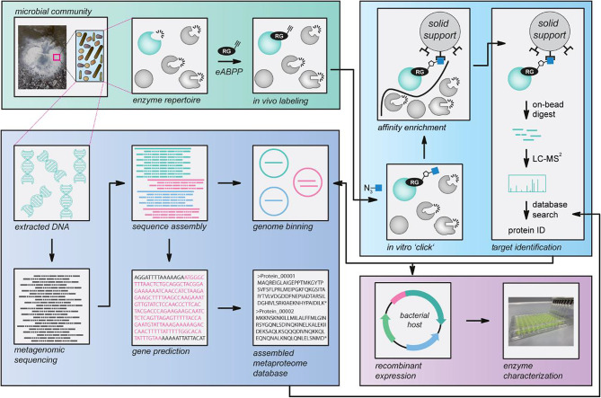
For establishing a function-based eABPP enzyme identification approach of an environmental microbial community, we designed a general workflow based on a combination of ABPP with metagenome sequencing (Fig. 1). This workflow starts with the collection of a microbial community sample, here a hot spring sediment. This sample is then split into seven aliquots and six of them are used for in vivo labeling with an enzyme- or enzyme class-specific two-step ABP; the downstream analysis is thereby focused on active enzymes with the desired function among the microbial enzyme repertoire already at the environmental sampling step. While three aliquots are treated with the respective probe, the other three replicates serve as solvent controls.
Fig. 1.
Environmental ABPP workflow. Workflow of the established environmental ABPP (eABPP) approach for the function-based identification of serine hydrolases. This approach can be divided into four different blocks, i.e., sampling and in vivo labeling of an environmental microbial community, metagenomics, target protein identification by LC-MS/MS, and enzyme characterization of a protein of interest
For further sample processing including ABP-target enzyme enrichment and MS-based analysis, these in vivo-labeled samples are then transferred to the laboratory along with the remaining sample aliquot. There, the untreated reference sample is used for metagenome analysis. Accordingly, the metagenomic DNA of this sample is extracted and subsequently submitted to metagenome sequencing, enabling the generation of the microbial community-specific metaproteome database via sequence assembly, genome binning, and gene prediction (Supplementary Material 1: Fig. S1). In parallel, the six eABPP samples are processed by protein extraction and two essential clean-up steps including phenol extraction and ammonium acetate precipitation, followed by an in vitro ‘click’ reaction with downstream affinity enrichment of labeled enzymes. The identification of microbial target enzymes is then achieved by on-bead digestion of captured enzymes with downstream LC-MS/MS analysis using the self-assembled in silico metaproteome as a reference database. Finally, the function of eABPP-identified enzymes can be confirmed by recombinant expression of these target proteins and subsequent biochemical enzyme characterization.
Of note, our eABPP approach relies on the availability of ABPs with sufficient target specificity and stability (e.g., for application in hot springs). Moreover, performing ABPP studies in microbial communities has some inherent complexities that require the development of distinct sample preparation procedures for a clean-up of proteins from the complex sample matrix prior to MS analysis, specific data analysis methodologies for detecting the usually only low-abundant single proteins from a metaproteome sample as well as the accurate construction of a metaproteome sequence database [42]. These aspects have already been addressed in prior studies that focused on the gut microbiome and combined ABPP with metagenomics [45–47]. However, profiling environmental communities for the functional identification of active enzymes within an ecosystem implies different prerequisites that are not fully covered by the methods employed in the previous studies. For instance, creating a database from publicly available sequence information, as presented in Mayers et al. [45], would likely suffer from an insufficient representation of the environmental sequences and might increase the chances of false positives due to many irrelevant targets in the database. In contrast to the study of Killinger et al. [46] that also relied on metagenome sequencing, the metaproteome generation in this study only allows the prediction of complete genes that are not gapped by ‘N’s within the assembly (prodigal -m), which results in a highly reliable protein prediction but might reduce the number of identified genes in the protein analysis. Consequently, the herein presented workflow represents the most comprehensive design available to date.
Microbial diversity of the sampled springs
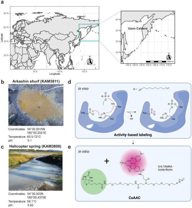
To showcase the applicability of this eABPP workflow, we sampled environmental microbial communities from two hot springs located in the Uzon Caldera (Kamchatka Peninsula, Russia; Fig. 2a), i.e., the ‘Arkashin shurf’ (KAM3811) and the ‘Helicopter spring’ (KAM3808; Fig. 2b, c) and performed ABPP of serine hydrolases using a FP-based ABP (Fig. 2d, e). These two sites were chosen due to their physicochemical properties (a temperature in the mesothermal range and a slightly acidic pH) that favor the development of an abundant yet unique microbial community. While KAM3811 is an artificial but for more than 30 years stable thermal pool [62] for which 16 metagenome-assembled genomes (MAGs) have been published recently [63], KAM3808 is a larger natural spring with no metagenome data available so far.
Fig. 2.
Location of the sampled springs and two-step chemical labeling of serine hydrolases. (a) The map shows the location of the two sampled springs ‘Arkashin shurf’ (KAM3811) and ‘Helicopter spring’ (KAM3808) in the Uzon Caldera region, Kamchatka Peninsula, Russia. (b, c) A representative picture is shown along with the exact coordinates and physicochemical properties of both springs. (d) Chemical structure of the employed FP-alkyne probe and overview of the reaction mechanism taking place at the active site of serine hydrolases during activity-based in vivo labeling of microbial communities from the two springs. (e) Attachment of the trifunctional reporter 5/6-TAMRA-azide-biotin via a copper-catalyzed azide-alkyne cycloaddition (CuAAC) for downstream target enzyme enrichment in a second in vitro step after protein extraction from sediments
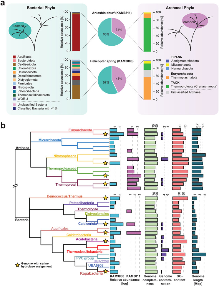
All sediment samples (i.e., two times seven aliquots) taken at the two hot springs were treated, processed, and analyzed according to our reported workflow. We then used the metagenome sequence data to gain insights into the community composition of both sediments at the domain and class level by analyzing the relative abundance of ribosomal protein S3 (rpS3) genes within the metagenomic datasets (relative abundance was determined via metagenomic read mapping; Fig. 3a, Supplementary Material 2). The two metagenomes displayed 99.9% and 99.4% of the microbial community of KAM3811 and KAM3808, respectively, with a sequencing effort of 30 Gbp. Both communities showed considerable differences in their distribution between Bacteria and Archaea as well as in the number of different classes present within each domain. For KAM3811, 66% of the assigned species belonged to the bacterial domain, with the Aquificota being the dominating phylum, while the archaeal domain was mainly represented by the Thermoproteota (Supplementary Material 1: Table S1). In the phenotypically and phylogenetically more diverse spring KAM3808, bacterial species account for 57% of all organisms, with the Caldisericota making up the largest fraction, followed by the Desulfobacterota and the Aquificota. In contrast to KAM3811, archaeal species comprised mainly Euryarchaeota, while Archaea from the DPANN group made up the smallest fraction (Supplementary Material 1: Table S2). In a more detailed analysis of these metagenomic datasets, again by relying on genome coverages, all phyla identified within the respective spring were displayed in a domain-specific phylogenetic tree with their corresponding abundancies (Fig. 3b; Supplementary Material 3). For KAM3808, which displayed more heteromorphic sediments, rpS3 genes of 49 microorganisms could be identified, with Aciduliprofundum sp. (relative abundance of 488.1), Caldisericum exile (408.2) and Caldimicrobium thiodismutans (139.9) as the most abundant species (Supplementary Material 1: Table S2). KAM3811, a distinctly smaller thermal pool with homogenous brown to red sediments, comprised 19 different organisms as determined from rpS3 gene analysis. Among them, Sulfurihydrogenibium sp. (2329.1) was identified as the dominating genus, followed by Pyrobaculum ferrireducens (411.3) and Caldisphaera sp. (389.6; Supplementary Material 1: Table S1).
Fig. 3.
Distribution of microorganisms across KAM3811 and KAM3808. (a) The proportion of Bacteria (cyan) and Archaea (magenta) within the sediments sampled for eABPP is depicted as pie diagrams for KAM3811 and KAM3808. An overview of the relative distribution of representative phyla from these domains is given respectively based on the GTDB taxonomy. (b) Phylogenetic tree displaying the relationship between the microorganisms found across both springs as calculated with GTDB-Tk based on the dereplicated, binned, and curated metagenomes from KAM3811 and KAM3808. Relative abundances of microorganisms based on the coverage of genomes are given for KAM3811 (magenta) and KAM3808 (light blue), respectively, along with their genome completeness (light green), contamination (purple), GC content (light red), and genome length (dark cyan) as calculated via checkM. The yellow stars indicate the genomes from which predicted serine hydrolases were confidently identified with the applied eABPP approach
Altogether, these experiments unambiguously show that the sampled communities are highly diverse in their microbial composition and should thus harbor diverse enzymes with serine hydrolase activity. The detected phyla largely comprise unexplored microorganisms with a functional potential yet to be uncovered.
Multifaceted annotation of serine hydrolases in the metaproteome database
The assembled metagenomes were then used to construct two metaproteome databases consisting of 45,649 (KAM3811) and 99,930 (KAM3808) protein-coding sequences, respectively. We relied on high-quality reads for assembly and only predicted full-length gene sequences (fragments were ignored). Combined with a fairly great sequencing depth, we provide enough coverage for a robust database for protein identification, yet avoiding the usage of public datasets, thereby decreasing the rate of false positives during detection. Annotation of the here-derived metaproteome databases against the UniRef100 database resulted in 35,323 genes encoding proteins with a predicted function for the KAM3811 dataset, while 8,575 proteins remained uncharacterized. Analogously, 85,393 genes encoded proteins with a predicted function for the KAM3808 dataset, whereas 17,923 genes encoded uncharacterized proteins. The discrepancy between the number of proteins with predicted function and hypothetical ones stemmed from the lack of represented similar genes within public databases or from the fragmentation of genes. Within the metaproteome dataset of KAM3811 and KAM3808, this annotation procedure surprisingly led to only two and sixteen proteins, respectively, which were assigned the term ‘serine hydrolase’. This is a consequence of the variety of terms used for describing the different members of the serine hydrolase superfamily. Predicting the actual number of serine hydrolase sequences present in the two datasets thus remains intricate, since it requires elaborate manual curation of relevant search strings. Although this difficulty is also addressed by our function-based eABPP approach, a bioinformatic solution to this problem would be beneficial for future studies.
The advantage of using a metagenome-derived metaproteome database relies on the exact blueprint of the genes and in silico predicted proteins for improved matching of newly generated peptide fragments. Public databases might not have the respective gene from the same organism, i.e., it might have differences in its amino acid sequence though conveying the same function. This might substantially decrease the number of assigned peptide fragments. Secondly, un(der)explored ecosystems often carry novel genes and thus proteins, which are not yet available in public databases and would thus not generate a proper match. In fact, recent analyses of public metagenomes demonstrated a continuous increase of novel protein families across new metagenomes [64, 65]. Thirdly, we resolved the metagenome at the genome level, which enables the direct assignment of enzymes to respective organisms, which is usually not possible with public databases (as general taxonomic assignments of functional genes are not stable in nature due to, e.g., horizontal gene transfer).
Identification of active serine hydrolases by eABPP
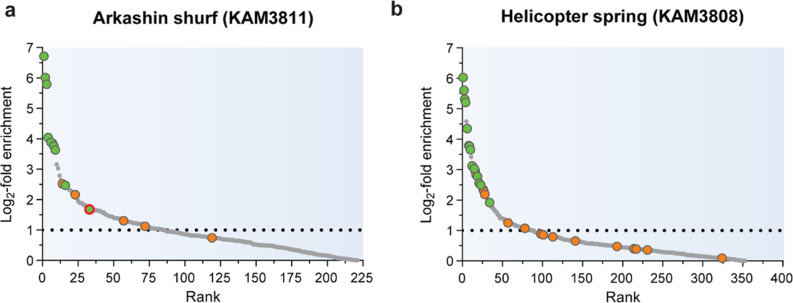
To identify active serine hydrolases from the sampled environmental communities, the self-constructed metaproteome databases were then employed as reference databases for the analysis of eABPP MS data resulting from an affinity enrichment of FP-alkyne-labeled target proteins via a biotin handle, on-bead digest, and subsequent LC-MS/MS analysis. Overall, 811 and 1,489 protein groups (comprising proteins that were not distinguishable based on the identified peptides) were identified for the KAM3811 and KAM3808 datasets, respectively, excluding hits from the implemented contaminants database. After filtering the initial data (see the methods section), a total of 385 protein groups for KAM3811 and 672 protein groups for KAM3808 remained for further analysis (Supplementary Material 4). Of these, 1.6% and 2.1% of protein groups for KAM3811 and KAM3808, respectively, comprised more than one protein. For KAM3811, 221 protein groups displayed a positive log2-fold change when comparing the group of FP-labeled samples against the DMSO-treated control group, while KAM3808 harbored 353 log2-fold enriched protein groups (Fig. 4). In contrast to the two large metaproteomes, these strongly reduced protein numbers now allowed a manual curation of serine hydrolase prediction by complementation of the UniRef100-based protein annotation with sequence searches against the Pfam, NCBI CCD and InterPro databases. Where necessary, additional searches against the SWISS-MODEL Template Library or with HHpred were also performed to identify structural homologs (Supplementary Material 5).
Fig. 4.
Predicted serine hydrolases identified from the sampled hot springs. Log2-fold enrichment of identified proteins labeled with FP-alkyne compared to the DMSO control for the sediments sampled from (a) KAM3811 and (b) KAM3808. Proteins predicted as serine hydrolases are displayed as colored dots (green: p-value ≤ 0.05, orange: p-value ≥ 0.05). The protein chosen for function validation is circled in red. Hits lying above the dotted line were more than two-fold enriched with the probe and were therefore considered primary hits. Each treatment group comprised three biological replicates
For KAM3811, this extended annotation procedure predicted 15 protein groups as serine hydrolases (Table 1). Most notably, these protein groups were predominantly found among the top enriched hits, with 11 protein groups found among the top 30 enriched hits. For KAM3808, which showed a larger microbial diversity, 31 predicted serine hydrolases were obtained (Table 2), with 19 protein groups found among the top 30 enriched hits. Further protein groups found among these top 30 enriched hits either lacked evidence for being potential serine hydrolases or remained uncharacterized as no conclusive information on their function was obtained based on sequence- or structure-based homology. However, their annotation from the UniRef100 database indicates that for both datasets, many of these are potential components of membrane transporters or other membrane-associated proteins (Supplementary Material 4). The top-ranked serine hydrolases likely represent the most active enzymes under the in vivo conditions of the hot springs due to either their higher abundances or their elevated activities compared to other serine hydrolases in the sampled environment. Although lower-ranked serine hydrolases not only feature a smaller fold change but often also a smaller p-value, this does not necessarily mean that they are less valid hits. The inhomogeneity of sediment and thus between samples as well as the necessity for imputation of missing data can add a bias to these computed parameters, especially for low abundant or inconsistently detected hits. Another thing to be aware of is that the use of a large metaproteome database increases the likelihood of false positives during peptide spectral matching. eABPP, however, provides a degree of inherent confirmation of correct identification through matching with the profiled function. Please note that we here report only those serine hydrolases as hits for both springs which were confidently identified by such a sequence- or structure-based homology analysis to demonstrate the robustness of the eABPP approach. It is however likely that there are several more enzymes displaying serine hydrolase features and activities across the two sets of enriched proteins that were not uncovered by this analysis.
Table 1.
Predicted serine hydrolases identified for KAM3811
| No. | Log2-fold change | -Log P | Identifier | Protein annotation# |
|---|---|---|---|---|
| 1 | 6.711 | 2.287 | ExploCarb_3811S_S4_2994_length_2665_cov_56_1 | Peptidase S8, subtilisin-related |
| 2 | 6.011 | 4.069 | ExploCarb_3811S_S4_103_length_35923_cov_48_7 | Penicillin/GL-7-ACA/AHL acylase |
| 3 | 5.787 | 3.448 | ExploCarb_3811S_S4_37_length_59942_cov_23_2 | Peptidase S8, subtilisin-related |
| 4 | 4.038 | 2.999 | ExploCarb_3811S_S4_179_length_24712_cov_181_27 | Penicillin/GL-7-ACA/AHL acylase |
| 6 | 3.881 | 3.141 | ExploCarb_3811S_S4_477_length_13181_cov_9_10 | Peptidase S8, subtilisin-related |
| 7 | 3.852 | 0.946 | ExploCarb_3811S_S4_782_length_9153_cov_146_4 | Penicillin/GL-7-ACA/AHL acylase |
| 8 | 3.760 | 1.690 | ExploCarb_3811S_S4_412_length_14764_cov_159_5 | Peptidase S8, subtilisin-related |
| 9 | 3.630 | 2.838 | ExploCarb_3811S_S4_5916_length_1495_cov_4_1 | Peptidase_S8/S53_dom |
| 14 | 2.520 | 1.043 | ExploCarb_3811S_S4_1380_length_5439_cov_180_2 | Peptidase_S9 |
| 16 | 2.470 | 3.204 | ExploCarb_3811S_S4_7165_length_1270_cov_6_1 | Peptidase S8, subtilisin-related |
| 23 | 2.162 | 0.734 | ExploCarb_3811S_S4_1591_length_4725_cov_42_1 | Protein of unknown function DUF915, hydrolase-like |
| 33 | 1.675 | 1.544 | ExploCarb_3811S_S4_483_length_13114_cov_941_5 | Uncharacterised protein family UPF0227/Esterase YqiA |
| 57 | 1.303 | 0.869 | ExploCarb_3811S_S4_1740_length_4311_cov_157_3 | Xaa-Pro-like_dom |
| 72 | 1.122 | 0.593 | ExploCarb_3811S_S4_259_length_20079_cov_121_4 | Peptidase_S49 |
| 119 | 0.745 | 0.826 | ExploCarb_3811S_S4_1428_length_5265_cov_777_6 | Peptidase_S49 |
# InterProScan annotation on either protein family or domain level
Table 2.
Predicted serine hydrolases identified for KAM3808
| No. | Log2-fold change | -Log P | Identifier | Protein annotation# |
|---|---|---|---|---|
| 1 | 6.028 | 2.903 | ExploCarb_3808S_S2_15464_length_1283_cov_2_1 | GDSL lipase/esterase |
| 2 | 5.607 | 1.416 | ExploCarb_3808S_S2_19633_length_1026_cov_55_1 | Putative S8A family peptidase† |
| 3 | 5.316 | 3.107 | ExploCarb_3808S_S2_5917_length_3034_cov_1_3 | Peptidase S8, subtilisin-related |
| 4 | 5.204 | 1.675 | ExploCarb_3808S_S2_470_length_27453_cov_4_11 | SGNH_hydro |
| 6 | 4.351 | 3.267 | ExploCarb_3808S_S2_593_length_22580_cov_176_21 | Esterase/lipase |
| 8 | 3.791 | 1.563 | ExploCarb_3808S_S2_12785_length_1520_cov_45_2 | Peptidase S8, subtilisin-related |
| 9 | 3.771 | 2.222 | ExploCarb_3808S_S2_10394_length_1832_cov_227_1 | Peptidase_S8/S53_dom_sf |
| 10 | 3.657 | 2.291 | ExploCarb_3808S_S2_1348_length_11661_cov_169_4 | PNPLA_dom |
| 12 | 3.124 | 2.085 | ExploCarb_3808S_S2_277_length_40468_cov_33_33 | Peptidase S8A, fervidolysin-like |
| 13 | 3.101 | 1.017 | ExploCarb_3808S_S2_2184_length_7533_cov_3_4 | Penicillin/GL-7-ACA/AHL/aculeacin-A acylase |
| 15 | 3.030 | 1.860 | ExploCarb_3808S_S2_12072_length_1604_cov_127_1 | Peptidase_S8/S53_dom_sf |
| 16 | 2.954 | 0.995 | ExploCarb_3808S_S2_1901_length_8595_cov_3_5 | Penicillin/GL-7-ACA/AHL acylase |
| 17 | 2.839 | 1.008 | ExploCarb_3808S_S2_5211_length_3405_cov_171_3 | Peptidase family S66 |
| 19 | 2.776 | 2.407 | ExploCarb_3808S_S2_84_length_75002_cov_30_5 | PNPLA_dom |
| 21 | 2.560 | 1.689 | ExploCarb_3808S_S2_12016_length_1611_cov_2_1 | AB_hydrolase_1 |
| 23 | 2.492 | 2.063 | ExploCarb_3808S_S2_1180_length_13195_cov_3_10 | SGNH_hydro_sf |
| 26 | 2.344 | 0.874 | ExploCarb_3808S_S2_5180_length_3427_cov_2_3 | Peptidase S8, subtilisin-related |
| 27 | 2.282 | 0.487 | ExploCarb_3808S_S2_14352_length_1370_cov_1_2 | AB_hydrolase_1 |
| 28 | 2.185 | 1.070 | ExploCarb_3808S_S2_1_length_789533_cov_14_559 | Peptidase S8, subtilisin-related |
| 34 | 1.914 | 1.385 | ExploCarb_3808S_S2_7708_length_2386_cov_3_1 | Lipase_bact_N |
| 57 | 1.253 | 0.146 | ExploCarb_3808S_S2_17847_length_1126_cov_1_2 | AB_hydrolase_1 |
| 78 | 1.068 | 0.956 | ExploCarb_3808S_S2_4160_length_4192_cov_19_2 | Penicillin/GL-7-ACA/AHL acylase |
| 98 | 0.884 | 0.766 | ExploCarb_3808S_S2_2577_length_6506_cov_183_2 | Peptidase S8, subtilisin-related |
| 101 | 0.856 | 0.606 | ExploCarb_3808S_S2_5424_length_3285_cov_7_1 | Peptidase S8, subtilisin-related |
| 113 | 0.792 | 0.541 | ExploCarb_3808S_S2_45_length_96604_cov_169_9 | PNPLA_dom |
| 141 | 0.657 | 0.331 | ExploCarb_3808S_S2_593_length_22580_cov_176_11 | Peptidase S8, subtilisin-related |
| 193 | 0.477 | 0.170 | ExploCarb_3808S_S2_1272_length_12299_cov_60_1 | AB_hydrolase_1 |
| 214 | 0.397 | 0.143 | ExploCarb_3808S_S2_12631_length_1536_cov_2_2 | Peptidase S1C, Do |
| 217 | 0.387 | 0.590 | ExploCarb_3808S_S2_858_length_17095_cov_20_14 | Hydrolase_4 (Serine aminopeptidase, S33) |
| 231 | 0.358 | 0.176 | ExploCarb_3808S_S2_21_length_132706_cov_167_47 | Peptidase S1C |
| 324 | 0.089 | 0.091 | ExploCarb_3808S_S2_2714_length_6206_cov_211_1 | Penicillin/GL-7-ACA/AHL acylase |
# InterProScan annotation on either protein family, homologous superfamily or domain level unless stated otherwise
† UniRef100 annotation
The eABPP-enriched active microbial community serine hydrolases comprised enzymes from the entire serine hydrolase superfamily, including proteases (or peptidases as synonymous term), lipases, amidases, and esterases [55]. Across both springs, we found proteins predicted as serine-type peptidases from the (super)families S1 (chymotrypsin family, subfamily S1C), S8/S53 (clan SB: S8 (subtilisin family, including subfamily S8A), S53 (type peptidase: sedolisin)), S9 (prolyl oligopeptidase family), S15 (type peptidase: Xaa-Pro dipeptidyl peptidase), S33 (type peptidase: prolyl aminopeptidase), S45 (type peptidase: penicillin G acylase precursor), S49 (protease IV family), and S66 (type peptidase: murein tetrapeptidase LD-carboxypeptidase). Moreover, we detected proteins predicted as esterases/lipases, including enzymes from the SGNH hydrolase superfamily or enzymes containing a GDSL, a PNPLA (patatin-like phospholipases), or a Lipase_bact_N domain, and a DUF915 family enzyme with structural similarity to esterases/lipases as well as a putative esterase from the UPF0227 family. Furthermore, enzymes predicted as alpha/beta fold-1 hydrolases without further classification but with esterases/lipases as structural homologs were identified from these datasets.
Altogether, these results show that our eABPP approach enabled the identification of functionally diverse serine hydrolases, ranging from various proteases to esterases/lipases up to serine hydrolases with uncharacterized functions (e.g., DUFs or UPFs), thus being superior over a simple bioinformatic metagenome annotation procedure. Of note, the majority of the identified active serine hydrolases are annotated as subtilases or penicillin acylases, which demonstrates that the applied eABPP screen indeed allows the detection of thermostable representatives of the functionally highly diverse family of serine hydrolases that already find application for industrial purposes [66–69].
Function validation of a selected serine hydrolase
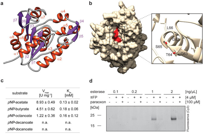
Finally, to confirm the suitability of eABPP for a function-based enzyme identification from an environmental microbial community, we chose the serine hydrolase ExploCarb_3811S_S4_483_length_13114_cov_941_5 (Table 1) from our list of eABPP-identified serine hydrolases for further bioinformatical and biochemical characterization. This 191 amino acid-containing enzyme was selected due to its relatively short and complete gene sequence as well as the availability of a straightforward enzyme kinetic assay. In addition, the differential annotation of this enzyme (see Supplementary Material 5) nicely demonstrates the caveats of bioinformatic-based protein function prediction when relying only on a single database, as it was predicted as an esterase based on the UniRef100 database, while the domain-predicting databases Pfam and InterPro revealed it as a UPF0227 (uncharacterized protein family 0227) protein. We therefore generated an AlphaFold-derived structural model for this protein that revealed a fold in which a GTSLG sequence previously associated with thioesterase activity [70] is accommodated at the functionally conserved GxSxG active site motif of serine hydrolases (Fig. 5a, b). The esterase sequence shows 100% identity to a predicted esterase (NCBI accession number: PMP76296.1) from a Sulfurihydrogenibium sp. MAG from a published metagenome, originating from the same spring [63]. BLAST-based sequence as well as HHpred-based structural homology searches further confirmed its similarity to other esterases (Supplementary Material 1: Table S3). These analyses together suggest that the UPF0227 domain-containing protein is most likely a serine esterase with however unknown substrate specificity.
Fig. 5.
Characterization of the putative esterase. (a) Predicted structure of the UPF0227 protein with the secondary structures visualized in red (alpha helices) and purple (beta strands). The five-stranded parallel beta-sheet consists of the strands β1, β2, β3, β6 and β7. (b) Surface-displaying structure of the putative esterase. The close-up displays the conserved serine hydrolase motif GxSxG, comprising the residues G63, T64, S65, L66, and G67, which is located in a substrate pocket (depicted in red). Structure prediction was performed with AlphaFold and the output was processed in Chimera. (c) Kinetic characterization of the esterase using the pNP-substrates pNP-acetate, -butyrate, -octanoate, -decanoate, and -dodecanoate at concentrations up to 0.7 mM at pH 8.0 and 70 °C and calculation of Vmax and Km. Values represent the mean of three technical replicates ± SD. (d) In vitro labeling of varying amounts of the esterase with FP-alkyne in the presence or absence of paraoxon
In order to biochemically validate the function of the putative esterase, E. coli Rosetta(DE3) cells were transformed with the codon-optimized gene sequence cloned into a pET-28b(+) vector for heterologous protein expression. The purified enzyme was biochemically characterized using para-nitrophenyl (pNP) substrates. The putative esterase showed highest activity at a pH of 8.0 (Supplementary Material 1: Fig. S2a) and a temperature of 70 °C (Supplementary Material 1: Fig. S2b) using pNP-butyrate as a substrate. Moreover, enzyme kinetics were determined using different pNP-esters (Fig. 5c). Effective hydrolysis was observed for pNP-acetate (Vmax = 8.93 U mg− 1, Km = 0.12 mM), pNP-butyrate (Vmax = 4.51 U mg− 1, Km = 0.18 mM) and pNP-octanoate (Vmax = 1.22 U mg− 1, Km = 0.16 mM), confirming its function as an esterase, whereas no activity was measured for pNP-esters with longer chain lengths, such as pNP-decanoate or pNP-dodecanoate. Furthermore, in vitro ABPP of the purified esterase with FP-alkyne, analogously to the in vivo ABPP experiment, revealed robust labeling over a range of esterase concentrations, which could be strongly diminished by pre-incubation with the generic serine esterase inhibitor paraoxon (Fig. 5d). Consequently, the esterase was proven a bona fide target of the covalent-acting FP probe. In line with this result, FP-alkyne was furthermore shown to reduce the esterase activity of the enzyme in correlation with the applied probe concentration (Supplementary Material 1: Figure S2c).
Conclusions
In this study, we established eABPP for the efficient function-based identification of active enzymes directly from the environment. The eABPP workflow relies on activity-based labeling of a microbial community sample in combination with metagenome sequencing. In this way, the bioinformatic annotation of the metagenome can be directly confirmed through the activity-dependent reaction of the ABP-targeted enzyme. This provides direct experimental evidence for the bioinformatically predicted enzyme function along with its protein identification. As a showcase, we profiled epi-sedimentary communities from two hot springs located in the Uzon Caldera region of Kamchatka (Russia), exploiting the broad range serine hydrolase target specificity of FP-alkyne for functional enzyme annotation. Thorough bioinformatic analysis of the eABPP-enriched and -identified proteins from the hot spring sediments revealed that the top-ranked hits mainly comprised different serine peptidases, mostly from the families S8 and S15, as well as esterases/lipases, thus demonstrating the applicability of the method. To further corroborate the versatility of our eABPP approach, we heterologously expressed an ‘uncharacterized protein family UPF0227’ enzyme found among the top eABPP-enriched hits and confirmed its esterase activity.
We therefore believe that our approach can be further extended to profile additional enzyme activities of microbial communities from diverse ecosystems, in particular as a large and constantly growing repertoire of probes with a wide variety of warheads for targeting different enzymes or enzyme classes is already available. These probes provide a broad coverage of enzymes from different classes of the Enzyme Commission (EC) number classification system. Although most of the probes have been designed for hydrolases (EC 3), several probes targeting different enzymes from subclasses of the oxidoreductases (EC 1), transferases (EC 2), mainly kinases, and ligases (EC 6) as well as ‘reverse-polarity’ probes engaging protein electrophiles instead of nucleophilic residues, which target enzymes across different classes, have been developed [22, 71, 72]. Despite the broad coverage of enzymes with different modes of action, ABPP is still limited to a few enzyme families in light of the great diversity of existing enzyme activities. As the probes available to date mainly address metabolic enzymes, eABPP may be adapted for ecological research, for instance to study different aspects of ecosystem functioning or to decipher enzyme activity patterns, e.g., from synergistic or mutual community interactions. Besides, we anticipate that our approach may open new avenues in enzyme discovery, especially for finding enzyme activities that harbor potential for application in biotechnological processes. These include, for example, cysteine proteases or glycoside hydrolases, for which a broad set of probes such as the well-established E64- or cyclophellitol-based ABPs, which we believe would also allow reliable enzyme identification in complex environmental samples with a rather low abundance of single proteins, is currently at hand [73, 74]. Glycoside hydrolases, for instance, are of particular interest with regard to biocatalyst discovery since they largely function in (lignocellulosic) biomass degradation [39–41, 75]. In addition, it might be reasonable to design more specific probes with activity towards serine hydrolases that function in the degradation of environmental pollutants (e.g., PETases and different biphenyl and meta-cleavage product hydrolases capable of cleaving mono- and bicyclic aromatic compounds), which would allow a more precise confirmation of function compared to broad-spectrum probes with a large and diverse target repertoire, as these enzymes are of interest for environmental and industrial applications.
Electronic supplementary material
Below is the link to the electronic supplementary material.
Acknowledgements
We acknowledge the use of the BioRender software (BioRender.com) for graphics preparation.
Author contributions
T.K. and T.V.K. performed in-field chemical labeling experiments, T.V.K. and I.V.K. performed DNA extraction and metagenomic sequencing, S.P.E. and A.J.P. performed refinement of metagenome data and metaproteome assembly, S.N., T.K. and L.S. performed protein extraction and affinity enrichment experiments, S.N. and F.K. performed MS analyses, T.K. performed protein expression and enzyme assays, B.S., M.K. and I.V.K. designed the workflow for the field ABPP, C.B., B.S. and M.K. supervised the study and S.N., T.K., B.S. and M.K. wrote the manuscript. S.N. and T.K. contributed equally to this paper.
Funding
Open Access funding enabled and organized by Projekt DEAL. B.S., M.K. and I.V.K. acknowledge funding within the DFG-RSF Cooperation for joint German-Russian projects by the DFG (SI 642/12 − 1, KA 2894/6 − 1) and RSF (18-44-04024). This work was also supported by the DFG grant INST 20876/322-1 FUGG (to M. K. and F.K.).
Data availability
The mass spectrometry proteomics datasets generated during this study have been deposited to the ProteomeXchange Consortium via the PRIDE [76] partner repository (https://www.ebi.ac.uk/pride/archive/) with the project accession number PXD025833. Processed mass spectrometry data is contained in Supplementary Material 4. All raw sequencing data was submitted to the NCBI (https://www.ncbi.nlm.nih.gov/) Sequence Read Archive (SRA) under the BioProject accession number PRJNA1013789 (SRR25937770 and SRR25937769). The accession numbers of the genomes can be found in Supplementary Material 3 with the respective statistical data on the genome quality. The rpS3 genes are available under 10.6084/m9.figshare.25805215.v1.
Declarations
Ethics approval and consent to participate
Not applicable.
Consent for publication
Not applicable.
Competing interests
The authors declare no competing interests.
Footnotes
Publisher’s Note
Springer Nature remains neutral with regard to jurisdictional claims in published maps and institutional affiliations.
Sabrina Ninck and Thomas Klaus contributed equally to this work.
Contributor Information
Sabrina Ninck, Email: sabrina.ninck@uni-due.de.
Bettina Siebers, Email: bettina.siebers@uni-due.de.
Markus Kaiser, Email: markus.kaiser@uni-due.de.
References
- 1.Locey KJ, Lennon JT. Scaling laws predict global microbial diversity. P Natl Acad Sci USA. 2016;113:5970–5. doi: 10.1073/pnas.1521291113. [DOI] [PMC free article] [PubMed] [Google Scholar]
- 2.Baquero F, Coque TM, Galan JC, Martinez JL. The origin of niches and species in the Bacterial World. Front Microbiol. 2021;12:657986. doi: 10.3389/fmicb.2021.657986. [DOI] [PMC free article] [PubMed] [Google Scholar]
- 3.Verstraete W. Microbial ecology and environmental biotechnology. Isme J. 2007;1:4–8. doi: 10.1038/ismej.2007.7. [DOI] [PubMed] [Google Scholar]
- 4.Calcagno V, Jarne P, Loreau M, Mouquet N, David P. Diversity spurs diversification in ecological communities. Nat Commun. 2017;8:15810. doi: 10.1038/ncomms15810. [DOI] [PMC free article] [PubMed] [Google Scholar]
- 5.Shu WS, Huang LN. Microbial diversity in extreme environments. Nat Rev Microbiol. 2022;20:219–35. doi: 10.1038/s41579-021-00648-y. [DOI] [PubMed] [Google Scholar]
- 6.Franzosa EA, Hsu T, Sirota-Madi A, Shafquat A, Abu-Ali G, Morgan XC, et al. Sequencing and beyond: integrating molecular ‘omics’ for microbial community profiling. Nat Rev Microbiol. 2015;13:360–72. doi: 10.1038/nrmicro3451. [DOI] [PMC free article] [PubMed] [Google Scholar]
- 7.Elleuche S, Schroder C, Sahm K, Antranikian G. Extremozymes - biocatalysts with unique properties from extremophilic microorganisms. Curr Opin Biotech. 2014;29:116–23. doi: 10.1016/j.copbio.2014.04.003. [DOI] [PubMed] [Google Scholar]
- 8.Sysoev M, Grotzinger SW, Renn D, Eppinger J, Rueping M, Karan R. Bioprospecting of Novel Extremozymes from prokaryotes-the Advent of Culture-Independent methods. Front Microbiol. 2021;12:630013. doi: 10.3389/fmicb.2021.630013. [DOI] [PMC free article] [PubMed] [Google Scholar]
- 9.Berini F, Casciello C, Marcone GL, Marinelli F. Metagenomics: novel enzymes from non-culturable microbes. FEMS Microbiol Lett. 2017;364:fnx211. doi: 10.1093/femsle/fnx211. [DOI] [PubMed] [Google Scholar]
- 10.Robinson SL, Piel J, Sunagawa S. A roadmap for metagenomic enzyme discovery. Nat Prod Rep. 2021;38:1994–2023. doi: 10.1039/D1NP00006C. [DOI] [PMC free article] [PubMed] [Google Scholar]
- 11.Kennedy J, Marchesi JR, Dobson ADW. Marine metagenomics: strategies for the discovery of novel enzymes with biotechnological applications from marine environments. Microb Cell Fact. 2008;7:27. doi: 10.1186/1475-2859-7-27. [DOI] [PMC free article] [PubMed] [Google Scholar]
- 12.DeCastro ME, Rodriguez-Belmonte E, Gonzalez-Siso MI. Metagenomics of thermophiles with a focus on Discovery of Novel Thermozymes. Front Microbiol. 2016;7:1521. doi: 10.3389/fmicb.2016.01521. [DOI] [PMC free article] [PubMed] [Google Scholar]
- 13.Harrington ED, Singh AH, Doerks T, Letunic I, von Mering C, Jensen LJ, et al. Quantitative assessment of protein function prediction from metagenomics shotgun sequences. P Natl Acad Sci USA. 2007;104:13913–8. doi: 10.1073/pnas.0702636104. [DOI] [PMC free article] [PubMed] [Google Scholar]
- 14.Mirete S, Morgante V, Gonzalez-Pastor JE. Functional metagenomics of extreme environments. Curr Opin Biotech. 2016;38:143–9. doi: 10.1016/j.copbio.2016.01.017. [DOI] [PubMed] [Google Scholar]
- 15.Newgas SA, Jeffries JWE, Moody TS, Ward JM, Hailes HC. Discovery of New Carbonyl Reductases using functional metagenomics and applications in Biocatalysis. Adv Synth Catal. 2021;363:3044–52. doi: 10.1002/adsc.202100199. [DOI] [PMC free article] [PubMed] [Google Scholar]
- 16.Wohlgemuth R, Littlechild J, Monti D, Schnorr K, van Rossum T, Siebers B, et al. Discovering novel hydrolases from hot environments. Biotechnol Adv. 2018;36:2077–100. doi: 10.1016/j.biotechadv.2018.09.004. [DOI] [PubMed] [Google Scholar]
- 17.Nasseri SA, Betschart L, Opaleva D, Rahfeld P, Withers SG. A mechanism-based Approach to Screening Metagenomic libraries for Discovery of unconventional glycosidases. Angew Chem Int Ed Engl. 2018;57:11359–64. doi: 10.1002/anie.201806792. [DOI] [PubMed] [Google Scholar]
- 18.Lam KN, Cheng JJ, Engel K, Neufeld JD, Charles TC. Current and future resources for functional metagenomics. Front Microbiol. 2015;6:1196. doi: 10.3389/fmicb.2015.01196. [DOI] [PMC free article] [PubMed] [Google Scholar]
- 19.Hatzenpichler R, Krukenberg V, Spietz RL, Jay ZJ. Next-generation physiology approaches to study microbiome function at single cell level. Nat Rev Microbiol. 2020;18:241–56. doi: 10.1038/s41579-020-0323-1. [DOI] [PMC free article] [PubMed] [Google Scholar]
- 20.Cravatt BF, Wright AT, Kozarich JW. Activity-based protein profiling: from enzyme chemistry to proteomic chemistry. Annu Rev Biochem. 2008;77:383–414. doi: 10.1146/annurev.biochem.75.101304.124125. [DOI] [PubMed] [Google Scholar]
- 21.Benns HJ, Wincott CJ, Tate EW, Child MA. Activity- and reactivity-based proteomics: recent technological advances and applications in drug discovery. Curr Opin Chem Biol. 2021;60:20–9. doi: 10.1016/j.cbpa.2020.06.011. [DOI] [PubMed] [Google Scholar]
- 22.Fang H, Peng B, Ong SY, Wu Q, Li L, Yao SQ. Recent advances in activity-based probes (ABPs) and affinity-based probes (AfBPs) for profiling of enzymes. Chem Sci. 2021;12:8288–310. doi: 10.1039/D1SC01359A. [DOI] [PMC free article] [PubMed] [Google Scholar]
- 23.Fonović M, Bogyo M. Activity-based probes as a tool for functional proteomic analysis of proteases. Expert Rev Proteomic. 2008;5:721–30. doi: 10.1586/14789450.5.5.721. [DOI] [PMC free article] [PubMed] [Google Scholar]
- 24.Böttcher T, Pitscheider M, Sieber SA. Natural products and their biological targets: proteomic and metabolomic labeling strategies. Angew Chem Int Edit. 2010;49:2680–98. doi: 10.1002/anie.200905352. [DOI] [PubMed] [Google Scholar]
- 25.van Bergen W, Hevler JF, Wu W, Baggelaar MP, Heck AJR. Site-specific activity-based protein profiling using Phosphonate handles. Mol Cell Proteom. 2023;22:100455. doi: 10.1016/j.mcpro.2022.100455. [DOI] [PMC free article] [PubMed] [Google Scholar]
- 26.Ha J, Park H, Park J, Park SB. Recent advances in identifying protein targets in drug discovery. Cell Chem Biol. 2021;28:394–423. doi: 10.1016/j.chembiol.2020.12.001. [DOI] [PubMed] [Google Scholar]
- 27.Barglow KT, Cravatt BF. Activity-based protein profiling for the functional annotation of enzymes. Nat Methods. 2007;4:822–7. doi: 10.1038/nmeth1092. [DOI] [PubMed] [Google Scholar]
- 28.Speers AE, Adam GC, Cravatt BF. Activity-based protein profiling in vivo using a copper(I)-catalyzed azide-alkyne [3 + 2] cycloaddition. J Am Chem Soc. 2003;125:4686–7. doi: 10.1021/ja034490h. [DOI] [PubMed] [Google Scholar]
- 29.Verhelst SHL, Bonger KM, Willems LI. Bioorthogonal reactions in activity-based protein profiling. Molecules. 2020;25:5994. doi: 10.3390/molecules25245994. [DOI] [PMC free article] [PubMed] [Google Scholar]
- 30.Geurink PP, Prely LM, van der Marel GA, Bischoff R, Overkleeft HS. Photoaffinity labeling in activity-based protein profiling. Top Curr Chem. 2012;324:85–113. doi: 10.1007/128_2011_286. [DOI] [PubMed] [Google Scholar]
- 31.Yang PY, Liu K. Activity-based protein profiling: recent advances in Probe Development and Applications. ChemBioChem. 2015;16:712–24. doi: 10.1002/cbic.201402582. [DOI] [PubMed] [Google Scholar]
- 32.Berger AB, Vitorino PM, Bogyo M. Activity-based protein profiling: applications to biomarker discovery, in vivo imaging and drug discovery. Am J Pharmacogenomics. 2004;4:371–81. doi: 10.2165/00129785-200404060-00004. [DOI] [PubMed] [Google Scholar]
- 33.Xu JQ, Li XQ, Ding K, Li ZQ. Applications of activity-based protein profiling (ABPP) and bioimaging in Drug Discovery. Chem-Asian J. 2020;15:34–41. doi: 10.1002/asia.201901500. [DOI] [PubMed] [Google Scholar]
- 34.Roberts AM, Ward CC, Nomura DK. Activity-based protein profiling for mapping and pharmacologically interrogating proteome-wide ligandable hotspots. Curr Opin Biotech. 2017;43:25–33. doi: 10.1016/j.copbio.2016.08.003. [DOI] [PMC free article] [PubMed] [Google Scholar]
- 35.Nomura DK, Dix MM, Cravatt BF. Activity-based protein profiling for biochemical pathway discovery in cancer. Nat Rev Cancer. 2010;10:630–8. doi: 10.1038/nrc2901. [DOI] [PMC free article] [PubMed] [Google Scholar]
- 36.Keller LJ, Babin BM, Lakemeyer M, Bogyo M. Activity-based protein profiling in bacteria: applications for identification of therapeutic targets and characterization of microbial communities. Curr Opin Chem Biol. 2020;54:45–53. doi: 10.1016/j.cbpa.2019.10.007. [DOI] [PMC free article] [PubMed] [Google Scholar]
- 37.Morimoto K, van der Hoorn RA. The increasing impact of activity-based protein profiling in Plant Science. Plant Cell Physiol. 2016;57:446–61. doi: 10.1093/pcp/pcw003. [DOI] [PubMed] [Google Scholar]
- 38.Sadler NC, Wright AT. Activity-based protein profiling of microbes. Curr Opin Chem Biol. 2015;24:139–44. doi: 10.1016/j.cbpa.2014.10.022. [DOI] [PubMed] [Google Scholar]
- 39.Klaus T, Ninck S, Albersmeier A, Busche T, Wibberg D, Jiang J, et al. Activity-based protein profiling for the identification of Novel carbohydrate-active enzymes involved in Xylan Degradation in the Hyperthermophilic Euryarchaeon Thermococcus sp. Strain 2319 × 1E. Front Microbiol. 2021;12:734039. doi: 10.3389/fmicb.2021.734039. [DOI] [PMC free article] [PubMed] [Google Scholar]
- 40.McGregor NGS, de Boer C, Santos M, Haon M, Navarro D, Schroder S, et al. Activity-based protein profiling reveals dynamic substrate-specific cellulase secretion by saprotrophic basidiomycetes. Biotechnol Biof Biop. 2022;15:6. doi: 10.1186/s13068-022-02107-z. [DOI] [PMC free article] [PubMed] [Google Scholar]
- 41.Liu Y, Fredrickson JK, Sadler NC, Nandhikonda P, Smith RD, Wright AT. Advancing understanding of microbial bioenergy conversion processes by activity-based protein profiling. Biotechnol Biofuels. 2015;8:156. doi: 10.1186/s13068-015-0343-7. [DOI] [PMC free article] [PubMed] [Google Scholar]
- 42.Whidbey C, Wright AT. Activity-based protein profiling—enabling Multimodal Functional studies of Microbial communities. In: Cravatt B, Hsu KL, Weerapana E, editors. Activity-based protein profiling. Current topics in Microbiology and Immunology. Cham: Springer; 2019. pp. 1–21. [DOI] [PMC free article] [PubMed] [Google Scholar]
- 43.Whidbey C, Sadler NC, Nair RN, Volk RF, DeLeon AJ, Bramer LM, et al. A probe-enabled Approach for the selective isolation and characterization of functionally active subpopulations in the gut Microbiome. J Am Chem Soc. 2019;141:42–7. doi: 10.1021/jacs.8b09668. [DOI] [PMC free article] [PubMed] [Google Scholar]
- 44.Reichart NJ, Steiger AK, Van Fossen EM, McClure R, Overkleeft HS, Wright AT. Selection and enrichment of microbial species with an increased lignocellulolytic phenotype from a native soil microbiome by activity-based probing. ISME Commun. 2023;3:106. doi: 10.1038/s43705-023-00305-w. [DOI] [PMC free article] [PubMed] [Google Scholar]
- 45.Mayers MD, Moon C, Stupp GS, Su AI, Wolan DW. Quantitative metaproteomics and activity-based Probe Enrichment reveals significant alterations in protein expression from a mouse model of inflammatory bowel disease. J Proteome Res. 2017;16:1014–26. doi: 10.1021/acs.jproteome.6b00938. [DOI] [PMC free article] [PubMed] [Google Scholar]
- 46.Killinger BJ, Whidbey C, Sadler NC, DeLeon AJ, Munoz N, Kim YM, et al. Activity-based protein profiling identifies alternating activation of enzymes involved in the bifidobacterium shunt pathway or mucin degradation in the gut microbiome response to soluble dietary fiber. Npj Biofilms Microbi. 2022;8:60. doi: 10.1038/s41522-022-00313-z. [DOI] [PMC free article] [PubMed] [Google Scholar]
- 47.Simpson JB, Sekela JJ, Graboski AL, Borlandelli VB, Barker NK, Sorgen AA, et al. Metagenomics combined with activity-based proteomics point to gut bacterial enzymes that reactivate mycophenolate. Gut Microbes. 2022;14:2107289. doi: 10.1080/19490976.2022.2107289. [DOI] [PMC free article] [PubMed] [Google Scholar]
- 48.Chauvigne-Hines LM, Anderson LN, Weaver HM, Brown JN, Koech PK, Nicora CD, et al. Suite of activity-based probes for cellulose-degrading enzymes. J Am Chem Soc. 2012;134:20521–32. doi: 10.1021/ja309790w. [DOI] [PMC free article] [PubMed] [Google Scholar]
- 49.Ansong C, Sadler NC, Hill EA, Lewis MP, Zink EM, Smith RD, et al. Characterization of protein redox dynamics induced during light-to-dark transitions and nutrient limitation in cyanobacteria. Front Microbiol. 2014;5:325. doi: 10.3389/fmicb.2014.00325. [DOI] [PMC free article] [PubMed] [Google Scholar]
- 50.Sadler NC, Melnicki MR, Serres MH, Merkley ED, Chrisler WB, Hill EA, et al. Live cell chemical profiling of temporal redox dynamics in a photoautotrophic cyanobacterium. ACS Chem Biol. 2014;9:291–300. doi: 10.1021/cb400769v. [DOI] [PubMed] [Google Scholar]
- 51.Bennett K, Sadler NC, Wright AT, Yeager C, Hyman MR. Activity-based protein profiling of Ammonia Monooxygenase in Nitrosomonas europaea. Appl Environ Microbiol. 2016;82:2270–9. doi: 10.1128/AEM.03556-15. [DOI] [PMC free article] [PubMed] [Google Scholar]
- 52.Zweerink S, Kallnik V, Ninck S, Nickel S, Verheyen J, Blum M, et al. Activity-based protein profiling as a robust method for enzyme identification and screening in extremophilic Archaea. Nat Commun. 2017;8:15352. doi: 10.1038/ncomms15352. [DOI] [PMC free article] [PubMed] [Google Scholar]
- 53.Sakoula D, Smith GJ, Frank J, Mesman RJ, Kop LFM, Blom P, et al. Universal activity-based labeling method for ammonia- and alkane-oxidizing bacteria. Isme J. 2022;16:958–71. doi: 10.1038/s41396-021-01144-0. [DOI] [PMC free article] [PubMed] [Google Scholar]
- 54.Liu YS, Patricelli MP, Cravatt BF. Activity-based protein profiling: the serine hydrolases. P Natl Acad Sci USA. 1999;96:14694–9. doi: 10.1073/pnas.96.26.14694. [DOI] [PMC free article] [PubMed] [Google Scholar]
- 55.Simon GM, Cravatt BF. Activity-based proteomics of enzyme superfamilies: serine hydrolases as a case study. J Biol Chem. 2010;285:11051–5. doi: 10.1074/jbc.R109.097600. [DOI] [PMC free article] [PubMed] [Google Scholar]
- 56.Chandra P, Enespa, Singh R, Arora PK. Microbial lipases and their industrial applications: a comprehensive review. Microb Cell Fact. 2020;19:169. doi: 10.1186/s12934-020-01428-8. [DOI] [PMC free article] [PubMed] [Google Scholar]
- 57.Panda T, Gowrishankar BS. Production and applications of esterases. Appl Microbiol Biot. 2005;67:160–9. doi: 10.1007/s00253-004-1840-y. [DOI] [PubMed] [Google Scholar]
- 58.Barzkar N, Sohail M, Jahromi ST, Gozari M, Poormozaffar S, Nahavandi R, et al. Marine Bacterial esterases: emerging biocatalysts for Industrial Applications. Appl Biochem Biotech. 2021;193:1187–214. doi: 10.1007/s12010-020-03483-8. [DOI] [PubMed] [Google Scholar]
- 59.Anobom CD, Pinheiro AS, De-Andrade RA, Aguieiras ECG, Andrade GC, Moura MV, et al. From structure to Catalysis: recent developments in the Biotechnological Applications of lipases. Biomed Res Int. 2014;2014:684506. doi: 10.1155/2014/684506. [DOI] [PMC free article] [PubMed] [Google Scholar]
- 60.Romano D, Bonomi F, de Mattos MC, Fonseca TD, de Oliveira MDF, Molinari F. Esterases as stereoselective biocatalysts. Biotechnol Adv. 2015;33:547–65. doi: 10.1016/j.biotechadv.2015.01.006. [DOI] [PubMed] [Google Scholar]
- 61.Wu ZM, Liu CF, Zhang ZY, Zheng RC, Zheng YG. Amidase as a versatile tool in amide-bond cleavage: from molecular features to biotechnological applications. Biotechnol Adv. 2020;43:107574. doi: 10.1016/j.biotechadv.2020.107574. [DOI] [PubMed] [Google Scholar]
- 62.Burgess EA, Unrine JM, Mills GL, Romanek CS, Wiegel J. Comparative geochemical and microbiological characterization of two thermal pools in the Uzon Caldera, Kamchatka, Russia. Microb Ecol. 2012;63:471–89. doi: 10.1007/s00248-011-9979-4. [DOI] [PubMed] [Google Scholar]
- 63.Wilkins LGE, Ettinger CL, Jospin G, Eisen JA. Metagenome-assembled genomes provide new insight into the microbial diversity of two thermal pools in Kamchatka, Russia. Sci Rep. 2019;9:3059. doi: 10.1038/s41598-019-39576-6. [DOI] [PMC free article] [PubMed] [Google Scholar]
- 64.Rodríguez Del Río Á. Giner-Lamia J, Cantalapiedra CP, Botas J, Deng Z, Hernández-Plaza A, et al. Functional and evolutionary significance of unknown genes from uncultivated taxa. Nature. 2024;626:377–84. doi: 10.1038/s41586-023-06955-z. [DOI] [PMC free article] [PubMed] [Google Scholar]
- 65.Pavlopoulos GA, Baltoumas FA, Liu S, Selvitopi O, Camargo AP, Nayfach S, et al. Unraveling the functional dark matter through global metagenomics. Nature. 2023;622:594–602. doi: 10.1038/s41586-023-06583-7. [DOI] [PMC free article] [PubMed] [Google Scholar]
- 66.Niehaus F, Gabor E, Wieland S, Siegert P, Maurer KH, Eck J. Enzymes for the laundry industries: tapping the vast metagenomic pool of alkaline proteases. Microb Biotechnol. 2011;4:767–76. doi: 10.1111/j.1751-7915.2011.00279.x. [DOI] [PMC free article] [PubMed] [Google Scholar]
- 67.De Oliveira Martinez JP, Cai GQ, Nachtschatt M, Navone L, Zhang ZY, Robins K, et al. Challenges and opportunities in identifying and characterising keratinases for value-added peptide production. Catalysts. 2020;10:184. doi: 10.3390/catal10020184. [DOI] [Google Scholar]
- 68.Li J, Cheng JH, Teng ZJ, Zhang X, Chen XL, Sun ML, et al. A Novel Gelatinase from Marine Flocculibacter collagenilyticus SM1988: characterization and potential application in Collagen Oligopeptide-Rich Hydrolysate Preparation. Mar Drugs. 2022;20:48. doi: 10.3390/md20010048. [DOI] [PMC free article] [PubMed] [Google Scholar]
- 69.Arroyo M, de la Mata I, Acebal C, Castillon MP. Biotechnological applications of penicillin acylases: state-of-the-art. Appl Microbiol Biotechnol. 2003;60:507–14. doi: 10.1007/s00253-002-1113-6. [DOI] [PubMed] [Google Scholar]
- 70.Thankachan D, Fazal A, Francis D, Song L, Webb ME, Seipke RF. A trans-acting cyclase offloading strategy for nonribosomal peptide synthetases. ACS Chem Biol. 2019;14:845–9. doi: 10.1021/acschembio.9b00095. [DOI] [PubMed] [Google Scholar]
- 71.Fuerst R, Breinbauer R. Activity-based protein profiling (ABPP) of Oxidoreductases. ChemBioChem. 2021;22:630–8. doi: 10.1002/cbic.202000542. [DOI] [PMC free article] [PubMed] [Google Scholar]
- 72.Lin Z, Wang X, Bustin KA, Shishikura K, McKnight NR, He L, et al. Activity-based Hydrazine Probes for protein profiling of Electrophilic functionality in therapeutic targets. ACS Cent Sci. 2021;7:1524–34. doi: 10.1021/acscentsci.1c00616. [DOI] [PMC free article] [PubMed] [Google Scholar]
- 73.Wu L, Armstrong Z, Schroder SP, de Boer C, Artola M, Aerts JM, et al. An overview of activity-based probes for glycosidases. Curr Opin Chem Biol. 2019;53:25–36. doi: 10.1016/j.cbpa.2019.05.030. [DOI] [PubMed] [Google Scholar]
- 74.Serim S, Haedke U, Verhelst SH. Activity-based probes for the study of proteases: recent advances and developments. ChemMedChem. 2012;7:1146–59. doi: 10.1002/cmdc.201200057. [DOI] [PubMed] [Google Scholar]
- 75.Suleiman M, Kruger A, Antranikian G. Biomass-degrading glycoside hydrolases of archaeal origin. Biotechnol Biofuels. 2020;13:153. doi: 10.1186/s13068-020-01792-y. [DOI] [PMC free article] [PubMed] [Google Scholar]
- 76.Vizcaíno JA, Csordas A, del-Toro N, Dianes JA, Griss J, Lavidas I, et al. 2016 update of the PRIDE database and its related tools. Nucleic Acids Res. 2016;44:D447–56. doi: 10.1093/nar/gkv1145. [DOI] [PMC free article] [PubMed] [Google Scholar]
Associated Data
This section collects any data citations, data availability statements, or supplementary materials included in this article.
Supplementary Materials
Data Availability Statement
The mass spectrometry proteomics datasets generated during this study have been deposited to the ProteomeXchange Consortium via the PRIDE [76] partner repository (https://www.ebi.ac.uk/pride/archive/) with the project accession number PXD025833. Processed mass spectrometry data is contained in Supplementary Material 4. All raw sequencing data was submitted to the NCBI (https://www.ncbi.nlm.nih.gov/) Sequence Read Archive (SRA) under the BioProject accession number PRJNA1013789 (SRR25937770 and SRR25937769). The accession numbers of the genomes can be found in Supplementary Material 3 with the respective statistical data on the genome quality. The rpS3 genes are available under 10.6084/m9.figshare.25805215.v1.


