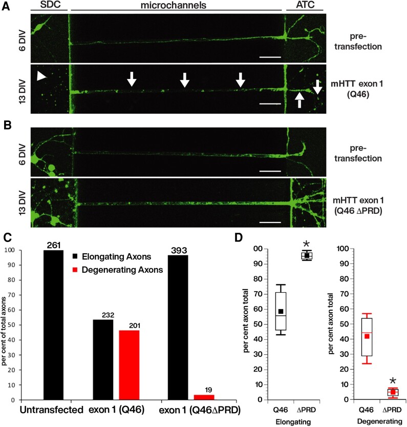
Figure 4.
Reduced outgrowth and degeneration of axons from primary cortical neurons following transfection with cherry-tagged mHTT exon 1, but not with cherry-tagged mHTT exon 1 lacking the proline-rich domain (PRD). Live imaging of axons elongating within the microchannels of our custom microfluidic chambers were visualized with the live neuronal marker, NeuO, beginning just prior to transfection at 6 days in vitro (DIV) after many axons have already entered the microchannels (Supplementary Fig. 2) and continuing until 13 DIV, or 7 days post-transfection. This design evaluates the effect of constructs on the growth and viability of pre-existing axons. Neurons were transfected with (A) mCherry-mHTT exon 1 (Q46) or with (B) mCherry-mHTT exon 1 (Q46 ΔPRD). The length of axons within each microchannel was measured as they entered the microchannel from the somatodendritic compartment (SDC) until it reached the axon terminal compartment (ATC). Cortical neurons expressing mCherry-mHTT exon 1 (Q46) developed axonal discontinuities, observed with the loss of NeuO labelling (A, arrows), illustrating axonal degeneration within the microchannel and in the ATC. There was an apparent reduction of cortical neuron somas in the SDC for cultured neurons transfected with mCherry-mHTT exon 1 (Q46) (A, arrowhead), but degeneration of neuronal soma was not quantitatively evaluated. Scale bar = 50 μm. (C) The total number of axons elongating and degenerating in cortical neurons expressing mHTT exon 1 (Q46) (n = 433) or mHTT exon 1 (Q46 ΔPRD) (n = 412) was quantified over time. Scale bars = 50 μm. Analysis of 261 axons of untransfected neurons had no degenerating axons. See also Supplementary Figs 2 and 3. (D) Comparison of axonal degeneration between neurons expressing mCherry-mHTT exon 1 (Q46) and mCherry-mHTT exon 1 (Q46 ΔPRD) indicates that the fraction of degenerating axons was reduced by >90% (P = 0.0067).

