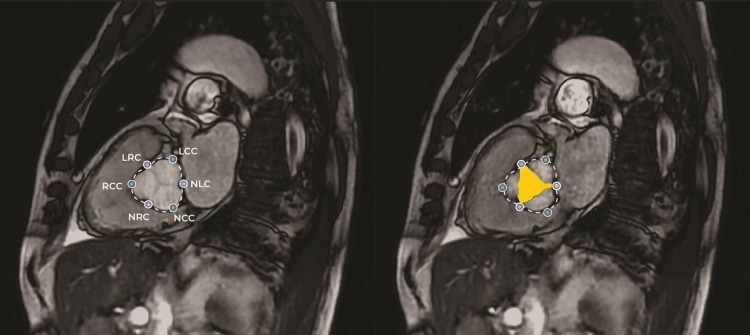
Figure 2. CMR imaging with AI-generated measurements and landmarks.
This figure presents an example of the cardiovascular magnetic resonance (CMR) imaging used in this study, featuring AI-generated measurements. The left panel illustrates the diastolic phase of the cardiac cycle, showcasing AI-identified anatomical landmarks including the left coronary cusp (LCC), non-coronary cusp (NCC), right coronary cusp (RCC), left-right coronary commissure (LRC), non-coronary-right commissure (NRC), and non-coronary-left commissure (NLC). The right panel depicts the systolic phase of the cardiac cycle, highlighting both the AI-detected landmarks and the area measurement of the aortic valve lumen, represented in yellow.

