Abstract
Overview:
Q fever is a zoonotic disease caused by Coxiella burnetii. Farm animals and pets are the main reservoirs of infection.
Infection:
Cats become infected by ingestion or inhalation of organisms from contaminated carcases of farm animals, or tick bites. Infection is common, as shown by several serological studies.
Clinical signs:
Experimentally, fever, anorexia and lethargy have been noted. In the field, infection usually remains subclinical. Abortion might occur. C burnetii has been isolated from the placenta of aborting cats, but also from cats experiencing normal parturition.
Diagnosis:
Infection with C burnetii can be diagnosed by isolation of the agent or serology.
Prevention:
Most important is the potential zoonotic risk. Cats suspected of having been exposed to C burnetii might shed organisms during parturition. Wearing gloves and a mask when attending parturient or aborting cats can minimise the risk of infection. Tick prevention is recommended.
Bacterial properties
Q fever is a zoonotic disease caused by Coxiella burnetii. This is a Gram-negative, obligate intracellular, small, pleomorphic bacterium belonging to the order Legionellales. The organism has a complicated life cycle with different morphological stadia. It may occur as a small-cell variant and a large-cell variant. The small-cell variants are the resistant spore-like forms that can survive for long periods in the environment, being resistant to several chemical and physical noxae. 1
Epidemiology and pathogenesis
Many species of mammals, birds and ticks can be infected with C burnetii. However, the most common reservoirs are cattle, sheep and goats. Since the bacterium has a tropism for the uterus and mammary gland, the placenta and fetal membranes may be heavily contaminated. Contaminated aerosols from fetal membranes, urine, faeces or milk of infected animals are considered the main reservoir of infection for humans. Especially during parturition, high numbers of the bacteria are excreted, thereby contaminating the environment.

Cats can also become infected and have been implicated as a source of infection for humans.2 –6 Cats most commonly become infected via tick bites, ingestion of contaminated carcases or after aerosol exposure. Exposure of cats is relatively common, as can be concluded from several serological studies.7 –10 In these studies, results for seropositivity in cats ranged from 2–19%. In one study, a significantly higher antibody positive rate was demonstrated in stray cats (41.7%) as compared with pet cats (14.2%). 10 In a study on the prevalence of C burnetii DNA in vaginal and uterine samples from healthy shelter or client-owned cats, 4/47 uterine biopsies were shown to be positive by polymerase chain reaction. 11 Like in farm animals, C burnetii colonises the placenta of infected cats during pregnancy in high numbers. C burnetii could be cultured from the uterus of cats for 10 weeks after parturition. 8 After experimental infection, C burnetii was cultured for 2 months from the urine of infected cats. 12

Studies have been published indicating an association between Q fever pneumonia in humans after exposure to placenta and amniotic fluid of aborting or apparently healthy cats.2,3,5,13 In a case-control study from Maritime Canada, several risk factors for developing Q fever in human patients were identified. The strongest association was documented for exposure to stillborn kittens and parturient cats. 6
In a seroepidemiological study among US veterinarians, contact with cats was not shown to be associated with C burnetii seropositivity. 14 In this study, risk factors associated with seropositivity included age >46 years, routine contact with ponds, and treatment of cattle, swine and wildlife. In another study, no relationship was found between cat and dog ownership and an increased incidence of seropositivity for C burnetii. 15
In conclusion, periparturient cats should be considered a potential source of infection. However, farm animals are by far the most important source of infection for humans.
Clinical signs
In animals the disease is usually subclinical, but abortion might occur. In experimentally infected cats, fever, anorexia and lethargy have been noted. Clinical signs started 2 days after inoculation and lasted for 3 days. 12
Diagnosis
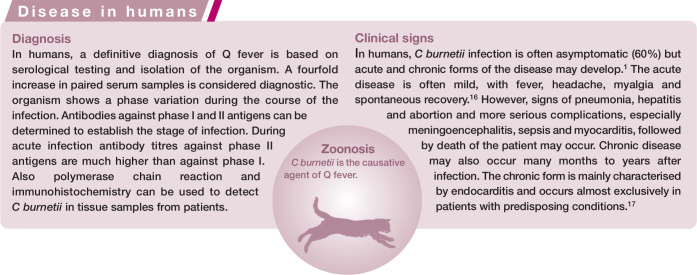
Serological testing and isolation of the organism might be used, as for humans (see box above); however, in cats diagnosis is not routinely performed.
Treatment
If a diagnosis has been established in a cat with clinical signs, tetracyclines and chloramphenicol can be used for treatment [EBM grade IV].
Prevention
Cats potentially exposed to C burnetii by contact with infected farm animals or recent tick infections may excrete bacteria during parturition. To minimise the risk of infection, gloves and a mask should be worn when attending parturient or aborting cats. Predation and ectoparasite exposure put the cat at risk of infection and tick prevention is recommended (see ESCCAP guideline on control of ectoparasites in dogs and cats) [EBM grade IV]. 18 Vaccines are not available for cats.
Footnotes
Funding: The authors received no specific grant from any funding agency in the public, commercial or not-for-profit sectors for the preparation of this article. The ABCD is supported by Merial, but is a scientifically independent body.
The authors do not have any potential conflicts of interest to declare.
Key Points
Infection of cats with C burnetii occurs frequently, as shown by seroprevalence studies.
Cats become infected by tick bites or contact with farm animals, by ingestion or inhalation of the bacteria.
The disease in cats is usually subclinical; abortion may occur.
After experimental infection, cats develop fever, anorexia and lethargy.
C burnetii causes Q fever in man.
Cats have been implicated as a source of infection for humans, in particular through contact with bacteria excreted during abortion or parturition.
References
- 1. Angelakis E, Raoult D. Q fever. Vet Microbiol 2010; 140: 297–309. [DOI] [PubMed] [Google Scholar]
- 2. Langley JM, Marrie TJ, Covert A, Waag DM, Williams JC. Poker players’ pneumonia. An urban outbreak of Q fever following exposure to a parturient cat. N Engl J Med 1988; 319: 354–356. [DOI] [PubMed] [Google Scholar]
- 3. Kosatsky T. Household outbreak of Q-fever pneumonia related to a parturient cat. Lancet 1984; 2: 1447–1449. [DOI] [PubMed] [Google Scholar]
- 4. Marrie TJ, Langille D, Papukna V, Yates L. Truckin’ pneumonia – an outbreak of Q fever in a truck repair plant probably due to aerosols from clothing contaminated by contact with newborn kittens. Epidemiol Infect 1989; 102: 119–127. [DOI] [PMC free article] [PubMed] [Google Scholar]
- 5. Marrie TJ, MacDonald A, Durant H, Yates L, McCormick L. An outbreak of Q fever probably due to contact with a parturient cat. Chest 1988; 93: 98–103. [DOI] [PubMed] [Google Scholar]
- 6. Marrie TJ, Durant H, Williams JC, Mintz E, Waag DM. Exposure to parturient cats: a risk factor for acquisition of Q fever in Maritime Canada. J Infect Dis 1988; 158: 101–108. [DOI] [PubMed] [Google Scholar]
- 7. Matthewman L, Kelly P, Hayter D, Downie S, Wray K, Bryson N, et al. Exposure of cats in southern Africa to Coxiella burnetii, the agent of Q fever. Eur J Epidemiol 1997; 13: 477–479. [DOI] [PubMed] [Google Scholar]
- 8. Higgins D, Marrie TJ. Seroepidemiology of Q fever among cats in New Brunswick and Prince Edward Island. Ann N Y Acad Sci 1990; 590: 271–274. [DOI] [PubMed] [Google Scholar]
- 9. Htwe KK, Amano K, Sugiyama Y, Yagami K, Minamoto N, Hashimoto A, et al. Seroepidemiology of Coxiella burnetii in domestic and companion animals in Japan. Vet Rec 1992; 131: 490. [DOI] [PubMed] [Google Scholar]
- 10. Komiya T, Sadamasu K, Kang MI, Tsuboshima S, Fukushi H, Hirai K. Seroprevalence of Coxiella burnetii infections among cats in different living environments. J Vet Med Sci 2003; 65: 1047–1048. [DOI] [PubMed] [Google Scholar]
- 11. Cairns K, Brewer M, Lappin MR. Prevalence of Coxiella burnetii DNA in vaginal and uterine samples from healthy cats of north-central Colorado. J Feline Med Surg 2007; 9: 196–201. [DOI] [PMC free article] [PubMed] [Google Scholar]
- 12. Greene CE. (ed). Francisella and Q fever. In: Infectious diseases of the dog and cat. St Louis: Elsevier Saunders, 2012, pp 482–484. [Google Scholar]
- 13. Pinsky RL, Fishbein DB, Greene CR, Gensheimer KF. An outbreak of cat-associated Q fever in the United States. J Infect Dis 1991; 164: 202–204. [DOI] [PubMed] [Google Scholar]
- 14. Whitney EA, Massung RF, Candee AJ, Ailes EC, Myers LM, Patterson NE, et al. Seroepidemiologic and occupational risk survey for Coxiella burnetii, antibodies among US veterinarians. Clin Infect Dis 2009; 48: 550–557. [DOI] [PubMed] [Google Scholar]
- 15. Skerget M, Wenisch C, Daxboeck F, Krause R, Haberl R, Stuenzner D. Cat or dog ownership and seroprevalence of ehrlichiosis, Q fever, and cat-scratch disease. Emerg Infect Dis 2003; 9: 1337–1340. [DOI] [PMC free article] [PubMed] [Google Scholar]
- 16. Caron F, Meurice JC, Ingrand P, Bourgoin A, Masson P, Roblot P, et al. Acute Q fever pneumonia: a review of 80 hospitalized patients. Chest 1998; 114: 808–813. [DOI] [PubMed] [Google Scholar]
- 17. Fenollar F, Fournier PE, Carrieri MP, Habib G, Messana T, Raoult D. Risks factors and prevention of Q fever endocarditis. Clin Infect Dis 2001; 33: 312–316. [DOI] [PubMed] [Google Scholar]
- 18. European Scientific Counsel for Companion Animal Parasites (ESCCAP). Control of ectoparasites in dogs and cats. http://www.esccap.org/uploads/file/ESCCAP%20Guidelines%20GL3%20Final%2029June2012(2).pdf


