Abstract
Background
Tarsal tunnel syndrome (TTS) is a condition in which the tibial nerve (TN) (or its terminal branches) is compressed by the flexor retinaculum (FR) and the deep fascia of the abductor hallucis muscle at the tarsal tunnel, causing symptoms that negatively impact the patient’s quality of life, including numbness, a sensation of a foreign object, coldness, and pain. FR release via microtrauma using needle-knife has proven to be effective in China and is widely used by clinicians. The traditional acupotomy, however, is the “blind knife” treatment, which cannot guarantee patient safety due to risk of injury to important structures, particularly the neurovascular bundle. Compared with the conventional treatments, ultrasound-guided percutaneous FR release possesses noteworthy advantages including high efficacy and safety.
Methods
Percutaneous release of the FR was performed on 51 formalin-fixed specimens. The specimens were divided into two groups: an ultrasound-guided acupotomy pushing group comprising 20 legs (group U) and a nonultrasound-guided acupotomy pushing group comprising 31 legs (group N). After high-frequency ultrasound exploration, those with clear vascular imaging were included in group U; otherwise, they were included in group N. The FR was released percutaneously, soft tissue was dissected layer by layer, and anatomical data were recorded.
Results
There no cases of injury in group U (0%) and four in group N (12.9%). Among the different intervention methods, there were no significant differences in tissue injury types (χ2=2.80; P=0.09). The percentage of released FR in group U was 80.00% while that in group N was 61.29% (χ2=1.977; P=0.16), which did not represent a significant difference between the two groups. However, group U had a significantly greater release length than that in the group N (t=3.359; P=0.002), indicating that the flexor release length guided by ultrasound is significantly greater than the unguided one.
Conclusions
Ultrasound-guided percutaneous release of the FR using a needle-knife can provide greater length and percentage of released FR while maintaining a comparable safety rate to the unguided procedure.
Keywords: Acupotome, anatomy, percutaneous release, tarsal tunnel syndrome (TTS), ultrasound-guided technique
Introduction
Tarsal tunnel syndrome (TTS) is a local entrapment neuropathy that affects the entire tibial nerve (TN) or one of its associated branches (1,2). Patients with TTS usually complain about poorly localized paraesthesia, such as pain and sensation of tightness, localized or radiating pain, burning pain, dysesthesias, and hyperesthesia, with Tinel sign often being present upon clinical examination (3-5). In conservative management, many techniques can be used, including anti-inflammatory drugs, resting the distal part of the lower limb, activity modification coupled with progressive rehabilitation, nerve block, acupuncture or local anesthetic or corticosteroid infiltration and physiotherapy (the use of bracing, stretching, icing, massage, transcutaneous electrical nerve stimulation, remedial massage therapy, and ultrasound are all incorporated into this treatment) (6). A local anesthetic or infiltration of corticosteroid can be used for treating the “algetic form” of TTS in order to reverse any intraneural edema, but these infiltrations may increase infection risks. Corticosteroid injections into the tarsal tunnel should be performed with considerable caution, and multiple injections are inadvisable because of their increased risk and limited chance of success (7). In addition to orthotic shoes, activity modifications, night splints, and immobilizing braces or removable boot walkers, it is possible to control symptoms by decreasing the pressure on the nerve (6), but in many cases, these treatments may exacerbate symptoms, and orthopedic treatment may be required.
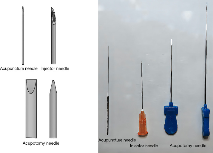
The needle-knife, also referred to as acupotome, consists of a bladed needle with a flat head, a cylindrical needle body, and a handle (8) (Figure 1). Acupotomy therapy is the method of treating abnormal, cicatricial, or contracted soft tissues with microtrauma using the needle-knife. Clinically, it has proven to be effective in China and is widely used by clinicians (9,10).
Figure 1.
Images of the acupuncture needle, injector needle, and acupotome needle.
Several disadvantages of percutaneous release techniques include the inability to visualize the release, the inability to determine its completeness, and the possibility of inflicting injuries (e.g., to the digital vessels, tendons, and nerves). Furthermore, it does not allow for the transmission and learning of new information (11). In clinical practice, ultrasound-guided techniques are highly valuable in allowing for visualization of the percutaneous release (12-14). Thus far, clinical trials on ultrasound guided percutaneous flexor retinaculum (FR) release or its anatomical research have not been published, and thus the promotion of this technology has been limited. This study was conducted for the purpose of describing the anatomic basis for ultrasound-guided percutaneous FR release by the small needle-knife and analyzing the efficacy and safety of percutaneously releasing the FR in 51 cadaveric feet fixed with 10% formalin.
Methods
A total of 51 specimens from 26 cadavers (14 males and 12 females) aged 47 to 98 years were obtained from a Laboratory at the School of Basic Medical Sciences, Peking University (Human Anatomy), and dissected between September and October of 2020. The study was conducted in accordance with the Declaration of Helsinki (as revised in 2013). The ethical approval was waived by the Institutional Review Board of Peking University (No. IRB00001052-21011-Exempt). Informed consent was obtained from all the body donors. In all 51 feet examined, no injuries were observed, and there was no history indicating previous operation or deformity. The 51 feet were then divided into 2 groups, with 20 feet in group U (ultrasound-guided small needle-knife pushing group) and another 31 feet in group N (nonultrasound-guided small needle-knife pushing group).
Groups
Group U
First, three imaginary reference lines were made for measurements: the first one was the line from point A (the tip of the medial malleolus) to point B (the calcaneus’ center, a calcaneal tubercle tip located at the greatest distance from the medial malleolus), which is called the Dellon-McKinnon malleolar-calcaneal line (DML) (15), while the other two lines consisted of one line that started 10 mm from the Dellon-McKinnon malleolar-calcaneal line proximally (DML-P) and one from it distally (DML-D) (Figure 2).
Figure 2.
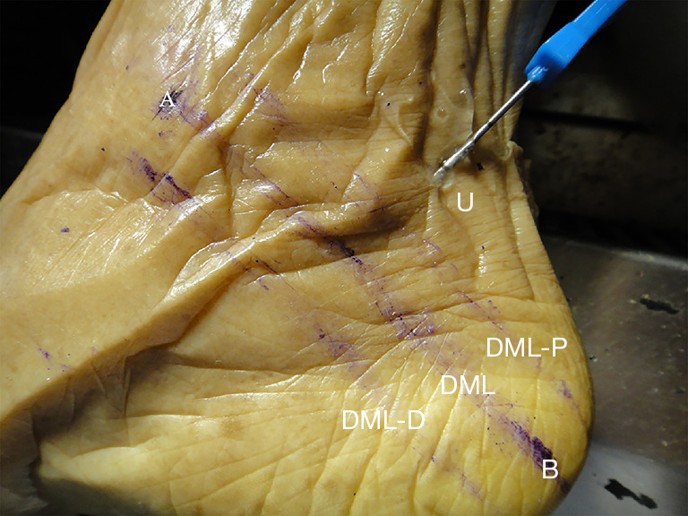
Actual anatomical diagram of the anatomical landmarks points, including three imaginary reference lines and the needle entry point. Group U, ultrasound-guided small needle-knife pushing group; DML, Dellon-McKinnon malleolar-calcaneal line; DML-P, a line beginning 10 mm proximal to the Dellon-McKinnon malleolar-calcaneal line; DML-D, a line which started 10 mm distal to the DML; A, the tip of the medial malleolus; B, the calcaneus’ center, a calcaneal tubercle’s tip located at the greatest distance from the medial malleolus.
In Figure 2, point A is the tip of the medial malleolus; point B is the calcaneus’ center; DML is the DML, which was from point A to the center of the calcaneus (the tip of the calcaneal tubercle at its greatest distance from the medial malleolus); DML-P is a line beginning 10 mm proximal to the DML; and DML-D is a line beginning 10 mm distal to the DML.
As a standard procedure, all surgical specimens were placed in a decubitus supinated with dorsiflexed-everted positions (“frog-leg position”), to ensure standardized measurement and image acquisition. Subsequently, a 12-MHz high-frequency ultrasound probe (Wisonic Navi, Shenzhen Wisonic Medical Technology Co., Ltd., China) was placed perpendicular to the FR on the medial side of the ankle (at a point parallel with the long axis of the blood vessel) to identify the medial malleus, the calcaneus, the posterior tibial vein (PTV), the posterior tibial artery (PTA). and the TN. Next, we rotated the ultrasonic probe to make it parallel to the FR, and then the needle knife was guided through the in-plane insertion proximal to the transducer via the high-frequency ultrasound probe. The shortest distance between the needle injection (point U) and point A, the shortest distance between point U and the DML-P line, and the angle of acupotome to the DML-P line and the angle of acupotome to the skin were measured, and the data were averaged to provide evidence for the nonultrasound-guided needle knife insertion group (Figure 3).
Figure 3.
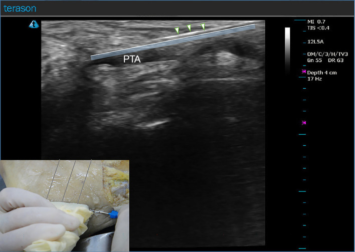
Ultrasound image of needle knife operation in group U (original figure). The shaded blue area is the flexor retinaculum. △: the cylindrical needle body of the needle-knife. Group U, ultrasound-guided small needle-knife pushing group; PTA, posterior tibial artery; MI, mechanical index; TIS, thermal index of soft tissue; OM, Omni; C, the third of all nine maps (A-I); H, high frequency; TV, TeraVision; Gn, gain; DR, dynamic range.
Group N
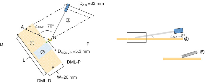
For the nonultrasound guided needle approach (i.e., the blind needle approach), the route was determined by taking reference to the average of group U, as shown in Table 1. The needle injection of this group was point N. The needle-knife was inserted 5.3 mm proximal to the DML-P line at a 6° angle to the skin (∠S-Z). The shortest distance between point N and point A was 33 mm; the angle ∠AB-Z between the needle knife and DML-P line was approximately 70° (Figure 4). After the needle knife passed through the skin and we slowly explored with the needle until it felt that the needle tip could touch the firm primary tissue under the needle in the FR. After four successive needle movements in a proximal-distal direction, the FR was released, and the injection depth was DN=30 mm.
Table 1. The data of group U.
| Items | Minimum value | Maximum value | Average value () |
|---|---|---|---|
| DU-DML-P (mm) | 12.13 | 1.12 | 5.38±2.41 |
| DA-U (mm) | 43.04 | 13.34 | 32.69±7.78 |
| DU (mm) | 35.65 | 21.83 | 30.42±4.11 |
| ∠AB-Z (°) | 85.00 | 36.00 | 69.75±13.92 |
| ∠S-Z (°) | 12 | 4 | 6.37±2.09 |
Group U, ultrasound-guided small needle-knife pushing group. DN-DML-P, the shortest distance between point U to the Dellon-McKinnon malleolar-calcaneal line; DA-U, the shortest distance between the point A and the point U; DU, the injection depth of group U; ∠AB-Z, the angle of acupotome to the DML-P line; ∠S-Z, the angle of acupotome to the skin; DML-P, a line beginning 10 mm proximal to the Dellon-McKinnon malleolar-calcaneal line.
Figure 4.
The image of the needle-knife operation of group N. ① The flexor retinaculum, ② the high-frequency ultrasound probe, ③ the needle knife, ④ the skin, and ⑤ the needle tip. Group N, nonultrasound-guided small needle-knife pushing group. A, the tip of the medial malleolus; B, center of the calcaneus; D, the distal end of the flexor retinaculum; L, the length of the flexor retinaculum; P, the proximal end of the flexor retinaculum; W, the width of the flexor retinaculum; S, skin; ∠S-Z, the angle of the needle to the skin; ∠AB-Z, the angle of the needle-knife and the line of the DML; DA-N, the shortest distance between the point A and the point N; DML-P, a line beginning 10 mm proximal to the Dellon-McKinnon malleolar-calcaneal line; DML-D, a line which started 10 mm distal to the DML; DN-DML-P, the shortest distance between point N to the line 10 mm proximal to the Dellon-McKinnon malleolar-calcaneal line.
After release operation was completed with the small needle-knife, a transverse incision was made on the skin surface along the needle-knife direction to expose the underlying adipose tissue and FR layer by layer. After acupotomy release, the structures of tarsal tunnel were observed and recorded.
Types of outcome measures
After the release of the FR, an incision was made that began 10 mm proximal to the DML line and ran toward the medial aspect of the navicular-cuneiform joint distally. Following this, each researcher assessed the injury of the tibialis posterior tendon (TP), flexor digitorum longus tendon (FDL), flexor hallucis long tendon (FHL), PTV, the PTA, and the TN and measured the length of the released the FR. A measurement error of 0.01 mm was measured using Vernier digital calipers (Pittsburgh, Harbor Freight Tools, USA).
Safety
Safety indexes were used to measure injuries outside the target (i.e., the FR). First, there were two options for assessing tarsal tunnel (TT) structures (the TN, the PTV, and the PTA): normal (if no cut had been made) or injured (if cut). The injuries (Figure 4) were classified as no injury, slight laceration (interruption of one edge of the tendons with the substance tendons maintained; slight scratches on the surface of blood vessels or nerve), or serious laceration (interrupted tendon continuity; blood vessels or nerve penetrated by injury).
The respective injuries to the TN, the PTV, the PTA were counted, as was the total number of injuries in each group. The injury rate was calculated as follows: injury rate (%) = number of ankle injuries/total cases × 100%.
Efficacy
In observing the needle-knife releasing mark, it was determined whether the releasing mark was within the FR range and whether the needle knife could completely release the FR.
The criteria for effective release were as follows: length of release = LR≥W/2 (where W is the width of the FR, defined as 20 mm) and the anatomical structure appearing by the naked eye to be effectively released after dissection. The full release rate of the FR was calculated as follows: full FR release rate (%) = the number of cases of full releases of the FR/total number of cases × 100%.
Statistical analysis
Data analysis was conducted using SPSS 20.0 software (IBM Corp., Armonk, NY, USA). The mean ± standard deviation () was used to express the anatomical measurement data, and independent-samples T-test was used if the data conformed to a normal distribution and the variance was homogeneous; otherwise, the nonparametric Mann-Whitney U test was conducted. The percentage (%) is used to express the count data injury rate and full FR release rate, and the rates were compared and analyzed with the Fisher exact test between the two groups. The inspection level was a =0.05.
Results
Ultrasound images of the tarsal tunnel structures
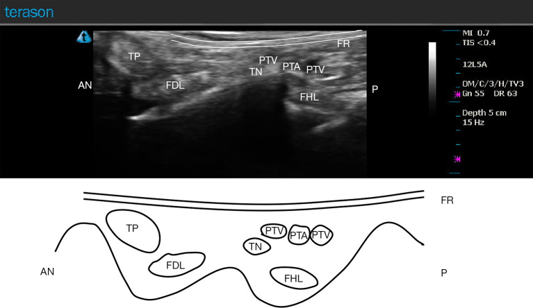
In the short axial section, the order of TT structures was as follows: the tibialis posterior tendon, the flexor digitorum longus tendon, the tibial nerve, the blood vessels, and the flexor hallucis longus tendon. The tendon present as oval, isoechoic internal echo textureand well-defined structures. And the tendon can be seen by a shuttling movement up and down on the ultrasound image along with the flexion and extension activity of the ankle joint. And the vessel had a lumen structure in the anechoic area. Color Doppler blood flow imaging showed blood flow signal fluctuation with vascular beating. The TN usually manifested as oval, hypoechoic fascicles, appearing as a typical honeycomb structure (Figure 5).
Figure 5.
The short-axis ultrasound image and schematic diagram of the tarsal tunnel. AN, ankle; TP, tibialis posterior tendon; FDL, flexor digitorum longus tendon; TN, tibial nerve; PTV, posterior tibial vein; PTA, posterior tibial artery; FR, flexor retinaculum; FHL, flexor hallucis long tendon; P, pelma; MI, mechanical index; TIS, thermal index of soft tissue; OM, Omni; C, the third of all nine maps (A-I); H, high frequency; TV, TeraVision; Gn, gain; DR, dynamic range.
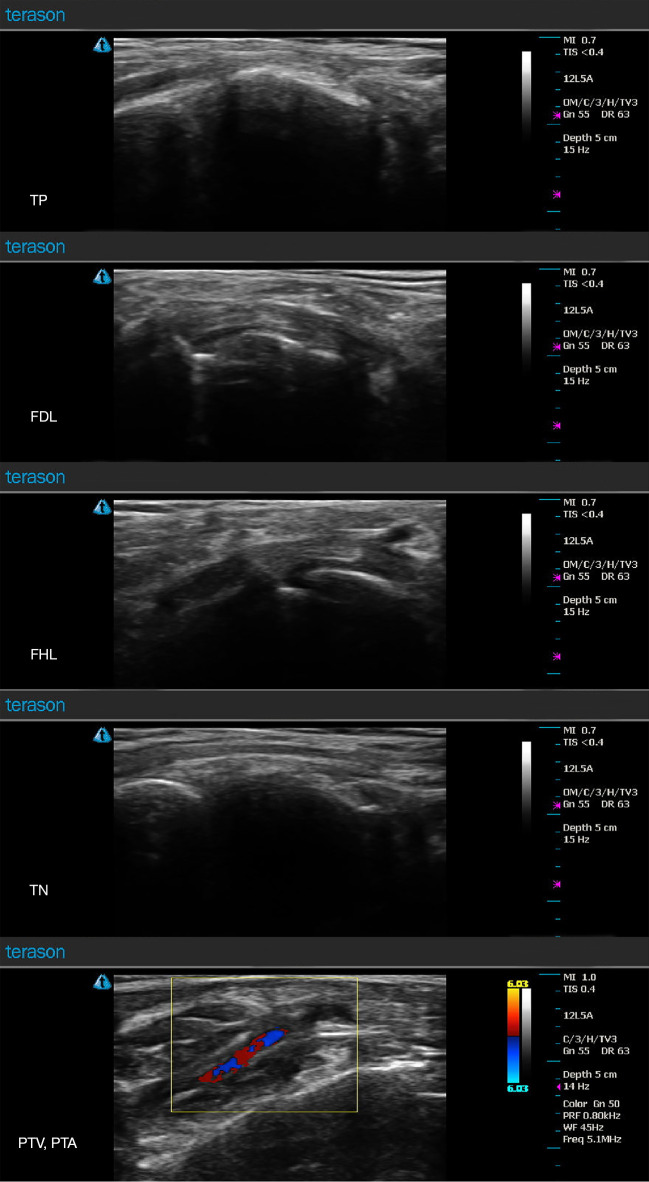
During the long axial section inspection, the bone part was hyperechoic, which gradually decayed down to hypoechoic, and the flexor retinaculum was visibly thin and moderately echoic. The tendon was strip-shaped, with a dense fine linear texture visible inside, and the tendon could be seen with a shuttling movement up and down on the ultrasound image along with a flexion and extension activity of the ankle joint. Moreover, the long nerve axis section had a “cable-like” structure with uniform thickness, the vascular structure involved an anechoic zone of the tubular strip, and the blood flow signal was filled the color Doppler blood flow imaging, which is basically consistent with previous reports (15) (Figure 6).
Figure 6.
The long-axis ultrasound image of the tarsal tunnel. TP, tibialis posterior tendon; FDL, flexor digitorum longus tendon; FHL, flexor hallucis long tendon; TN, tibial nerve; PTV, posterior tibial vein; PTA, posterior tibial artery; MI, mechanical index; TIS, thermal index of soft tissue; OM, Omni; C, the third of all nine maps (A-I); H, high frequency; TV, TeraVision; Gn, gain; DR, dynamic range.
Safety
In group U, there were no injuries to the TT structures, including vessels, nerve, or tendons. The injury rate was 0%. Meanwhile, in group N, there were four (12.90%) cases of injury but not to the nerve or tendons. Of these, two had a serious blood vessel injury with penetration by the needle. In the four injured blood vessels, there were three PTVs and one PTA (Table 2).
Table 2. The injury cases and injury rate of the two groups.
| Items | Injury of the TP | Injury of the FDL | Injury of the FHL | Injury of the TN | Injury of the PTV | Injury of the PTA | Total |
|---|---|---|---|---|---|---|---|
| Group U (n=20) | 0/0 | 0/0 | 0/0 | 0/0 | 0/0 | 0/0 | 0/0 |
| Group N (n=31) | 0/0 | 0/0 | 0/0 | 1/3.23 | 3/9.68 | 0/0 | 4/12.90 |
| P value | – | – | 0.41 | 0.15 | – | 0.09 | |
| χ2 | – | – | – | 0.658 | 2.056 | – | 2.800 |
Data are represented as cases/percentage. Group U, ultrasound-guided small needle-knife pushing group; Group N, nonultrasound-guided small needle-knife pushing group. TP, tibialis posterior tendon; FDL, flexor digitorum longus tendon; FHL, flexor hallucis long tendon; TN, tibial nerve; PTV, posterior tibial vein; PTA, posterior tibial artery.
According to the Fisher exact test, the number of injured TT elements was greater in group N than that in group U (χ2=2.80, P=0.09), but not significantly so. Despite group N having a higher injury rate, there was no statistically significant difference between the two groups in terms of the total injury rate.
Efficacy
Table 3 summarizes the released length of the FR and the percentage of released FRs om the two groups.
Table 3. The cases of different types of release and full release rate of the two groups.
| Items | Intermittent release | Straight release | Missed release | Surface release | Full-layer release |
|---|---|---|---|---|---|
| Group U (n=20) | 20/100.00 | 0/0.00 | 0/100.00 | 4/20.00 | 16/80.00 |
| Group N (n=31) | 31/100.00 | 0/0.00 | 3/9.68 | 9/29.03 | 19/61.29 |
| Total (n=51) | 51/100 | 0/0.00 | 3/5.88 | 13/25.49 | 35/68.63 |
Data are represented as cases/percentage. Group U, ultrasound-guided small needle-knife pushing group; Group N, nonultrasound-guided small needle-knife pushing group.
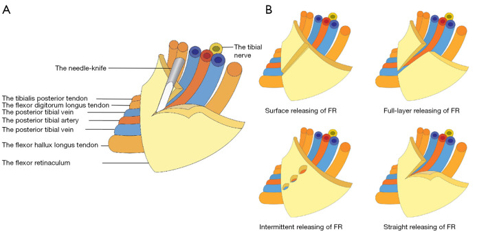
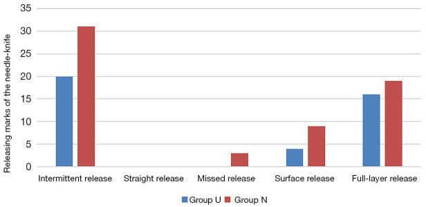
In group U, the releasing trace of the needle-knife was greater than 10.00 mm, the average length of the releasing trace needle-knife was 18.29±1.18 mm, and the range of length was 15.43–20.00 mm. Four cases were considered to be failed because the release was too superficial on the FR, and 16 cases demonstrated successful release by achieving a full-layer release on the FR, with an overall success rate of release of 80.00%. According to the data from the transverse releasing marks, it was apparent that all 20 specimens had intermittent releasing, with no straight releasing (Figures 7,8).
Figure 7.
Schematic diagram of the flexor retinaculum release via microtrauma with the needle-knife. (A) Schematic diagram of desirable needle knife operation; (B) the different types of release. FR, flexor retinaculum.
Figure 8.
Percutaneous release distribution in group U and group N. Group U, ultrasound-guided small needle-knife pushing group; Group N, nonultrasound-guided small needle-knife pushing group.
From the longitudinal release trace, surface release occurred in four cases, accounting for 20.00% of cases; meanwhile, the needle-knife released the full-layer of the FR in 16 cases, which accounted for 80.00% of cases, as shown in Table 3.
In group N, releasing trace of the needle knife was also more than 10.00 mm, the average length of the releasing trace of the needle knife was approximately 16.67±2.25 mm, and the range of length was approximately 12.46–20.00 mm. According to the data from the transverse releasing marks, the 31 cases of specimens all had intermittent releasing, with no straight releasing. Regarding the longitudinal releasing trace, in all 20 specimens, there were nine cases of surface release, accounting for 29.03% of the cases, and 19 cases (61.29%) of full-layer release, and 3 cases of missed release, accounting for 9.68% of cases (Tables 3,4).
Table 4. The release length, actual width of the FR, and the percentage of released FR of two groups.
| Items | L1: the release length of FR (, mm) | L: the specified width of FR (, mm) | The percentage of released FR (L1/L) |
|---|---|---|---|
| Group U (n=20) | 18.29±1.18 | 20.00±0.00 | 0.91±0.06 |
| Group N (n=31) | 16.67±2.25 | 20.00±0.00 | 0.83±0.11 |
| P value | 0.002 | – | 0.002 |
| t | 3.359 | – | 3.344 |
Group U, ultrasound-guided small needle-knife pushing group; Group N, nonultrasound-guided small needle-knife pushing group. FR, flexor retinaculum.
According to the independent-samples t, the release length of group U was significantly greater than that of group N (t=3.359, P=0.002<0.05), indicating that the release length is greater under ultrasound guidance than under no guidance.
According to the Fisher exact test, the response efficiency in group U was greater than that of group N, but not significantly so (χ2=1.977; P=0.16).
Discussion
In cases where strict conservative treatment has failed, surgical treatment should be considered, but the rate of success ranges from 44% to 96% for these operative treatments (16). Related postoperative complications may be apparent, including impaired wound healing, infection, and keloid formation (17).
Although having only a small percutaneous incision may produce a benefit, it is also limited, as it involves restricting the operation area and impeding the operator and assistant surgeon’s visibility during the operation. Undoubtedly, these operations require immense levels of training and may also be highly operator dependent.
However, needle-knife treatment leaves no scar and is relatively cost-effective. One study reported that the skin incision of endoscopic TT syndrome surgery via the universal subcutaneous endoscope system was only 1 mm, but our incision measured only 0.8–1.0 mm and was more cost-efficient (18). In addition to this, the technology of the needle-knife is relatively easy to learn for clinicians training in similar procedures and even more safe under ultrasound guidance.
In traditional acupotomy treatment, a “blind knife” treatment is performed without direct visualization, including cutting, peeling, and dredging without the use of an internal image guidance system or direct visualization. In order to determine the position of the needle-knife in the tissue, the clinician is required to rely on their own clinical experience (such as the “needle sense” or “empty feeling”) of the different levels of resistance they encounter during acupotomy and depend on intraoperative sensory feedback from the patients to avoid nerves, blood vessels, and reach the target tissue. Yan-hong et al. (19) used ultrasound guidance technology combined with needle = knife to treat knee osteoarthritis, and the complication rate of the ultrasound-guided arm was significantly lower than that of the pain point positioning arm (P<0.05). There were no hematoma cases and one nerve injury case in the ultrasound guidance group, while there were five hematoma cases and three nerve injury cases in the pain point location group. The ankle joint is complicated, with the TN being closely adjacent to the PTV and the PTA under the FR. Clinicians who are unfamiliar with anatomical structures and needle-knife techniques can potentially injure important structures, particularly blood vessels. Moreover, due to the complex structure of the human body and the possible abnormalities in the physiological and pathological structure in some patients, the chances of injury to the vital structures during surgery are never zero.
In our controlled trial, the injury rates between the two groups were not significantly different (P>0.05). Of these injury cases, several cases in group N had vascular injury. According to our study, both needle-knife methods of percutaneously releasing the FR were highly safe. Moreover, in other studies of cadavers (20,21), ultrasound-guided release of the TN, its branches, and the FR was completed without any signs of compromise or injury into the neurovascular structures. By using ultrasound guidance, it is possible to locate the FR with great accuracy and to precisely identify tendons and blood vessels to avoid damage to the them. Naturally, this makes it easier for novices to understand and master the procedure.
It is advantageous to use ultrasound guidance to locate the TN and identify tendons and blood vessels prior to treatment. In the surgery, local anesthesia is used for localizing acupuncture points, for visualizing the operation in real time, and for guiding the process of percutaneous FR release to improve accuracy and safety. An ultrasound scan can clearly show anatomical structures; help to avoid blood vessels, nerves, organ tissues; detect diseased tissues; assist with diagnosis; and accurately locate the therapeutic targets (22). In addition, ultrasound guidance allows for accurate acupotomy release in the anesthetized position. In addition to monitoring drug distribution after injection, it improves the safety of release targets and can facilitate assesses of the FR’s condition. As a result of the visualization of the procedure, ultrasound guidance is also easier to follow and promote, resulting in a lower psychological burden on patients (23-25).
This study was based on cadaveric samples, which makes extrapolating clinical results difficult. As a result of postmortem soft tissue shrinkage or fluid movements, cadavers may have altered landmarks and tissue turgor, which directly impacts ultrasound imaging quality. A live patient’s released FR is difficult to measure, however. Further studies are needed to determine the correlation between clinical symptoms and the percentage of the FR released. Another limitation of the experiment was that it was not possible to diagnose whether the cadavers all had TTS, so we could only compare the safety and efficacy of needle-knife FR release guided by ultrasound with that not guided by ultrasound. In the future, we will compare needle-knife release of FR in TTS cadavers with those in ordinary cadavers.
Based on the data from this study, ultrasound-guided percutaneous release of the FR using a needle knife results in longer lengths and greater percentages of retinacula release than does the nonguided procedure while providing a comparable injury rate. Those whose conservative treatments have failed for TTS should consider ultrasound-guided percutaneous release of the FR using a needle knife as a safer and more effective procedure. While the needle knife is a minimal surgical procedure, once a blood vessel has been injured, it becomes a likely source of nosocomial infection, resulting in secondary TTS.
We further found that even with ultrasound guidance, there was no successful straight releases of the FR. This may be because the thinness of FR, at only about 1 mm, and the nerves and vessels being under the FR make it soft in texture. This structure is not able to provide as much support for the needle-knife cutting by the bottom bone as the needle insertion at the bone margin. Moreover, FR is pliable but strong, sandwiched between the fatty layer and the contents of the tarsal tunnel, and it is nearly impossible to accurately cut a complete tear through this rigid layer of fascia. From the longitudinal releasing trace, although the flexor retinaculum is very thin, it still has thickness and stratification, and ultrasound-guided release still cannot achieve 100% longitudinal full-layer release.
Some limitations to the study should be considered. The presence of nerve impingement under the deep fascia of the abductor hallucis muscle or other etiologies of TTS were not evaluated. This is important to note as the tarsal tunnel itself is complex and because the FR is not the only etiology known to cause TTS. Clinically, many patients with TTS may continue to have pain despite release of the FR (26,27).
Conclusions
Our study simulated the operation of percutaneous FR via acupotomy in the treatment of TTS on human specimens via clinical anatomy and compared the safety and accuracy of an ultrasound-guided procedure with those of a nonultrasound one. We observed the puncture path of the percutaneous FR release and its adjacent neurovascular tissue, which may provide the anatomical basis for the treatment of TTS via ultrasound-guided percutaneous FR release through acupotomy.
Supplementary
The article’s supplementary files as
Acknowledgments
The study work was a part of postgraduate (Master’s) thesis of the first author Xiaojie Sun.
Funding: None.
Ethical Statement: The authors are accountable for all aspects of the work in ensuring that questions related to the accuracy or integrity of any part of the work are appropriately investigated and resolved. The study was conducted in accordance with the Declaration of Helsinki (as revised in 2013), and ethical approval was waived by the Institutional Review Board of Peking University (No. IRB00001052-21011-Exempt). Informed consent was obtained from all the body donors.
Footnotes
Conflicts of Interest: All authors have completed the ICMJE uniform disclosure form (available at https://qims.amegroups.com/article/view/10.21037/qims-24-81/coif). The authors have no conflicts of interest to declare.
References
- 1.Vega-Zelaya L, Iborra Á, Villanueva M, Pastor J, Noriega C. Ultrasound-Guided Near-Nerve Needle Sensory Technique for the Diagnosis of Tarsal Tunnel Syndrome. J Clin Med 2021;10:3065. 10.3390/jcm10143065 [DOI] [PMC free article] [PubMed] [Google Scholar]
- 2.Sun XJ, Shi C, Li YN, Lan YJ, Wang JW, Zhang WG, Li SL. Clinical anatomical study on the treatment of tarsal tunnel syndrome with four-point vertical acupotomy. Zhongguo Gu Shang 2022;35:543-7. [DOI] [PubMed] [Google Scholar]
- 3.McSweeney SC, Cichero M. Tarsal tunnel syndrome-A narrative literature review. Foot (Edinb) 2015;25:244-50. 10.1016/j.foot.2015.08.008 [DOI] [PubMed] [Google Scholar]
- 4.Franson J, Baravarian B. Tarsal tunnel syndrome: a compression neuropathy involving four distinct tunnels. Clin Podiatr Med Surg 2006;23:597-609. 10.1016/j.cpm.2006.04.005 [DOI] [PubMed] [Google Scholar]
- 5.Reichert P, Zimmer K, Wnukiewicz W, Kuliński S, Mazurek P, Gosk J. Results of surgical treatment of tarsal tunnel syndrome. Foot Ankle Surg 2015;21:26-9. 10.1016/j.fas.2014.08.013 [DOI] [PubMed] [Google Scholar]
- 6.Alshami AM, Souvlis T, Coppieters MW. A review of plantar heel pain of neural origin: differential diagnosis and management. Man Ther 2008;13:103-11. 10.1016/j.math.2007.01.014 [DOI] [PubMed] [Google Scholar]
- 7.Antoniadis G, Scheglmann K. Posterior tarsal tunnel syndrome: diagnosis and treatment. Dtsch Arztebl Int 2008;105:776-81. [DOI] [PMC free article] [PubMed] [Google Scholar]
- 8.Qiu Z, Li H, Shen Y, Jia Y, Sun X, Zhou Q, Li S, Zhang W. Safety and efficacy of ultrasound-guided percutaneous A1 pulley release using a needle knife: An anatomical study. Front Surg 2022;9:967400. 10.3389/fsurg.2022.967400 [DOI] [PMC free article] [PubMed] [Google Scholar]
- 9.Pan M, Sheng S, Fan Z, Lu H, Yang H, Yan F, E Z. Ultrasound-Guided Percutaneous Release of A1 Pulley by Using a Needle Knife: A Prospective Study of 41 Cases. Front Pharmacol 2019;10:267. 10.3389/fphar.2019.00267 [DOI] [PMC free article] [PubMed] [Google Scholar]
- 10.Peng G, Zheng Y, Luo D. Effects of Acupuncture and Moxibustion Combined with Needle-Knife on Pain and Lumbar Function in Patients with Lumbar Disc Herniation. J Healthc Eng 2022;2022:9185384. 10.1155/2022/9185384 [DOI] [PMC free article] [PubMed] [Google Scholar] [Retracted]
- 11.Rajeswaran G, Healy JC, Lee JC. Percutaneous Release Procedures: Trigger Finger and Carpal Tunnel. Semin Musculoskelet Radiol 2016;20:432-40. 10.1055/s-0036-1594283 [DOI] [PubMed] [Google Scholar]
- 12.Chern TC, Kuo LC, Shao CJ, Wu TT, Wu KC, Jou IM. Ultrasonographically Guided Percutaneous Carpal Tunnel Release: Early Clinical Experiences and Outcomes. Arthroscopy 2015;31:2400-10. 10.1016/j.arthro.2015.06.023 [DOI] [PubMed] [Google Scholar]
- 13.Lapègue F, André A, Meyrignac O, Pasquier-Bernachot E, Dupré P, Brun C, Bakouche S, Chiavassa-Gandois H, Sans N, Faruch M. US-guided Percutaneous Release of the Trigger Finger by Using a 21-gauge Needle: A Prospective Study of 60 Cases. Radiology 2016;280:493-9. 10.1148/radiol.2016151886 [DOI] [PubMed] [Google Scholar]
- 14.Wang PH, Wu PT, Jou IM. Ultrasound-guided percutaneous carpal tunnel release: 2-year follow-up of 641 hands. J Hand Surg Eur Vol 2021;46:305-7. 10.1177/1753193420948824 [DOI] [PubMed] [Google Scholar]
- 15.Moroni S, Zwierzina M, Starke V, Moriggl B, Montesi F, Konschake M. Clinical-anatomic mapping of the tarsal tunnel with regard to Baxter's neuropathy in recalcitrant heel pain syndrome: part I. Surg Radiol Anat 2019;41:29-41. 10.1007/s00276-018-2124-z [DOI] [PMC free article] [PubMed] [Google Scholar]
- 16.Ahmad M, Tsang K, Mackenney PJ, Adedapo AO. Tarsal tunnel syndrome: A literature review. Foot Ankle Surg 2012;18:149-52. 10.1016/j.fas.2011.10.007 [DOI] [PubMed] [Google Scholar]
- 17.Reade BM, Longo DC, Keller MC. Tarsal tunnel syndrome. Clin Podiatr Med Surg 2001;18:395-408. 10.1016/S0891-8422(23)01201-6 [DOI] [PubMed] [Google Scholar]
- 18.Yoshida A, Okutsu I, Hamanaka I. Endoscopic tarsal tunnel syndrome surgery using the Universal Subcutaneous Endoscope system. Asia Pac J Sports Med Arthrosc Rehabil Technol 2016;3:1-5. 10.1016/j.asmart.2015.09.001 [DOI] [PMC free article] [PubMed] [Google Scholar]
- 19.Ma YH, Ding K, Wang SX. Efficacy of Ultrasound-guided technique in acupotomy therapy for knee osteoarthritis. Modern Journal of Integrated Traditional Chinese and Western Medicine 2012;21:1074-5. [Google Scholar]
- 20.Iborra Á, Villanueva-Martínez M, Barrett SL, Rodríguez-Collazo ER, Sanz-Ruiz P. Ultrasound-Guided Release of the Tibial Nerve and Its Distal Branches: A Cadaveric Study. J Ultrasound Med 2019;38:2067-79. 10.1002/jum.14897 [DOI] [PubMed] [Google Scholar]
- 21.Fernández-Gibello A, Moroni S, Camuñas G, Montes R, Zwierzina M, Tasch C, Starke V, Sañudo J, Vazquez T, Konschake M. Ultrasound-guided decompression surgery of the tarsal tunnel: a novel technique for the proximal tarsal tunnel syndrome-Part II. Surg Radiol Anat 2019;41:43-51. 10.1007/s00276-018-2127-9 [DOI] [PMC free article] [PubMed] [Google Scholar]
- 22.Jia Y, Qiu Z, Sun X, Shen Y, Zhou Q, Zhu X, Li S. Ultrasound-guided acupuncture of the sphenopalatine ganglion for the treatment of vasomotor rhinitis: a case report. Acupunct Med 2020;38:361-3. 10.1177/0964528420906414 [DOI] [PubMed] [Google Scholar]
- 23.Oteri V, Occhipinti F, Gribaudo G, Marastoni F, Chisari E. Integration of ultrasound in medical School: Effects on Physical Examination Skills of Undergraduates. Med Sci Educ 2020;30:417-27. 10.1007/s40670-020-00921-4 [DOI] [PMC free article] [PubMed] [Google Scholar]
- 24.Griswold-Theodorson S, Hannan H, Handly N, Pugh B, Fojtik J, Saks M, Hamilton RJ, Wagner D. Improving patient safety with ultrasonography guidance during internal jugular central venous catheter placement by novice practitioners. Simul Healthc 2009;4:212-6. 10.1097/SIH.0b013e3181b1b837 [DOI] [PubMed] [Google Scholar]
- 25.Osborn SR, Borhart J, Antonis MS. Medical students benefit from the use of ultrasound when learning peripheral IV techniques. Crit Ultrasound J 2012;4:2. 10.1186/2036-7902-4-2 [DOI] [PMC free article] [PubMed] [Google Scholar]
- 26.Snyder SB, Maslowski DJ, Zdilla MJ, Lambert HW. Incision of the flexor retinaculum of the foot fails to relieve symptoms of tarsal tunnel syndrome due to presence of a variant flexor digitorum accessorius longus (FDAL) muscle. Faseb J 2019;33:769.3.
- 27.Yalcinkaya M, Ozer UE, Yalcin MB, Bagatur AE. Neurolysis for failed tarsal tunnel surgery. J Foot Ankle Surg 2014;53:794-8. 10.1053/j.jfas.2014.05.012 [DOI] [PubMed] [Google Scholar]
Associated Data
This section collects any data citations, data availability statements, or supplementary materials included in this article.
Supplementary Materials
The article’s supplementary files as


