Although a recent history of a painful foot may indicate gout or cellulitis, a diagnosis of severe ischaemia should always be suspected, even when the foot is erythematous. Failure to recognise severe foot ischaemia can have an adverse impact on the outcome for the patient—and may have legal consequences for the doctor—as the three cases discussed below illustrate.
Case reports
Case 1
—A 72 year old woman presented with a three day history of pain in her left foot. The pain was worse at night and was described as “throbbing.” The family doctor, who visited the woman, diagnosed cellulitis and prescribed antibiotics. Three days later the condition had not improved. The woman was eventually admitted to hospital by another general practitioner, who believed that she might have ischaemia as he had difficulty detecting a pulse. Physical examination showed that the foot was erythematous while dependent but cool to the touch and pale when it was raised. The patient was investigated by Doppler ultrasonography and arteriography, and ischaemia was confirmed. Femoropopliteal artery bypass surgery was carried out, and this relieved the pain.
Case 2
—A 52 year old man presented to his general practitioner with a three day history of pain in his left foot. At that time the forefoot looked red and inflamed. The man was treated with oral antibiotics, but after four days there was no improvement and his toes had become blue and lacked sensation. The patient was admitted to a medical ward as tests had shown glycosuria. Investigation of his mild diabetes resulted in a further delay in referral to the vascular surgeon. At the time of referral no pulses were detected below the femoral, and the foot, although red while dependent, was pale when it was elevated. An aortogram was carried out, and this showed a thrombosis of the popliteal artery. The man was treated successfully by popliteal artery thrombectomy and vein patch graft and by amputation of the forefoot.
Case 3
—A 60 year old man developed increasing pain and tenderness in the right forefoot. A provisional diagnosis of gout was made as the foot looked red and inflamed. The man was treated with non-steroidal anti-inflammatory drugs, and a blood sample was taken so that his serum uric acid concentration could be measured. At review four days later, his uric acid concentration was normal, and he was referred to a vascular surgeon. Physical examination showed that he had no ankle pulses, and a Doppler signal was inaudible. An aortogram showed a femoral and distal artery thrombosis. This was treated by percutaneous intrarterial thrombolysis. Despite full heparin treatment and initial success, the artery rethrombosed. A femorotibial bypass graft was unsuccessful and the patient’s leg was amputated below the knee.
Discussion
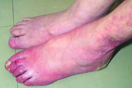
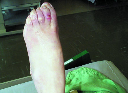
Critical leg ischaemia presents with a characteristic tight or burning pain, usually across the dorsum of the foot, but sometimes affecting the whole foot. Sitting or hanging the foot out of bed can often relieve the pain.1 In chronic ischaemia there is often a history of previous intermittent claudication. Acute thromboses of the distal arteries can occur de novo, and in some patients the acute phase of the pulseless, cold, pale foot is followed by an improvement in collateral blood supply and a cold, red foot. This can also follow an embolus. Examination findings can be misleading in that the foot is red when dependent and mimics cellulitis or gout (figure).
The temperature of the foot provides one diagnostic clue—in cellulitis or gout the affected area is warm or even hot, while in ischaemia the foot feels cold.2 However, even this can be misleading. In patients with diabetes, the temperature of an ischaemic foot can be normal or even warm. Raising the foot provides another clue. In cellulitis or gout, the foot will remain red when it is raised, but in ischaemia it will become pale. This so called Buerger’s sign has been questioned, however, and should be interpreted with care.1
Most patients with ischaemic pain who are visited at home by their general practitioner will be sitting in a chair, since this is the only position that relieves their pain. In patients who have been in a sitting position for a long time, the foot and ankle will often be swollen, and ischaemia may be mistaken for cellulitis or gout. Furthermore, physical examination of the pulses can be unreliable3; pulses may be present even in cases of severe ischaemia.4
To avoid these numerous diagnostic pitfalls all family doctors will need to have a high index of suspicion for ischaemia when consulted by any patient complaining of a painful foot. A missed diagnosis can be disastrous for a patient—it can lead to loss of a limb. It may also result in litigation, as happened with one of the cases described here. In another recent case quoted in the Journal of the Medical Defence Union, the settlement costs were £200 000.5
Family doctors cannot be blamed for not suspecting severe foot ischaemia. Although the condition is increasing in incidence, it is still uncommon—the annual incidence in the United Kingdom is 1 per 2500 people.6 Thus, a general practitioner can expect to see one or fewer patients with the condition each year. After hospital training in medical departments, young general practitioners may have seen only three cases in their career.
A solution to the problem is that all surgeries should have hand held Doppler equipment (cost £300-£400) for ankle pulse examination—and staff should learn how to use it. With the advent of health gain strategies in leg ulcer care this equipment is already available to most surgeries in the United Kingdom. The practice nurses can easily be taught pressure measurement. They will need to determine the blood pressure at the ankle and compare it with the systolic pressure at the arm (ankle to arm systolic pressure index).7 In normal patients, the pressure is marginally higher in the arm, but in patients with critical limb ischaemia the ankle pressure is half or less that of the arm. Sounds in the dorsalis pedis artery can be unreliable; pressure measurements should always be taken. Our clinical nurse specialist in vascular surgery helps to train practice nurses and community nurses in the surrounding area.
A note of caution is needed in patients with diabetes. The ankle to arm systolic pressure index can be normal in these patients, even in those with ischaemia. The diabetic foot can be confusing, even to the expert. The only guidance must be to refer the patient with a swollen and red foot for urgent assessment; they may need immediate surgery rather than antibiotics.
Vascular surgery cannot always save limbs, but it does so successfully in at least 70% of cases. A difficult question that patients (or their relatives) often ask me is, “Would it have made any difference if I had been referred earlier?” I would far rather that more of these patients were referred to me early than answer the question in rather vague evasive terms.
Figure.
In ischaemia the foot can be red while dependent (top) but pale when it is raised (bottom)
References
- 1.McGee SR, Boyko EJ. Physical examination and chronic lower-extremity ischaemia. A critical review. Arch Intern Med. 1998;158:1357–1364. doi: 10.1001/archinte.158.12.1357. [DOI] [PubMed] [Google Scholar]
- 2.Stoffers HEJH, Kester ADM, Saiser V, Rindens PELM. Diagnostic value of signs and symptoms associated with peripheral arterial occlusive disease seen in general practice: a multivariate approach. Med Decision Making. 1997;17:61–70. doi: 10.1177/0272989X9701700107. [DOI] [PubMed] [Google Scholar]
- 3.Brearley S, Shearman CP, Simms MH. Peripheral pulse palpation: an unreliable physical sign. Ann R Coll Surg Engl. 1982;74:169–171. [PMC free article] [PubMed] [Google Scholar]
- 4.Boylco EJ, Ahroni JH, Davignon O, Stensel V, Prigeon RL, Smith DG. Diagnostic utility of the history and physical examination for peripheral vascular disease among patients with diabetes mellitus. J Clin Epidemiol. 1997;50:659–668. doi: 10.1016/s0895-4356(97)00005-x. [DOI] [PubMed] [Google Scholar]
- 5.Case histories. J MDU. 1998;14:15–16. [Google Scholar]
- 6.Vascular Surgical Society of Great Britain and Ireland. Critical limb ischaemia management and outcome. Report of a national survey. Eur J Vasc Endovasc Surg. 1995;10:108–113. doi: 10.1016/s1078-5884(05)80206-0. [DOI] [PubMed] [Google Scholar]
- 7.Clements DL, Claes R. Diagnostic performance of segmental plethysmography and Doppler ultrasound in obliterating vascular diseases in the lower limbs. Angiology. 1980;31:272–282. doi: 10.1177/000331978003100407. [DOI] [PubMed] [Google Scholar]


