Abstract
Background The methylation of SHOX2 and RASSF1A shows promise as a potential biomarker for the early screening of lung cancer, offering a solution to remedy the limitations of morphological diagnosis. The aim of this study is to diagnose lung adenocarcinoma by measuring the methylation levels of SHOX2 and RASSF1A, and provide an accurate pathological diagnosis to predict the invasiveness of lung cancer prior to surgery.
Material and methods The methylation levels of SHOX2 and RASSF1A were quantified using a LungMe® test kit through methylation-specific PCR (MS-PCR). The diagnostic efficacy of SHOX2 and RASSF1A and the cutoff values were validated using ROC curve analysis. The hazardous factors influencing the invasiveness of lung adenocarcinoma were calculated using multiple regression.
Results: The cutoff values of SHOX2 and RASSF1A were 8.3 and 12.0, respectively. The sensitivities of LungMe® in IA, MIA and AIS patients were 71.3% (122/171), 41.7% (15/36), and 16.1% (5/31) under the specificity of 94.1% (32/34) for benign lesions. Additionally, the methylation level of SHOX2, RASSF1A and LungMe® correlated with the high invasiveness of clinicopathological features, such as age, gender, tumor size, TNM stage, pathological type, pleural invasion and STAS. The tumor size, age, CTR values and LungMe® methylation levels were identified as independent hazardous factors influencing the invasiveness of lung adenocarcinoma.
Conclusion: SHOX2 and RASSF1A combined methylation can be used as an early detection indicator of lung adenocarcinoma. SHOX2 and RASSF1A combined (LungMe®) methylation is significantly correlated to age, gender, tumor size, TNM stage, pathological type, pleural invasion and STAS. The SHOX2 and RASSF1A methylation levels, tumor size and CTR values could predict the invasiveness of the tumor prior to surgery, thereby providing guidance for the surgical procedure.
Keywords: Lung adenocarcinoma, SHOX2, RASSF1A, Methylation, Invasiveness
Introduction
Lung cancer is the leading cause of cancer deaths, and the high mortality rate of lung cancer can largely be attributed to late diagnosis, underscoring the critical importance of early diagnosis in mitigating cancer progression [1]. Currently, conventional diagnostic techniques employed for lung cancer encompass computed tomography imaging and morphological examination. The increasing sensitivity of CT imaging has facilitated the early detection of adenocarcinoma, resulting in smaller lesion sizes and fewer samples could be taken. While the implementation of screening has significantly enhanced the detection of early-stage lung cancer, it has also led to a rise in cases of overdiagnosis and overtreatment. Meanwhile, the morphological diversity of early lesions poses challenges in directly distinguishing between benign and malignant conditions. The identification of non-invasive early lung cancer or precancerous lesions imposes a significant psychological burden on patients and poses challenges for clinical surgeons in decision-making processes. Diagnosing small benign and malignant lesions poses a challenge, as does localizing malignant lesions during surgical procedures. Patients’ continuing observation presents a dilemma: on the one hand, the patient’s psychological pressure is heightened; on the other hand, the doctor is not completely sure whether the lesion is indolent or rapidly malignant progressing.
As we all know, the pathological evolution of early lung adenocarcinoma goes through the process of atypical adenomatous hyperplasia (AAH) to adenocarcinoma in situ (AIS) to micro invasive adenocarcinoma (MIA) to invasive adenocarcinoma (IA). In 2021, lung adenocarcinoma in situ (AIS) was excluded from the diagnostic category of lung adenocarcinoma by WHO and redefined as Precursor glandular lesion. Several studies have demonstrated the feasibility of sublobectomy in cases of AIS and minimal MIA. Sublobectomy, including wedge resection or segmentectomy, has been shown to maximize the preservation of lung function while ensuring the oncological effect, shorten the operation time, reduce postoperative complications, and yield favorable health and economic outcomes. The diverse imaging characteristics of lung adenocarcinoma nodules do not provide sufficient information to accurately predict histopathological features or patient prognosis. Comprehensive pathological judgment requires complete resection of the nodule. Secondly, the accurate pathological diagnosis of lung cancer poses a significant challenge in clinical practice. Conventional morphological diagnostic methods, such as cytological and histological examination, are susceptible to variations in specimen quality and the proficiency of pathologists.
The advancement of epigenetic research has garnered significant interest in the role of DNA methylation in the pathogenesis of cancer [2, 3]. Extensive research conducted in recent years has demonstrated that aberrant DNA methylation in these specific regions is a highly established epigenetic alteration in human cancers [4]. This characteristic not only holds the potential for distinguishing cancer cells from normal tissue but also presents opportunities for its application in early cancer detection.
Short Stature Homeobox 2 (SHOX2) methylation pattern has been employed for the purpose of diagnosing lung cancer. The expression of SHOX2 is significantly elevated in the majority of cancer types. Through a comparative analysis of SHOX2 methylation in lung cancer and normal tissues, it was observed that 96% of tumor tissues exhibited an elevated level of methylation [5]. Furthermore, the presence of high SHOX2 expression or hypomethylation is indicative of inferior differentiation and an unfavorable prognosis [6]. Ras-association domain family member 1 A (RASSF1A), a tumor suppressor gene, is usually missing in several cancers [7]. RASSF1A has been extensively investigated as an adjunctive DNA methylation biomarker in the context of lung cancer. The promoter region of RASSF1A demonstrated hypermethylation in 63% of non-small cell lung cancer (NSCLC) cells, while remaining unaffected in normal epithelial cells [8]. Additionally, RASSF1A methylation level could predict the disease progression in non-small cell lung cancer patients receiving pemetrexed-based chemotherapy [9]. The combination diagnosis of SHOX2 and RASSF1A has demonstrated utility in diagnosing a diverse range of tumors. To enhance its applicability in a diagnostic context, an in vitro diagnostic test kit, known as the LungMe® Assay (Tollgen, Shanghai, China), has been developed and validated for NMPA marking by the China National Medical Products Administration. The utilization of SHOX2 and RASSF1A methylation has been shown to enhance the sensitivity of early lung cancer detection, as substantiated by multiple scholarly literature [10, 11]. The sensitivity of the combined methylation for SHOX2 and RASSF1A in bronchoalveolar lavage fluid for NSCLC was found to range from 71.5 to 83.2%, while the specificity ranged from 90.0 to 97.4% [12, 13]. The combined promoter methylation assay for lung cancer using SHOX2 and RASSF1A demonstrated a sensitivity of 89.8% and a specificity of 90.4% in FFPE specimens [14]. The objective of this research is to examine the involvement of SHOX2 and RASSF1A methylation in the early detection of lung adenocarcinoma, specifically in distinguishing between AIS, MIA and IA. Additionally, the study aims to investigate the potential of SHOX2 and RASSF1A methylation as a supplementary diagnostic tool for early lung adenocarcinoma cases with uncertain pathological diagnoses. Early-stage lung adenocarcinoma exhibits several invasive characteristics, such as pleural invasion, lymph node invasion, airway dissemination, and pathological subtyping, which influence the selection of surgical procedures. This study delves into the association between methylation and lung adenocarcinoma pathological evolution, with the hope that more information can be provided by methylation in diagnosing early lung adenocarcinoma to assist in the selection of surgical strategies. Therefore, there is a pressing necessity to establish a comprehensive system that integrates non-invasive imaging characteristics with minimally invasive diagnostic sampling to enhance the assessment of lung cancer invasiveness with greater precision and efficacy. This advancement will facilitate clinicians in making more informed and accurate decisions regarding disease management prior to initiating treatment.
Materials and methods
Patients and specimens
The FFPE resection specimens were collected from 272 patients who visited the Affiliated Hospital of Nantong University. Out of these specimens, a total of 238 cases of lung adenocarcinoma were diagnosed. This included 171 cases of invasive adenocarcinoma, 36 cases of minimally invasive adenocarcinoma, 31 cases of carcinoma in situ, and 34 cases of benign lesions that served as controls. The FFPE samples had not been stored for a duration exceeding 2 years. The age, gender, and other pertinent information of the patients were shown in Table 1. The study was approved by the Ethics Committee of Affiliated Hospital of Nantong University. All the patients/participants provided written informed consent to partake in this study.
Table 1.
Baseline characteristics of patients
| Lung adenocarcinoma | ||||
|---|---|---|---|---|
| Clinicopathological index |
BC (n = 34) |
AIS (n = 31) |
MIA (n = 36) |
IA (n = 171) |
| Age (years) | ||||
| Median ± SEM | 61.0 ± 9.1 | 57.0 ± 13.5 | 67.0 ± 12.5 | 64.0 ± 11.2 |
| Range | 44–83 | 24–75 | 30–78 | 26–81 |
| Gender and Smoking | ||||
| Female (%) | 8(23.5) | 28(90.3) | 22(61.1) | 106(62.0) |
| Non-smoking | 8 | 28 | 22 | 105 |
| Smoking | 0 | 0 | 0 | 1 |
| Male (%) | 26(76.5) | 3(9.4) | 14(38.9) | 65(38.0) |
| Non-smoking | 8 | 3 | 11 | 51 |
| Smoking | 5 | 0 | 3 | 14 |
Note AIS: Adenocarcinoma in situ; MIA: Microinvasive adenocarcinoma; IA: Invasive adenocarcinoma; BC: benign lesion
DNA extraction and processing
The paraffin-embedded tissue material was lysed using the FFPE DNA extraction kit (CWY009S, CW Biotech Co., Ltd., China). The Qubit dsDNA HS Assay Kit (Life Technologies, Carlsbad, CA) was conducted to assess the DNA concentration on a Qubit® 3.0 fluorometer. Subsequently, 50 ng of DNA underwent sodium bisulfite treatment with the Tellgen DNA Purification Kit (Tellgen, Shanghai, China) to convert unmethylated cytosine to uracil.
Detection of DNA methylation levels in FFPE specimens
The China National Medical Products Administration (NMPA) approved in vitro diagnostic (IVD) test LungMe® (20,173,403,354, Tellgen, Shanghai, China) was utilized to ascertain the DNA methylation levels in FFPE specimens. The bisulfite-converted DNA that had been purified was utilized directly for MS-PCR using the commercially available LungMe® Real-time PCR kit (Tellgen, Shanghai, China). The PCR process was conducted on an ABI 7500 Real-Time PCR instrument (Applied Biosystems, CA, UAS). The primers were as follows: SHOX2 F: 5’- TTGTTTTTGGGTTCGGGTT-3’, R: 5’- CATAACGTAAACGCCTATACTC-3’; RASSF1A F: 5’- CGGGGTTCGTTTTGTGGTTTC-3’, R: 5’- CCGATTAAATCCGTACTTCGC-3’. The corresponding channels of amplification of methylated SHOX2, RASSF1A, and ACTB were VIC, FAM, and CY5, respectively. The calculation of the methylation level for each specific gene was determined by employing the subsequent formula: ΔCt = Ct − Ctβ−ACTB.
ROC curve
Receiver operator characteristic (ROC) curves were employed to assess the diagnostic effects of RASSF1A and SHOX2. All the specimens were divided into cancer and non-cancer groups. Carcinoma in situ was classified as a precancerous lesion. Therefore, the non-cancerous group induced patients with AIS and benign lesions. The cancer group covered patients with MIA and IA. The Youden indexes and areas under the curve (AUC) were detected using the ROC curve.
Statistical analysis
IBM SPSS Statistics 21.0 software (SPSS Inc., Chicago, IL) was conducted to perform the statistical analyses, and GraphPad Prism 8.0 was utilized to generate graphics. The ROC curve was employed to ascertain the ΔCt cutoff values of SHOX2 and RASSF1A, with the objective of assessing the diagnostic efficacy. The sensitivity and specificity of methylation were evaluated in diagnosing lung adenocarcinoma. Additionally, the methylation examination and the examination of tumor imaging features and clinicopathological characteristics were analyzed using the chi-square test. The hazardous factors influencing the invasiveness of lung adenocarcinoma were calculated using multiple regression. P-value < 0.05 was deemed statistically significant.
Results
The methylation levels of SHOX2 and RASSF1A in surgical FFPE specimens
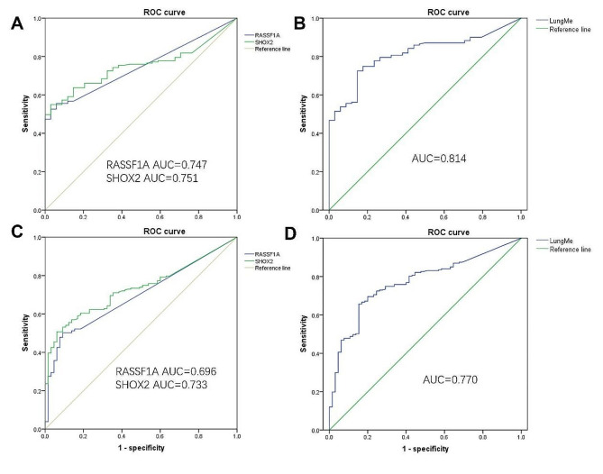
To explore the diagnostic efficiency, the methylation levels of SHOX2 and RASSF1A in surgical FFPE specimens were measured using MS-PCR. To calculate the cutoff values of SHOX2 and RASSF1A, ROC curves were performed using two methods of grouping. On one hand, the patients with invasive adenocarcinoma are in the cancer group and benign lesions are in the non-cancer group. At this time, the AUCs of SHOX2 and RASSF1A methylation were 0.747 and 0.75 (Fig. 1A), while the AUC of LungMe® was 0.814 (Fig. 1B). On the other hand, all the specimens were divided into cancer and non-cancer groups. Due to adenocarcinoma in situ being classified as a precancerous lesion, AIS together with benign lesions control (BC) was classified into non-cancerous group. For another, MIA and IA were classified into cancer group. Through ROC curve, the AUCs of SHOX2 and RASSF1A methylation were 0.696 and 0.733 (Fig. 1C), while the AUC of LungMe® was 0.770 (Fig. 1D). The calculated cutoff values of RASSF1A methylation, determined by employing two distinct methods, were both found to be 13.94. However, considering our prior research, which indicated that a cutoff value exceeding 12.0 resulted in inadequate PCR stability, we decided to modify the cutoff value to 12.0. For the cutoff of SHOX2 methylation, the calculated cutoff values of SHOX2 using the two methods were 8.27 and 8.28, respectively. Ultimately, we opted for the rounded value of 8.3.
Fig. 1.
ROC curve determines the cutoff values of SHOX2 and RASSF1A methylation. A: The ROC curves of SHOX2 and RASSF1A methylation in distinguishing IA and BC. B: The ROC curve of LungMe® methylation in distinguishing IA and BC. C: The ROC curve of SHOX2 and RASSF1A methylation in distinguishing cancer group (IA and MIA) and non-cancer groups (AIS and BC). D: The ROC curve of LungMe® methylation in distinguishing cancer group (IA and MIA) and non-cancer groups (AIS and BC)
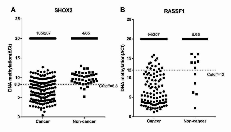
All the specimens were divided into cancer and non-cancer groups. In this case, cancer group contains MIA and IA, while non-cancerous group contains AIS and BC. The ΔCt values of SHOX2 and RASSF1A methylation were utilized to plot scatter plots (Fig. 2). Based on the cutoff values, the positive rate of SHOX2 and RASSF1A methylation were 50.7% (105/207) and 45.4% (94/207) in cancer group. The specificities of SHOX2 and RASSF1A methylation were 93.8% (61/65) and 92.3% (60/65). The sensitivity of LungMe® was 66.2% (137/207), and the specificity was 89.2% (58/65).
Fig. 2.
The methylation levels of SHOX2 and RASSF1A in surgical FFPE specimens. A: The methylation levels of SHOX2 in cancer and non-cancer FFPE specimens. B: The methylation levels of RASSF1A in cancer and non-cancer FFPE specimens. Caner group: microinvasive adenocarcinoma (MIA) and invasive adenocarcinoma (IA); Non-cancer group: adenocarcinoma in situ (AIS) and benign lesions
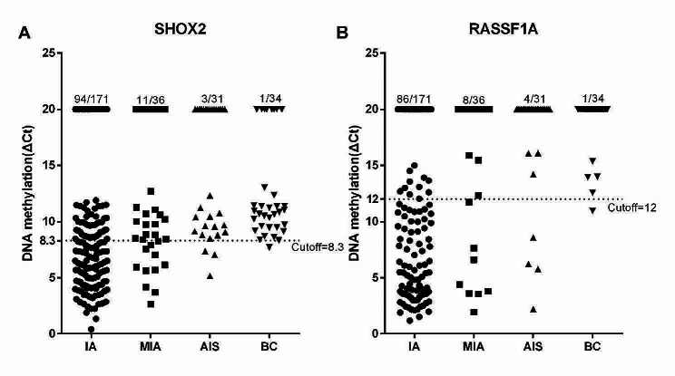
To further explore the diagnostic value of SHOX2 and RASSF1A methylation in subtypes of lung adenocarcinoma, the methylation levels were calculated in IA, MIA, AIS and BC, respectively. Scatter plots were utilized to represent the ΔCt values of SHOX2 and RASSF1A methylation (Fig. 3). The positive rates of SHOX2 methylation were 55.0% (94/171), 30.6% (11/36) and 9.7% (3/31) in IA, MIA and AIS patients. Meanwhile, the positive rates of RASSF1A methylation were 50.3% (86/171), 22.2% (8/36) and 12.9% (4/31) in IA, MIA and AIS patients. The sensitivities of LungMe® in IA, MIA and AIS patients were 71.3% (122/171), 41.7% (15/36), and 16.1% (5/31). The specificities of SHOX2, RASSF1A and LungMe® methylation in diagnosing lung adenocarcinoma from benign lesions were 97.1% (33/34), 97.1% (33/34) and 94.1% (32/34) (Table 2).
Fig. 3.
The methylation levels of SHOX2 and RASSF1A in different pathological types of lung adenocarcinoma. A: The methylation levels of SHOX2 in different pathological types of lung adenocarcinoma. B: The methylation levels of RASSF1A in different pathological types of lung adenocarcinoma. AIS: adenocarcinoma in situ; MIA: microinvasive adenocarcinoma; IA: invasive adenocarcinoma; BC: benign control
Table 2.
The sensitivity and specificity of SHOX2 and RASSF1A methylation in diagnosing lung adenocarcinoma
| Methylation indexes | Cutoff | Sensitivity for IA | Sensitivity for MIA | Sensitivity for AIS | Specificity for BC |
|---|---|---|---|---|---|
| SHOX2 | 8.3 | 55.0% (94/171) | 30.6% (11/36) | 9.7%(3/31) | 97.1% (33/34) |
| RASSF1A | 12 | 50.3% (86/171) | 22.2% (8/36) | 12.9%(4/31) | 97.1% (33/34) |
| LungMe® | - | 71.3% (122/171) | 41.7% (15/36) | 16.1%(5/31) | 94.1% (32/34) |
Note LungMe® is a combination of SHOX2 and RASSF1A methylation
MIA: microinvasive adenocarcinoma; IA: invasive adenocarcinoma
Relationship between tumor imaging features and clinicopathological characteristics and methylation in patients with lung adenocarcinoma
Imaging examinations were utilized to identify various characteristics of tumors, including their size, location, and GGO traits. Tumor traits were divided into pure ground-glass opacity (GGO), mixed ground-glass opacity and solid nodules through imaging examination. The Cavitary-to-Tumor Ratio (CTR) of pure GGO exhibited a value below 0.25, accompanied by a negligible presence of solid components. In contrast, the CTR of mixed GGO fell within the range of 0.25 to 1, whereas solid nodules demonstrated a CTR of 1. Most patients were mixed GGO, accounting for 57.1%, while the other two traits, pure GGO and solid nodules, account for 16.8% and 26.1%, respectively. We found that the tumor traits in patients younger than 65 years have no difference with patients older or equal to 65 years (p > 0.05). The number of females was more than that of males, but there was no significant difference (p > 0.05). For the tumor size, an overwhelming majority of the tumors were 1–3 cm, accounting for 68.1%. As the size of the tumor increased, there was a gradual progression in the types of nodules observed, transitioning from pure GGO to solid nodules (p < 0.001). 78.2% of the patients were classified as stage IA, in which stage the proportion of pure GGO, mixed GGO, and solid nodules were 80.0%, 80.9% and 71.0%. The incidence of solid nodules increased proportionally with the progression of the disease stage (p < 0.001). A majority of IA patients, precisely 93.5%, exhibited solid nodules, and only 6.4% of patients with AIS and MIA (p < 0.001). According to the World Health Organization’s classification of lung adenocarcinoma, invasive adenocarcinoma can be categorized into three sub-grades. A majority of patients in II grade, accounting for 70.2%. As the stage increased, there was a gradual progression in the types of nodules observed, transitioning from pure GGO to solid nodules. The occurrence of early adenocarcinoma was closely linked to three high-risk factors: lymphnode metastasis, pleural invasion, and airway dissemination. The positive rates of lymph node metastasis, pleural invasion, and airway dissemination are extremely low, whose overall proportions were 3.4%, 11.0% and 7.2%. A significant proportion of patients diagnosed with lymphnode metastasis, pleural invasion and airway dissemination exhibited solid nodules, while being infrequently identified in the pure GGO group (p < 0.001). For LungMe® methylation, the positive rates were progressively increased from pure GGO to solid nodules. The positive rate of LungMe® methylation in solid nodule group was 79.0%, which was significantly higher than the rates of the pure GGO and mixed GGO groups, respectively (p < 0.05) (Table 3).
Table 3.
Relationship between tumor imaging features and clinicopathological characteristics in patients with lung adenocarcinoma
| Clinicopathological index | n = 238 | Tumor imaging features [n (%)] | P | ||
|---|---|---|---|---|---|
| Pure GGO n = 40 |
Mixed GGO n = 136 |
Solid n = 62 |
|||
| Age (Years) | 0.945 | ||||
| <65 | 116 | 20(50.0) | 65(47.8) | 31(50.0) | |
| ≥ 65 | 122 | 20(50.0) | 71(52.2) | 31(50.0) | |
| Gender | 0.138 | ||||
| Male | 82 | 10(25.0) | 45(33.1) | 27(43.5) | |
| Female | 156 | 30(75.0) | 91(66.9) | 35(56.5) | |
| Tumor size | < 0.001* | ||||
| ≤ 1 cm | 59 | 16(40.0) | 36(26.5) | 7(11.3) | |
| >1,≤3 cm | 162 | 24(60.0) | 96(70.6) | 42(67.7) | |
| >3 cm | 14 | 0(0.0) | 4(2.9) | 10(16.1) | |
| TNM stage | < 0.001* | ||||
| T0 stage | 31 | 8(20.0) | 20(14.7) | 3(4.8) | |
| IA stage | 186 | 32(80.0) | 110(80.9) | 44(71.0) | |
| IB stage | 11 | 0(0.0) | 5(3.7) | 6(9.7) | |
| II ~ IV stage | 10 | 0(0.0) | 1(0.7) | 9(3.8) | |
| Pathological type | < 0.001* | ||||
| AIS | 31 | 8(20.0) | 20(14.7) | 3(4.8) | |
| MIA | 36 | 9(22.5) | 26(19.1) | 1(1.6) | |
| IA | 171 | 23(57.5) | 90(66 2) | 58(93.5) | |
| IA WHO subtype | < 0.001* | ||||
| I grade | 23 | 10(43.5) | 13(14.4) | 1(1.7) | |
| II grade | 120 | 13(56.5) | 74(82.2) | 33(56.9) | |
| III grade | 16 | 0(0.0) | 3(3.3) | 13(22.4) | |
| Pleural invasion | 0.001* | ||||
| No | 210 | 37(94.9) | 126(93.3) | 47(75.8) | |
| Yes | 26 | 2(5.1) | 9(6.7) | 15(24.2) | |
| STAS | < 0.001* | ||||
| No | 218 | 39(100) | 131(97.8) | 48(77.4) | |
| Yes | 17 | 0(0.0) | 3(2.2) | 14(22.6) | |
| Lymphnode metastasis | < 0.001* | ||||
| No | 227 | 39(100) | 134(99.3) | 54(88.5) | |
| Yes | 8 | 0(0.0) | 1(0.7) | 7(11.5) | |
| LungMe® methylation | 0.0011* | ||||
| Negative | 96 | 21(52.5) | 62(45.6) | 13(21.0) | |
| Positive | 142 | 19(47.5) | 74(54.4) | 49(79.0) | |
Note GGO: ground-glass opacity; AIS: Adenocarcinoma in situ; MIA: Microinvasive adenocarcinoma; IA: Invasive adenocarcinoma; IMA: Invasive mucinous adenocarcinoma; STAS: spread through air space. LungMe® is a combination of SHOX2 and RASSF1A methylation
Relationship between SHOX2 and RASSF1A methylation and clinicopathological characteristics in patients with lung adenocarcinoma
The relationship between the SHOX2 and RASSF1A methylation levels and clinicopathological characteristics of all the patients were detected. We found that the positive rate of SHOX2 methylation in patients younger than 65 years was significantly lower than that in patients older than or equal to 65 years (p < 0.05), while there was no significant difference in RASSF1A methylation (p > 0.05). The positive rate of LungMe® methylation in males was higher than in females (p < 0.01). For the tumor imaging features, the positive rates of LungMe® methylation in patients with solid nodules were higher than in the other two groups (p < 0.05). The positive rates of SHOX2, RASSF1A and LungMe® methylation in patients with solid nodules were 62.9%, 58.1% and 79.0%, respectively. The majority of tumor sizes are greater than 1 cm to smaller than 3 cm. The positive rate of LungMe® methylation exhibits an upward trend as tumor size increases (p < 0.001). The majority of patients were in IA stage. The methylation positive rates of LungMe® methylation exhibited a gradual increase with the progression of TNM stage, yet experienced a decline in stages II-IV stages, potentially attributable to the error caused by fewer patients (p < 0.001). The positive rates of LungMe® methylation exhibit an increase in conjunction with advancements in invasiveness (both p < 0.001). For the sub-grades of invasive adenocarcinoma, a majority of patients were in II stage, accounting for 70.2%. As the stage increased, the positive rates of SHOX2, RASSF1A and LungMe® methylation exhibited an upward trend, with a significant difference observed in LungMe® methylation (p < 0.001). The occurrence of early adenocarcinoma is closely linked to three high-risk factors, namely lymphnode metastasis, pleural invasion, and airway dissemination, all of which exhibit relatively low positive rates. Notably, patients with lymphnode metastasis, pleural invasion, and airway dissemination displayed a higher incidence of SHOX2, RASSF1A and LungMe® methylation compared to those without pleural invasion (p < 0.05). SHOX2, RASSF1A and LungMe® methylation significantly differ among patients with pleural invasion (both p < 0.01). SHOX2 and LungMe® methylation have differences among patients with spread through air space (STAS) (p < 0.05). However, no correlation was observed between SHOX2, RASSF1A, and LungMe® and lymph node metastasis (p > 0.05) (Table 4).
Table 4.
Relationship between SHOX2 and RASSF1A methylation and clinicopathological characteristics in patients with lung adenocarcinoma
| Clinicopathological index | n = 238 | SHOX2 positive [n (%)] n = 108 | P | RASSF1A positive [n (%)] n = 98 | P | LungMe® positive [n (%)] n = 142 | P |
|---|---|---|---|---|---|---|---|
| Age (Years) | < 0.001* | 0.062 | 0.011* | ||||
| <65 | 116 | 37(31.9) | 42(36.2) | 60 (51.7) | |||
| ≥ 65 | 122 | 71(58.2) | 56(45.9) | 82(67.2) | |||
| Gender | < 0.001* | 0.004* | 0.001* | ||||
| Male | 82 | 50(61.0) | 44(53.7) | 63(76.8) | |||
| Female | 156 | 58(37.2) | 54(34.6) | 79(50.6) | |||
| Tumor imaging features | 0.003* | 0.005* | 0.001* | ||||
| Pure GGO | 40 | 13(32.5) | 12(30.0) | 19(47.5) | |||
| Mixed GGO | 136 | 56(41.2) | 50(36.8) | 74(54.4) | |||
| Solid | 62 | 39(62.9) | 36(58.1) | 49(79.0) | |||
| Tumor size | < 0.001* | < 0.001* | < 0.001* | ||||
| ≤ 1 cm | 59 | 13(22.0) | 10(16.9) | 18(30.5) | |||
| >1,≤3 cm | 162 | 82(50.6) | 76(46.9) | 109(67.3) | |||
| >3 cm | 14 | 12(85.7) | 11(78.6) | 13(92.9) | |||
| TNM stage | < 0.001* | < 0.001* | < 0.001* | ||||
| T0 stage | 31 | 3(9.7) | 4(12.9) | 5(16.1) | |||
| IA stage | 186 | 90(48.4) | 80(43.0) | 119(64.0) | |||
| IB stage | 11 | 10(90.9) | 10(90.9) | 11(100) | |||
| II ~ IV stage | 10 | 5(50.0) | 4(40.0) | 7(70.0) | |||
| Pathological type | < 0.001* | < 0.001* | < 0.001* | ||||
| AIS | 31 | 3(9.7) | 4(12.9) | 5(16.1) | |||
| MIA | 36 | 11(30.6) | 8(22.2) | 15(41.7) | |||
| IA | 171 | 94(55.0) | 86(50.3) | 122(71.3) | |||
| IA WHO subtype | 0.175 | 0.251 | 0.026* | ||||
| I grade | 23 | 9(39.1) | 10(43.5) | 12(52.2) | |||
| II grade | 120 | 59(49.2) | 65(54.2) | 85(70.8) | |||
| III grade | 16 | 11(68.8) | 11(68.8) | 15(93.8) | |||
| Pleural invasion | 0.001* | 0.008* | < 0.001* | ||||
| No | 210 | 88(41.9) | 81(38.6) | 118(56.2) | |||
| Yes | 26 | 20(76.9) | 17(65.4) | 24(92.3) | |||
| STAS | 0.031* | 0.110 | 0.044* | ||||
| No | 218 | 96(44.0) | 88(40.4) | 128(58.7) | |||
| Yes | 17 | 12(70.6) | 10(58.8) | 14(82.4) | |||
| Lymphnode metastasis | 0.276 | 0.565 | 0.312 | ||||
| No | 227 | 103(45.4) | 94(41.4) | 135(59.5) | |||
| Yes | 8 | 5(62.5) | 3(37.5) | 6(75.0) | |||
Note LungMe® is a combination of SHOX2 and RASSF1A methylation
GGO: ground-glass opacity; AIS: Adenocarcinoma in situ; MIA: Microinvasive adenocarcinoma; IA: Invasive adenocarcinoma; IMA: Invasive mucinous adenocarcinoma; STAS: spread through air space
Analysis of high-risk factors for invasiveness of lung adenocarcinoma
All the patients with lung adenocarcinoma and benign lesion were divided into 11 groups through pathological types, pathological subtype, lymph node metastasis, pleural invasion, airway dissemination and TNM stage. Detailedly, the participants were classified into eight grades, namely BN, AIS, MIS, IA1, IA2, IA3, IB, and II-III, according to the TNM staging system. The three grades of IA (IA1, IA2, IA3) were categorized as high or low invasiveness based on the invasiveness of the pathology, resulting in a total of 11 grades. The low invasiveness group were patients with anchorage type or acinar type or papillary type, while the high invasiveness group were patients with micropapillary type or solid type or complex acinar type or pleural invasion or lymph node metastasis or STAS (Table 5).
Table 5.
The criteria for grouping
| Groups | Criteria for grouping | Number | Percentage (%) |
|---|---|---|---|
| 1 | Benign lesions patients | 34 | 12.8 |
| 2 | AIS patients in T0 stage | 31 | 11.7 |
| 3 | MIA patients in IA1 stage | 36 | 13.5 |
| 4 | IA patients in IA1 stage with low invasiveness | 17 | 6.4 |
| 5 | IA patients in IA1 stage with high invasiveness | 0 | 0.0 |
| 6 | IA patients in IA2 stage with low invasiveness | 85 | 32.0 |
| 7 | IA patients in IA2 stage with high invasiveness | 7 | 2.6 |
| 8 | IA patients in IA3 stage with low invasiveness | 19 | 7.1 |
| 9 | IA patients in IA3 stage with high invasiveness | 18 | 6.8 |
| 10 | IA patients in IB stage | 10 | 3.8 |
| 11 | Patients in II + III stages | 10 | 3.8 |
Note The low invasiveness groups: anchorage type or acinar type or papillary type; the high invasiveness groups: micropapillary type or solid type or complex acinar type or pleural invasion or lymph node metastasis or spread through air space
Multivariate linear regression was performed to analyze the hazardous factors affecting the invasiveness of lung adenocarcinoma. Prior to the surgical procedure, pertinent information including age, gender, methylation level, tumor size, and imaging features was available and subsequently utilized for conducting univariate analysis. Univariate analysis results indicated that when increased by one year, the grade increased by 0.061 (p < 0.05). For gender, the grades of males were lower by 0.142 than females, but the difference was not statistically significant (p > 0.05). Tumor size increased by one year, and the grade increased by 0.987 (p < 0.05). LungMe® methylation positively increased by 2.857 on grade compared to negative methylation (p < 0.05). The grade of patients with mixed GGO was lower by 0.454 than pure GGO, but the difference was not statistically significant (p > 0.05). The grade of patients with solid nodules was higher by 1.221 than pure GGO (p < 0.05). Age, tumor size and solid nodules, which were statistically significant in univariate analysis, were included in the multivariate regression analysis. Through multivariate analysis, the age, methylation level, and tumor size were identified as independent hazardous factors influencing the invasiveness of lung adenocarcinoma (p < 0.05) (Table 6).
Table 6.
The hazardous factors influencing the invasiveness of lung adenocarcinoma
| Variables | Univariate analysis | Multivariate analysis | ||
|---|---|---|---|---|
| HR (95% CI) | P | HR (95% CI) | P | |
| Age (Years) | 0.061(0.032,0.090) | <0.001 | 0.031(0.006,0.056) | 0.015 |
| Gender(Male/Female) | -0.142(-0.851,0.567) | 0.693 | - | - |
| Tumor size | 0.987(0.694,1.280) | <0.001 | 0.549(0.270,0.827) | <0.001 |
| LungMe methylation | 2.857 (2.245,3.470) | <0.001 | 2.398 (1.806,2.990) | <0.001 |
| Tumor imaging features | ||||
| Mixed GGO | -0.454(-1.146,0.238) | 0.198 | - | - |
| Solid nodules | 1.221(0.474,1.968) | 0.001 | 0.887(0.234,1.540) | 0.008 |
Note HR, hazard ratio; 95% CI: 95% Confidence Interval. GGO: ground-glass opacity, Mixed GGO: 0.25 < CTR < 1; Solid nodules: CTR = 1. *P < 0.005
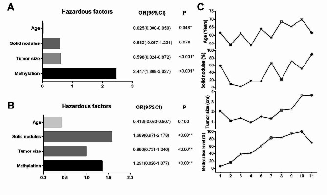
The inclusion of patients with benign lesions revealed that LungMe® methylation positive exerted a significant influence on the invasiveness of lung adenocarcinoma, as indicated by a hazard ratio (HR) value of 2.447 (Fig. 4A). Including patients without benign lesions revealed that solid nodules had the greatest impact on the invasiveness of lung adenocarcinoma, with an HR value of 1.689, followed by a positive LungMe® methylation of 1.291. However, age does not affect the grade when the patient has already identified the tumor (Fig. 4B). In addition, the average levels of age and tumor size, the positive rate of methylation level and proportions of solid nodules in lung adenocarcinoma were shown in Fig. 4C. It can be found that as the lung adenocarcinoma invasiveness increases, there is an upward trend in both the four indexes, especially methylation level.
Fig. 4.
Analysis of high-risk factors for invasiveness of lung adenocarcinoma. A: The hazardous factors of benign and malignant lung adenocarcinoma. B: The hazardous factors influencing the invasiveness of lung adenocarcinoma. C: The average levels of age and tumor size, the proportions of methylation level and solid nodule in lung adenocarcinoma. HR, hazard ratio; 95% CI: 95% Confidence Interval. *P < 0.005
Relationship between surgical procedure and risk factors associated with invasiveness of lung adenocarcinoma
Three surgical methods were wedge resection, segmentectomy and lobectomy, with lobectomy representing over 50% of the cases. A total of 178 patients were diagnosed with tumors measuring less than 2 cm, whereas 57 patients presented tumors exceeding 2 cm in size. The treatment approach for patients with tumors smaller than 2 cm primarily involved segmentectomy and lobectomy, whereas tumors larger than 2 cm were predominantly subjected to lobectomy (p < 0.05). Similar to the situation of tumor size, patients presenting a CTR below 0.5 predominantly underwent segmentectomy and lobectomy, while the majority of patients with a CTR exceeding 0.5 underwent lobectomy (p < 0.05). A total of 62.4% of patients exhibiting positive methylation underwent lobectomy, and patients with lobectomy were more than segmentectomy. In contrast, among patients with negative methylation, the majority of patients underwent segmentectomy more than lobectomy (p < 0.05)((Table 7).
Table 7.
Relationship between surgical procedure and risk factors associated with invasiveness of lung adenocarcinoma
| Index | N = 235 | Wedge resection(35) | Segmentectomy(77) | Lobectomy(123) | P |
|---|---|---|---|---|---|
| Tumor size | < 0.0001* | ||||
| ≤ 2 cm | 178 | 28(15.7) | 70(39.3) | 80(44.9) | |
| > 2 cm | 57 | 7(12.3) | 7(12.3) | 43(75.4) | |
| Tumor imaging features | 0.011* | ||||
| CTR ≤ 0.5 | 173 | 25(14.5) | 66(38.2) | 82(47.4) | |
| CTR > 0.5 | 62 | 10(16.1) | 11(17.7) | 41(66.1) | |
| LungMe® methylation | < 0.0001* | ||||
| Positive | 141 | 20(14.2) | 33(23.4) | 88(62.4) | |
| Negative | 94 | 15(16.0) | 44(46.8) | 35(37.2) | |
Note LungMe® is a combination of SHOX2 and RASSF1A methylation. CTR: Cavitary-to-tumor ratio
Discussion
The widespread adoption of low-dose computed tomography (LDCT) has facilitated the early detection of lung adenocarcinoma. Nevertheless, LDCT’s high sensitivity may lead to the identification of numerous, smaller early lesions, necessitating the assessment of benign and malignant nodules. Increasing evidence indicated that DNA methylation alteration is regarded as an early prognosticator of cancer and can be identified during the initial phases of tumorigenesis [15–18]. The combination detection of SHOX2 and RASSF1A methylation has been utilized in diagnosing lung cancer, with a sensitivity of 71.5–96.0%, and a specificity of 82.3–100% [13, 19].
In this study, SHOX2 and RASSF1A gene methylation were utilized to diagnose early lung adenocarcinoma, while surgical tissue samples were employed to mitigate the potential impact of sampling variability. The majority of prior investigations were conducted using bronchoalveolar lavage fluid, thus necessitating the establishment of novel threshold values in tissue specimens. To ascertain the threshold values for SHOX2 and RASSF1A, two distinct grouping methodologies were employed to draw ROC curves, resulting in similar cutoff values of 8.3 and 12.0. Considering prior research, the cutoff value of SHOX2 was 7.5 in surgical tissues, which was influenced by the inclusion of a substantial number of granulomas [14]. However, in the current study, surgical samples of tuberculosis were deliberately excluded from the initial stage of the experimental data, resulting in improved specificity and a revised cutoff value of 8.3. The combined AUCs of SHOX2 and RASSF1A were 0.814 and 0.770, respectively, exceeding the threshold of 0.75, indicating that the two methods all have diagnostic values. In this study, the sensitivity and specificity of LungMe® methylation in diagnosing lung adenocarcinoma were 66.2% and 94.1%. Our results were consistent with the previous research that the sensitivity and specificity of LungMe® methylation were 69.6% and 97.4 [13]. A prior investigation demonstrated that the diagnostic sensitivity of SHOX2 and RASSF1A combined methylation in lung adenocarcinoma was 52.5%, whereas cytology exhibited a sensitivity of 13.3%. Furthermore, the combined utilization of methylation and cytology yielded a sensitivity of 55.8% [20]. The sensitivity of DNA methylation detection in diagnosing lung cancer, particularly LUAD, was found to be higher compared to cytology detection, while also compensating for the limitations of cytology [21]. Methylation detection is a molecular method that utilizes PCR to greatly enhance the methylation signal by a factor of 1 million, exhibiting remarkable sensitivity and amplification specificity. In comparison to cytology, methylation demonstrates significantly higher sensitivity while maintaining equivalent specificity. Nevertheless, this study did not undertake a comparative analysis between cytology and methylation, which will be addressed in the subsequent article.
The classification of early adenocarcinoma was based on the extent of invasion, resulting in the categorization of IA, MIA, and AIS. The reclassification of AIS prompts an inquiry into the necessity of surgical intervention, specifically determining when surgery is warranted and when surgery is unnecessary for AIS. From a pathological diagnostic standpoint, AIS and MIA exhibit overlapping characteristics. AIS can be likened to a dormant intruder within the body, and it becomes imperative to ascertain the moment when this intruder may become active. Our contemplation revolves around the potential utility of methylation as a reliable triage indicator, and whether positive methylation can serve as a determinant for the impending activation of the intruder. The sensitivities of LungMe® in IA, MIA and AIS patients were 71.3% (122/171), 41.7% (15/36), and 16.1% (5/31), which was very consistent with our expectation. The specificities of LungMe® methylation in diagnosing lung adenocarcinoma from benign lesions was 94.1% (32/34). The methylation of LungMe® demonstrates significant diagnostic efficacy in the identification of lung adenocarcinoma, while also exhibiting varying discriminatory potential across distinct pathological stages of early lung adenocarcinoma. Research has demonstrated a positive association between the combined methylation levels of SHOX2 and RASSF1A and the expression of Ki-67 in early-stage lung adenocarcinoma [22]. Ki-67 is known to be involved in the proliferation of cancer cells, with higher values indicating accelerated tumor growth and development, ultimately leading to a poorer prognosis for patients [23–25]. It can be inferred that individuals who test positive for SHOX2 and RASSF1A methylation may experience more rapid tumor progression. Our results indicated that patients with IA had the highest sensitivity compared to MIA and AIS, which was consistent with the previous findings. Also, the association between RASSF1A and the heightened invasiveness of lung cancer has been reported. Suppression of the RASSF1A gene facilitated the invasion and migration of lung cancer cells [26]. Thus, there is a positive correlation between the invasiveness of the tumor and the rate of LungMe® methylation, with higher invasiveness resulting in a higher positive rate. The presence of positive LungMe® methylation in patients with AIS suggests a potential for invasion, which could progress to MIA, thereby recommending surgical intervention. This is the first time to propose that positive LungMe® methylation in patients with AIS may illustrate a risk for invasion.
Furthermore, the pathological characteristics associated with high invasiveness include tumor size, TNM stage, pathological type, lymph node metastasis, pleural invasion and STAS. Imaging serves as the primary clinical examination for discerning between benign and malignant conditions prior to surgical intervention, and its significance as a preoperative indicator is of utmost importance. Prior to assessing methylation, we initially explored the relationship between imaging features and highly aggressive attributes. A Previous study found that solid components are a negative prognostic factor in lung adenocarcinoma [27]. Especially, we discovered that a LungMe® methylation positive status was observed in 79% of solid nodules patients, which is consistent with prior research. The methylation of LungMe® exhibited correlations with lung adenocarcinoma high-risk factors, pleural invasion and STAS, except for lymph node metastasis. Despite a lack of statistical significance, patients with positive lymph node metastasis exhibited a higher degree of LungMe® methylation. The methylation level of SHOX2 was found to be significantly higher in patients with positive STAS, whereas no significant association was observed between the methylation level of RASSF1A and STAS. Considering the different pathological subtypes of adenocarcinoma, invasive adenocarcinoma can be categorized into three sub-grades according to the World Health Organization’s classification. The anchorage type was designated in I grade, while the acinar and papillary types were classified in II grade. Additionally, the solid, micropapillary, and complex acinar types were categorized in III grade. Previous research illuminated that lung adenocarcinoma at TNM I stage with minimal solid or micropapillary have higher invasiveness, thus predicting poor prognosis [28]. Consistent with the findings, we found that LungMe® methylation positive rates exhibited a gradual increase with the progression of IA subtype. The methylation rate in III grade, specifically in the subtype characterized by solid, micropapillary, and complex acinar features, was determined to be 93.8%. LungMe® methylation exhibited correlations with all pathological characteristics associated with high invasiveness, with the exception of lymph node metastasis, indicating a close association between methylation and the attributes of heightened invasiveness.
The controversy surrounding preoperative lobectomy or segmentectomy persists within academic discourse. Segmentectomy is a comparatively smaller surgical procedure than lobectomy for treating small NSCLC lesions. However, due to the presence of pathologically aggressive tumor features, there is a potential risk of positive surgical margins for cancer and subsequent recurrence. The research of JCOG0802/WJOG4607L also identified a comparatively elevated incidence of local recurrence associated with segmentectomy in contrast to lobectomy [29]. Consequently, the careful selection of appropriate surgical methods holds significant potential in enhancing the recurrence outcomes for early lung adenocarcinoma [30].
We want to make an initial assessment of the invasiveness of nodules based on the available information prior to surgery, in order to determine the most suitable surgical procedure and treatment approach. All the lung adenocarcinoma and benign lesions patients were categorized into 11 cohorts based on the invasiveness, and an analysis was conducted of the risk factors influencing the invasiveness of lung adenocarcinoma. Incorporating patients with benign lesions, the positive rate of LungMe® methylation was increasing, which serves as compelling evidence for assessing the level of invasiveness. However, the observed decline in group 11 may be attributed to the limited patient sample size. We also found that as the lung adenocarcinoma invasiveness increases, there is an upward trend in all the indexes, especially LungMe® methylation level. Consequently, wedge resection emerges as a viable treatment option for AIS and MIA. The research of JCOG0802/WJOG4607L first proposed that segmentectomy exhibits greater efficacy in terms of overall survival for early-stage lung cancer. They ultimately found that segmentectomy ought to be considered as the prevailing surgical approach for patients diagnosed with peripheral non-small cell lung cancer, characterized by a diameter of ≤ 2 cm and a CTR exceeding 0.5 [29]. When a patient is diagnosed with a tumor, early diagnostic indicators such as size, solid nodule (CTR = 1) and LungMe® methylation, exhibit a strong correlation with the invasiveness of the tumor, thereby enabling the establishment of a predictive model. Specifically, we incorporated a new highly sensitive indicator, LungMe® methylation, which was novel to this study. The calculated score not only assesses the invasiveness of the tumor but also offers supplementary information for determining the appropriate surgical approach based on tumor size.
Conclusion
LungMe® methylation can be used as an early detection indicator of lung adenocarcinoma, as it is associated with highly aggressive subtypes. The potential risk factors for the invasiveness of lung adenocarcinoma include age, methylation level, and tumor size. The SHOX2 and RASSF1A methylation levels, tumor size and CTR values could predict the invasiveness of the tumor prior to surgery, thereby providing guidance for the surgical procedure.
Acknowledgements
We would like to thank the patient and her family for their participation and support of this study.
Abbreviations
- NSCLC
Non-small cell lung cancer
- SHOX2
Short stature homeobox 2
- RASSF1A
Ras-association domain family member 1 A
- MS-PCR
Methylation-specific PCR
- FFPE
Formalin fixed paraffin-embedded
- AIS
Adenocarcinoma in situ
- MIA
Microinvasive adenocarcinoma
- IA
Invasive adenocarcinoma
- BC
Benign lesions control
- NMPA
National Medical Products Administration
- IVD
In vitro diagnostic
- ROC
Receiver operator characteristic
- AUC
Areas under the curve
- GGO
Ground-glass opacity
- CTR
Cavitary-to-tumor ratio
- STAS
Spread through air space
- HR
Hazard ratio
- LDCT
Low-dose computed tomography
- LUAD
Lung adenocarcinoma
Author contributions
All authors contributed to the study conception and design. Designed the study, provided the sources of all the samples and provided funding supports were performed by Honggang Ke and Yifei Liu. Material preparation, data collection and analysis were performed by Jiaping Zhao and Yu Lu. The first draft of the manuscript was written by Jiaping Zhao, Yu Lu and Xiaosha Ren, and all authors commented on previous versions of the manuscript. Helped collected data, helped analyze the data and final manuscript review were performed by Tingting Bian, Jia Feng, Hui Sun, Lei Liu and Bin She. Jiaping Zhao and Yu Lu contributed equally and were co-first authors. All authors read and approved the final manuscript.
Funding
This work was supported by Shanghai Pudong New District Foundation for Development of Science and Technology (PKX2021-S09).
Data availability
All data can be obtained from the corresponding author.
Declarations
Ethics approval and consent to participate
This study was performed in line with the principles of the Declaration of Helsinki. Approval was granted by the Ethics Committee of Affiliated Hospital of Nantong University. Informed consent was obtained from all individual participants included in the study.
Consent for publication
Not Applicable.
Competing interests
The authors declare no competing interests.
Footnotes
Publisher’s Note
Springer Nature remains neutral with regard to jurisdictional claims in published maps and institutional affiliations.
Contributor Information
Yifei Liu, Email: ntdxliuyifei@sina.com.
Honggang Ke, Email: tranquillity_z@163.com.
References
- 1.Xia C, et al. Cancer statistics in China and United States, 2022: profiles, trends, and determinants. Chin Med J (Engl) 2022;135(5):584–90. doi: 10.1097/CM9.0000000000002108. [DOI] [PMC free article] [PubMed] [Google Scholar]
- 2.Bird A. DNA methylation patterns and epigenetic memory. Genes Dev. 2002;16(1):6–21. doi: 10.1101/gad.947102. [DOI] [PubMed] [Google Scholar]
- 3.Jones PA. Functions of DNA methylation: islands, start sites, gene bodies and beyond. Nat Rev Genet. 2012;13(7):484–92. doi: 10.1038/nrg3230. [DOI] [PubMed] [Google Scholar]
- 4.Lister R, et al. Human DNA methylomes at base resolution show widespread epigenomic differences. Nature. 2009;462(7271):315–22. doi: 10.1038/nature08514. [DOI] [PMC free article] [PubMed] [Google Scholar]
- 5.Schneider KU, et al. Correlation of SHOX2 gene amplification and DNA methylation in lung cancer tumors. BMC Cancer. 2011;11:102. doi: 10.1186/1471-2407-11-102. [DOI] [PMC free article] [PubMed] [Google Scholar]
- 6.Li N, et al. Analysis of the Prognostic Value and Gene expression mechanism of SHOX2 in Lung Adenocarcinoma. Front Mol Biosci. 2021;8:688274. doi: 10.3389/fmolb.2021.688274. [DOI] [PMC free article] [PubMed] [Google Scholar]
- 7.Dammann R, et al. Epigenetic inactivation of the ras-association domain family 1 (RASSF1A) gene and its function in human carcinogenesis. Histol Histopathol. 2003;18(2):665–77. doi: 10.14670/HH-18.665. [DOI] [PubMed] [Google Scholar]
- 8.Burbee DG, et al. Epigenetic inactivation of RASSF1A in lung and breast cancers and malignant phenotype suppression. J Natl Cancer Inst. 2001;93(9):691–9. doi: 10.1093/jnci/93.9.691. [DOI] [PMC free article] [PubMed] [Google Scholar]
- 9.Deng Q, et al. Predictive value of unmethylated RASSF1A on disease progression in non-small cell lung cancer patients receiving pemetrexed-based chemotherapy. Cancer Biomark. 2020;27(3):313–23. doi: 10.3233/CBM-190258. [DOI] [PubMed] [Google Scholar]
- 10.Li N, Zeng Y, Huang J. Signaling pathways and clinical application of RASSF1A and SHOX2 in lung cancer. J Cancer Res Clin Oncol. 2020;146(6):1379–93. doi: 10.1007/s00432-020-03188-9. [DOI] [PubMed] [Google Scholar]
- 11.Wu J, Li P. Detection of short stature homeobox 2 and RAS-associated domain family 1 subtype A DNA methylation in interventional pulmonology. World J Clin Cases. 2021;9(20):5391–7. doi: 10.12998/wjcc.v9.i20.5391. [DOI] [PMC free article] [PubMed] [Google Scholar]
- 12.Ren M, et al. Methylation analysis of SHOX2 and RASSF1A in bronchoalveolar lavage fluid for early lung cancer diagnosis. Ann Diagn Pathol. 2017;27:57–61. doi: 10.1016/j.anndiagpath.2017.01.007. [DOI] [PubMed] [Google Scholar]
- 13.Zhang C, et al. DNA methylation analysis of the SHOX2 and RASSF1A panel in Bronchoalveolar Lavage Fluid for Lung Cancer diagnosis. J Cancer. 2017;8(17):3585–91. doi: 10.7150/jca.21368. [DOI] [PMC free article] [PubMed] [Google Scholar]
- 14.Shi J, et al. Performance evaluation of SHOX2 and RASSF1A methylation for the aid in diagnosis of Lung Cancer based on the analysis of FFPE Specimen. Front Oncol. 2020;10:565780. doi: 10.3389/fonc.2020.565780. [DOI] [PMC free article] [PubMed] [Google Scholar]
- 15.Zhang Y, et al. Comparison and combination of blood DNA methylation at smoking-associated genes and at lung cancer-related genes in prediction of lung cancer mortality. Int J Cancer. 2016;139(11):2482–92. doi: 10.1002/ijc.30374. [DOI] [PubMed] [Google Scholar]
- 16.Dirks RA, Stunnenberg HG, Marks H. Genome-wide epigenomic profiling for biomarker discovery. Clin Epigenetics. 2016;8:122. doi: 10.1186/s13148-016-0284-4. [DOI] [PMC free article] [PubMed] [Google Scholar]
- 17.Wang Y, et al. CDH13 promoter methylation regulates cisplatin resistance of non-small cell lung cancer cells. Oncol Lett. 2018;16(5):5715–22. doi: 10.3892/ol.2018.9325. [DOI] [PMC free article] [PubMed] [Google Scholar]
- 18.Srisuttee R, et al. Evaluation of NID2 promoter methylation for screening of oral squamous cell carcinoma. BMC Cancer. 2020;20(1):218. doi: 10.1186/s12885-020-6692-z. [DOI] [PMC free article] [PubMed] [Google Scholar]
- 19.Zhang N et al. DNA Methylation Analysis of the SHOX2 and RASSF1A Panel Using Cell-Free DNA in the Diagnosis of Malignant Pleural Effusion J Oncol, 2023. 2023: p. 5888844. [DOI] [PMC free article] [PubMed]
- 20.Zhang J, et al. A comprehensive diagnostic scheme of morphological combined molecular methylation under bronchoscopy. Front Oncol. 2023;13:1133675. doi: 10.3389/fonc.2023.1133675. [DOI] [PMC free article] [PubMed] [Google Scholar]
- 21.Zhong Q, et al. Combined methylation of SHOX2 and RASSF1A genes in diagnosing malignant pleural effusion. Discov Med. 2023;35(178):845–52. doi: 10.24976/Discov.Med.202335178.79. [DOI] [PubMed] [Google Scholar]
- 22.Gao H, et al. The diagnostic potential of SHOX2 and RASSF1A DNA methylation in early lung adenocarcinoma. Front Oncol. 2022;12:849024. doi: 10.3389/fonc.2022.849024. [DOI] [PMC free article] [PubMed] [Google Scholar]
- 23.Zhang C, et al. USP7 promotes cell proliferation through the stabilization of Ki-67 protein in non-small cell lung cancer cells. Int J Biochem Cell Biol. 2016;79:209–21. doi: 10.1016/j.biocel.2016.08.025. [DOI] [PubMed] [Google Scholar]
- 24.Jayaraman S, et al. PCNA and Ki67: prognostic proliferation markers for oral cancer. Oral Oncol. 2022;130:105943. doi: 10.1016/j.oraloncology.2022.105943. [DOI] [PubMed] [Google Scholar]
- 25.Juríková M, et al. Ki67, PCNA, and MCM proteins: markers of proliferation in the diagnosis of breast cancer. Acta Histochem. 2016;118(5):544–52. doi: 10.1016/j.acthis.2016.05.002. [DOI] [PubMed] [Google Scholar]
- 26.Dubois F, et al. RASSF1A suppresses the Invasion and metastatic potential of human non-small cell Lung Cancer cells by inhibiting YAP activation through the GEF-H1/RhoB pathway. Cancer Res. 2016;76(6):1627–40. doi: 10.1158/0008-5472.CAN-15-1008. [DOI] [PubMed] [Google Scholar]
- 27.Li J, et al. A comprehensive evaluation of clinicopathologic characteristics, molecular features and prognosis in lung adenocarcinoma with solid component. J Cancer Res Clin Oncol. 2018;144(4):725–34. doi: 10.1007/s00432-018-2588-6. [DOI] [PubMed] [Google Scholar]
- 28.Choi SH, et al. Clinical implication of minimal presence of solid or micropapillary subtype in early-stage lung adenocarcinoma. Thorac Cancer. 2021;12(2):235–44. doi: 10.1111/1759-7714.13754. [DOI] [PMC free article] [PubMed] [Google Scholar]
- 29.Saji H, et al. Segmentectomy versus lobectomy in small-sized peripheral non-small-cell lung cancer (JCOG0802/WJOG4607L): a multicentre, open-label, phase 3, randomised, controlled, non-inferiority trial. Lancet. 2022;399(10335):1607–17. doi: 10.1016/S0140-6736(21)02333-3. [DOI] [PubMed] [Google Scholar]
- 30.Okada M, et al. Radical sublobar resection for small-sized non-small cell lung cancer: a multicenter study. J Thorac Cardiovasc Surg. 2006;132(4):769–75. doi: 10.1016/j.jtcvs.2006.02.063. [DOI] [PubMed] [Google Scholar]
Associated Data
This section collects any data citations, data availability statements, or supplementary materials included in this article.
Data Availability Statement
All data can be obtained from the corresponding author.


