Abstract
Background
Urosepsis is a serious complication after percutaneous nephrolithotomy (PCNL). This study aimed to develop and validate a nomogram model that can effectively predict urosepsis following PCNL.
Methods
A total of 839 patients who underwent PCNL at General Hospital of Southern Theater Command from January 2018 to January 2023 and a total of 609 patients who underwent PCNL at Guangdong Second Provincial General Hospital from January 2020 to January 2023 were retrospectively analyzed in this study. The center with 839 patients was used to develop the model, and another center with 609 patients was used as an external validation group. Multivariate analysis was used to determine the optimal variables. The validation of the nomogram was assessed using the receiver operating characteristic (ROC) curve, calibration curve and decision curve analysis (DCA).
Results
Urosepsis was observed in 47 (5.6%) and 33 (5.4%) patients in the two centers. Four variables were selected to establish the nomogram through multivariate analysis, including operative time [P<0.001, odds ratio (OR): 1.035, 95% confidence interval (CI): 1.019–1.051], accumulated time of renal pelvic pressure ≥30 mmHg (0 vs. 0–60 s, P=0.011, OR: 3.180, 95% CI: 1.300–7.780; 0–60 vs. ≥60 s, P<0.001, OR: 6.389, 95% CI: 2.603–15.685), bladder urine culture (P<0.001, OR: 6.045, 95% CI: 2.454–14.891) and hydronephrosis (none or light vs. moderate, P=0.003, OR: 3.403, 95% CI: 1.509–7.674; moderate vs. several, P=0.002, OR: 4.704, 95% CI: 1.786–12.391). The calibration results showed that the model was well calibrated and ROC curve demonstrated excellent discrimination of the nomogram. In addition, the DCA showed that the nomogram had a positive net benefit.
Conclusions
A prediction nomogram was developed and validated to assist clinicians in assessing the probability of urosepsis after PCNL.
Keywords: Percutaneous nephrolithotomy (PCNL), nomogram, urosepsis, renal pelvic pressure (RPP), multivariate analysis
Highlight box.
Key findings
• This study is the first time that accumulated time of renal pelvic pressure (RPP) ≥30 mmHg is shown to be an independent predictor of urosepsis after percutaneous nephrolithotomy (PCNL).
What is known and what is new?
• Currently, there is a nomogram to predict the urosepsis after PCNL, but they did not include the renal pelvis pressure.
• We developed a nomogram including operative time, accumulated time of RPP ≥30 mmHg, bladder urine culture and hydronephrosis to assist clinicians in assessing the probability of urosepsis after PCNL through a dual center retrospective study.
What is the implication, and what should change now?
• We constructed the nomogram, including operative time, accumulated time of RPP ≥30 mmHg, bladder urine culture and hydronephrosis. Therefore, controlling operative time, maintaining low RPP during surgery, controlling urinary tract infections and improving hydronephrosis before surgery are key factors in preventing urosepsis.
Introduction
Urolithiasis is one of the most common diseases of the urinary system, and its incidence is still increasing worldwide in recent years (1,2). The treatment of urolithiasis has gradually evolved from traditional open surgery to various kinds of minimally invasive surgery with the development of science and technology (3). One of the minimally invasive surgical treatments, percutaneous nephrolithotomy (PCNL), has become the preferred treatment option for patients with kidney stones larger than 2 cm in diameter or upper urinary tract stones of any size that have failed or that are unsuitable for retrograde intrarenal surgery or shock wave lithotripsy (1,4).
PCNL has the advantages of quick postoperative recovery and high stone removal rate, but serious complications may occur after surgery, such as urosepsis, with an incidence of 0.3–4.7% (5). If we cannot detect and treat it in time, it can develop into septic shock, which threatens the patient’s life (6). Therefore, early and rapid identification of patients at potential risks of urosepsis is absolutely imperative.
However, in the beginning, the patients with urosepsis often lack typical clinical symptoms, and delay in treatment tends to lead to worsening of the condition (7). We can build a prediction model of urosepsis based on the risk factors of urosepsis, and evaluate the probability of urosepsis in post-PCNL patients at an early stage. Some risk factors have been determined in previous studies (8-11), however, most studies ignore the renal pelvic pressure (RPP), an important factor for urosepsis. Therefore, this study aims to develop and validate an objective and easily recognisable nomogram model that can effectively predict urosepsis following PCNL, which can guide urologists to perform early prevention and intervention of urosepsis in clinical practice. We present this article in accordance with the TRIPOD reporting checklist (available at https://tau.amegroups.com/article/view/10.21037/tau-23-616/rc).
Methods
Patient data
A total of 839 patients who underwent PCNL at General Hospital of Southern Theater Command between January 2018 and January 2023 and a total of 609 patients who underwent PCNL at Guangdong Second Provincial General Hospital between January 2020 and January 2023 were retrospectively analyzed in this study. Exclusion criteria included age younger than 18 years, incomplete medical records, kidney anatomical abnormality, uremia, urinary tract tuberculosis, history of tumor, blood disease, chemotherapy, or kidney transplantation. During the same period, a total of 1,713 patients underwent PCNL at the two hospitals, of whom 1,448 were included in the final analysis based on the aforementioned exclusion criteria. The procedures used in this study adhere to the tenets of the Declaration of Helsinki (as revised in 2013). Approval was obtained from the Research Ethics Committees of General Hospital of Southern Theater Command (No. NZLLKZ2023067) and Guangdong Second Provincial General Hospital (No. 2023-KY-KZ-203-02). Individual consent for this retrospective analysis was waived.
We collected the preoperative relevant clinical data of the patients, including age, gender, body mass index (BMI), history of urolithiasis surgery, hydronephrosis, bladder urine culture, computed tomography (CT) value, diabetes, urine white blood cell (WBC), urine nitrite, staghorn stone, stone burden, antibiotics before surgery and Guy’s stone score. And the postoperative information included number of channels, residual stone, mean RPP, accumulated time of RPP ≥30 mmHg, staghorn stone and operative time. The stone burden was calculated in mm2 using the formula: Σ(0.785 * length (max) * width (max)). Guy’s stone score used as follows: Grade 1—A solitary stone in the middle/lower pole of the kidney or renal pelvis located in patients with normal anatomy. Grade 2—A solitary stone in the upper pole of the kidney or multiple stones in patients with normal anatomy, a solitary stone in patients with abnormal anatomy. Grade 3—Multiple stones in patients with abnormal anatomy, stones in a calyceal diverticulum, partial staghorn stones; Grade 4—Complete staghorn stone, any stone in a patient with spinal malformation or spinal injury (12). Patients with positive urine WBC or urine nitrite were immediately given antibiotics (second-generation cephalosporins) for 3–7 days. Antibiotic prophylaxis (second-generation cephalosporins) was given to all patients 30 minutes prior to surgery and was continued for 48 hours after surgery if the urine culture was positive.
PCNL
After the patient underwent general anesthesia induction, a catheter that was self-designed (Figure S1) for RPP measurement was retrogradely inserted into the renal pelvis through a ureteroscope in the lithotomy position. Then the tail connector was connected to the pressure measurement module of the external anesthesia machine through pressure transducer to monitor changes in RPP in real time during the surgery for collecting RPP data every second (Figure S2). The way of measuring the RPP as presented in previous studies (13,14). The patient was then placed in the prone position and a puncture was made under ultrasound guidance at the 12th intercostal space, between the posterior axillary line and the scapular line. After successful puncture, a zebra guide wire was inserted and the puncture needle was removed. The skin was incised approximately 8 mm and a fascial dilator was used to gradually expand the channel to 24 F (1 F≈0.33 mm). After establishing a standard channel, a ureteroscope was inserted for PCNL with holmium laser lithotripsy to remove the stones. The stones were washed out with water and grasped or clamped with forceps. RPP was monitored during the operation, the surgeon adjusted the pressure according to the intraoperative situation and tried to control it within a safe range for the operation. After the operation, the catheter was removed, and a routine double-J stent was left in place for drainage. A 22-F nephrostomy tube was placed in the PCNL channel for drainage, and the nephrostomy tube was removed 3–5 days after surgery, while the double-J stent was usually removed one month after surgery, depending on the specific situation of the patient.
The definition of urosepsis
According to the Sepsis-3 criteria, urosepsis was defined as the quick sepsis-related organ failure assessment (qSOFA) score ≥2 consequent upon suspected or confirmed urinary system infection, the qSOFA score included: (I) respiratory rate ≥22/min; (II) alteration in mental status [Glasgow Coma Scale (GCS) score <13]; (III) systolic blood pressure ≤100 mmHg (15). We also measured the procalcitonin (PCT), C-reactive protein (CRP), and lactate to assist with screening, and blood cultures were performed if necessary.
Statistical analysis
Data were statistically analyzed using SPSS software (version 26.0) and R software (version 4.3.1). Continuous variables with a normal distribution were expressed as mean plus or minus standard deviation (SD), continuous variables with a non-normal distribution were expressed as median with interquartile range, and categorical variables were expressed as frequencies (percentages). T test was used to compare normally distributed continuous variables. Chi-squared test or Fisher’s exact test was used for analyzing categorical variables. Two-sided P<0.05 indicated a statistically significant difference. Univariate and multivariate analyses were used in this training cohort to select independent risk factors that affect the occurrence of urosepsis following PCNL, and a nomogram model was constructed according to the selected variables. The relative risk (odds ratio, OR) was used in multivariable backward stepwise logistic regression to determine risk factors for urosepsis. The validity of the model was tested internally and externally using calibration, receiver operating characteristic (ROC) curve and decision curve analysis (DCA).
Results
Table 1 shows the comparison of clinical parameters between the training cohort and the validation cohort. Age (P=0.03) and operative time (P<0.001) were significantly different between the training and validation cohorts, which might be attributed to patient hospital preference and surgeon surgical experience. Based on the definition of urosepsis, patients were divided into the urosepsis group and no-urosepsis group, the incidence of urosepsis after PCNL surgery was 5.6% and 5.4% in the training cohort and external validation cohort, respectively. In urine cultures, the most common type was Escherichia coli (61.7%) in the training cohort, including 56.7% of ESBL-producing bacteria and 1.1% of carbapenem-resistant bacteria. A total of 19 cases of urosepsis patients showed a positive bladder urine culture in the training cohort, among which the most common pathogenic bacteria were Escherichia coli (57.9%), followed by Enterococci (10.5%). The incidence of septic shock and bacteria was 2.2% and 2.4%, respectively. All urosepsis patients were treated with active antibiotic therapy (piperacillin-tazobactam or carbapenem antibiotics). Seven patients were admitted to the intensive care unit, and no death events were observed.
Table 1. Comparison of clinical parameters between the training cohort and validation cohort.
| Parameters | Training cohort (n=839) | Validation cohort (n=609) | t/χ2 | P |
|---|---|---|---|---|
| Age (years), mean ± SD | 55.16±14.80 | 53.79±14.25 | 2.185 | 0.03 |
| Gender, n (%) | 0.568 | 0.45 | ||
| Female | 343 (40.9) | 237 (38.9) | ||
| Male | 496 (59.1) | 372 (61.1) | ||
| BMI/ (kg/m2), mean ± SD | 25.57±3.06 | 25.30±3.52 | 1.541 | 0.12 |
| Urosepsis, n (%) | 0.023 | 0.88 | ||
| No | 792 (94.4) | 576 (94.6) | ||
| Yes | 47 (5.6) | 33 (5.4) | ||
| History of urolithiasis surgery, n (%) | 1.519 | 0.22 | ||
| Absent | 696 (83.0) | 507 (83.3) | ||
| Present | 143 (17.0) | 102 (16.7) | ||
| Number of channels, n (%) | 1.824 | 0.18 | ||
| Single | 727 (86.7) | 513 (84.2) | ||
| Multitude | 112 (13.3) | 96 (15.8) | ||
| Residual stone, n (%) | 1.670 | 0.20 | ||
| No | 706 (84.1) | 492 (80.8) | ||
| Yes | 133 (15.9) | 117 (19.2) | ||
| Hydronephrosis, n (%) | 2.566 | 0.28 | ||
| None or light | 666 (79.4) | 481 (79.0) | ||
| Moderate | 121 (14.4) | 100 (16.4) | ||
| Several | 52 (6.2) | 28 (4.6) | ||
| Mean RPP ≥20 mmHg, n (%) | 1.670 | 0.20 | ||
| No | 590 (70.3) | 413 (67.8) | ||
| Yes | 249 (29.7) | 196 (32.2) | ||
| RPP ≥30 mmHg, n (%) | 1.058 | 0.59 | ||
| 0 seconds | 421 (50.2) | 290 (47.6) | ||
| 0–60 seconds | 261 (31.1) | 195 (32.0) | ||
| >60 seconds | 157 (18.7) | 124 (20.4) | ||
| Bladder urine culture, n (%) | 1.587 | 0.21 | ||
| Negative | 698 (83.2) | 491 (80.6) | ||
| Positive | 141 (16.8) | 118 (19.4) | ||
| Operative time (min), mean ± SD | 112.45±18.01 | 106.04±17.88 | 6.713 | <0.001 |
| Stone burden, n (%) | 1.311 | 0.25 | ||
| <600 mm2 | 660 (78.7) | 494 (81.1) | ||
| ≥600 mm2 | 179 (21.3) | 115 (18.9) | ||
| Diabetes, n (%) | 1.689 | 0.19 | ||
| Absent | 745 (88.8) | 527 (86.5) | ||
| Present | 94 (11.2) | 82 (13.5) | ||
| Urine WBC, n (%) | 0.151 | 0.70 | ||
| Negative | 406 (48.4) | 301 (49.4) | ||
| Positive | 433 (51.6) | 308 (50.6) | ||
| Urine nitrite, n (%) | 2.878 | 0.09 | ||
| Negative | 764 (91.1) | 538 (88.3) | ||
| Positive | 75 (8.9) | 71 (11.7) | ||
| Staghorn stone, n (%) | 0.961 | 0.62 | ||
| Normal | 350 (41.7) | 269 (44.2) | ||
| Part staghorn stone | 320 (38.1) | 219 (35.9) | ||
| Complete staghorn stone | 169 (20.2) | 121 (19.9) | ||
| CT value, n (%) | 0.020 | 0.99 | ||
| <500 HU | 463 (55.2) | 336 (55.2) | ||
| 500–1,000 HU | 291 (34.7) | 210 (34.5) | ||
| >1,000 HU | 85 (10.1) | 63 (10.3) | ||
| Guy’s stone score | 1.593 | 0.66 | ||
| Grade 1 | 133 (15.8) | 94 (15.4) | ||
| Grade 2 | 202 (24.1) | 164 (26.9) | ||
| Grade 3 | 333 (39.7) | 229 (37.6) | ||
| Grade 4 | 171 (20.4) | 122 (20.0) |
SD, standard deviation; BMI, body mass index; RPP, renal pelvic pressure; WBC, white blood cell; CT, computed tomography.
Univariate and multivariate analyses were used to identify potentially important prognostic factors, and multivariate analysis was performed on the variables that reached the significance level of P<0.05 in the univariate analysis. Multivariate analysis showed that operative time [P<0.001, OR: 1.035, 95% confidence interval (CI): 1.019–1.051], accumulated time of RPP ≥30 mmHg (0 vs. 0–60 s, P=0.01, OR: 3.180, 95% CI: 1.300–7.780; 0–60 vs. ≥60 s, P<0.001, OR: 6.389, 95% CI: 2.603–15.685), bladder urine culture (P<0.001, OR: 6.045, 95% CI: 2.454–14.891) and hydronephrosis (none or light vs. moderate, P=0.003, OR: 3.403, 95% CI: 1.509–7.674; moderate vs. several, P=0.002, OR: 4.704, 95% CI: 1.786–12.391) were independent prognostic factors (Table 2).
Table 2. Univariate and multivariate analyses for predictive factors of urosepsis in the training cohort.
| Variables | Univariate analysis | Multivariate analysis | |||||
|---|---|---|---|---|---|---|---|
| OR | 95% CI | P | OR | 95% CI | P | ||
| Age | 1.005 | 0.984–1.025 | 0.65 | ||||
| Gender | |||||||
| Male | Reference | ||||||
| Female | 0.590 | 0.327–1.065 | 0.08 | ||||
| BMI | 1.001 | 1.000–1.002 | 0.14 | ||||
| Mean RPP ≥20 mmHg | |||||||
| No | Reference | Reference | |||||
| Yes | 3.466 | 1.905–6.307 | <0.001 | 1.181 | 0.477–2.927 | 0.72 | |
| Hydronephrosis | <0.001 | <0.001 | |||||
| None or light | Reference | Reference | |||||
| Moderate | 3.191 | 1.571–6.484 | 0.001 | 3.403 | 1.509–7.674 | 0.003 | |
| Several | 7.442 | 3.395–16.314 | <0.001 | 4.704 | 1.786–12.391 | 0.002 | |
| Operative time | 1.038 | 1.024–1.051 | <0.001 | 1.035 | 1.019–1.051 | <0.001 | |
| Accumulated time (RPP ≥30 mmHg) | <0.001 | <0.001 | |||||
| 0 seconds | Reference | Reference | |||||
| 0–60 seconds | 3.391 | 1.500–7.666 | 0.003 | 3.180 | 1.300–7.780 | 0.01 | |
| >60 seconds | 6.683 | 2.973–15.024 | <0.001 | 6.389 | 2.603–15.685 | <0.001 | |
| Bladder urine culture | |||||||
| Negative | Reference | Reference | |||||
| Positive | 8.855 | 4.784–16.390 | <0.001 | 6.045 | 2.454–14.891 | <0.001 | |
| Stone burden | |||||||
| <600 mm2 | Reference | ||||||
| ≥600 mm2 | 1.442 | 0.744–2.794 | 0.28 | ||||
| Diabetes | |||||||
| Absent | Reference | Reference | |||||
| Present | 2.278 | 1.093–4.747 | 0.03 | 2.018 | 0.831–4.899 | 0.12 | |
| Urine WBC | |||||||
| Negative | Reference | ||||||
| Positive | 0.977 | 0.542–1.760 | 0.94 | ||||
| Urine nitrite | |||||||
| Negative | Reference | ||||||
| Positive | 0.211 | 0.029–1.552 | 0.13 | ||||
| Staghorn stone | 0.12 | ||||||
| Normal | Reference | ||||||
| Part staghorn stone | 0.610 | 0.295–1.262 | 0.18 | ||||
| Complete staghorn | 1.415 | 0.701–2.857 | 0.33 | ||||
| CT value | 0.46 | ||||||
| <500 HU | Reference | ||||||
| 500–1,000 HU | 0.961 | 0.498–1.855 | 0.91 | ||||
| >1,000 HU | 1.672 | 0.698–4.006 | 0.25 | ||||
| History of urolithiasis surgery | |||||||
| Absent | Reference | ||||||
| Present | 1.730 | 0.875–3.421 | 0.12 | ||||
| Number of channels | |||||||
| Single | Reference | ||||||
| Multiple | 1.145 | 0.500–2.623 | 0.75 | ||||
| Residual stone | |||||||
| No | Reference | ||||||
| Yes | 1.901 | 0.960–3.767 | 0.07 | ||||
| Guy’s stone score | 0.22 | ||||||
| Grade 1 | Reference | ||||||
| Grade 2 | 0.729 | 0.274–1.939 | 0.53 | ||||
| Grade 3 | 0.737 | 0.305–1.782 | 0.50 | ||||
| Grade 4 | 1.502 | 0.617–3.658 | 0.37 | ||||
OR, odds ratio; CI, confidence interval; BMI, body mass index; RPP, renal pelvic pressure; WBC, white blood cell; CT, computed tomography.
Nomogram development and validation
Univariate analysis of the training cohort showed that the statistically significant risk factors were operative time, mean RPP ≥20 mmHg, accumulated time of RPP ≥30 mmHg, bladder urine culture, hydronephrosis and diabetes (P<0.05), statistically significant variables screened from the univariate analysis were included in the multivariate logistic regression. The four factors of operative time, accumulated time of RPP ≥30 mmHg, bladder urine culture and hydronephrosis were independent risk factors of ureteral calculi associated with urosepsis (Table 2) (P<0.05). We conducted collinearity diagnostics for the above independent risk factors, and the variance inflation factors (VIFs) were 1.009, 1.005, 1.022 and 1.015 respectively, suggesting that there was no multiple collinearity among the four independent risk factors.
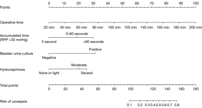
Four variables were selected to establish the nomogram to predict urosepsis. The probability of urosepsis was calculated by summing the scores of all selected factors (Figure 1).
Figure 1.
Nomogram with operative time, accumulated time of RPP ≥30 mmHg, bladder urine culture and hydronephrosis predicts the probability of urosepsis which were selected by multivariate analysis. RPP, renal pelvic pressure.
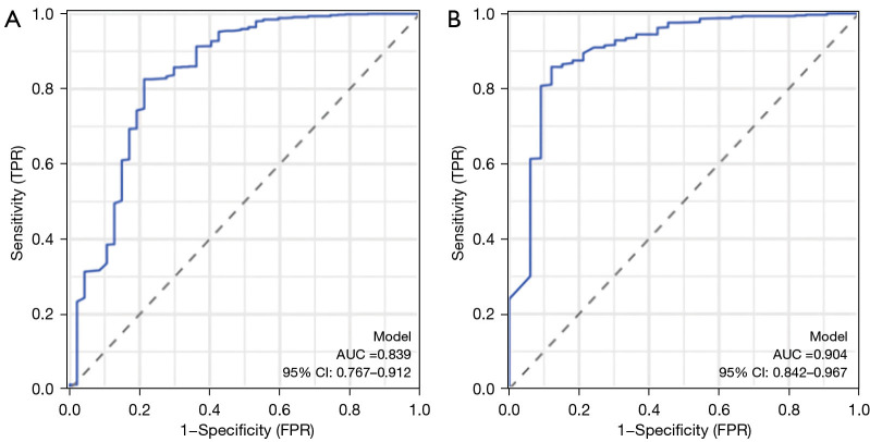
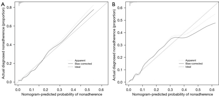
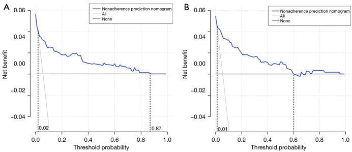
To validate the effectiveness of the nomogram in the prediction of the likelihood of urosepsis following PCNL, we drew the discrimination and calibration curves (Figures 2,3). The calibration results showed that the model was well calibrated and the ROC curves demonstrated outstanding discrimination of the nomogram. The DCA demonstrated a significant area threshold probability (Figure 4), signalling that clinical interventions guided by our nomogram had a higher net benefit when the threshold probability was between 0.02 and 0.87 in the training cohort and between 0.01 and 0.6 in the validation cohort.
Figure 2.
ROC curves for the prognostic nomogram model validation. (A) Internal validation; ROC curves showed AUC values of 0.839 (95% CI: 0.767–0.912). (B) External validation. The nomogram showed good predictive accuracy with an AUC of 0.904 (95% CI: 0.842–0.967). ROC, receiver operating characteristic; AUC, area under ROC curve; CI, confidence interval; TPR, true positive rate; FPR, false positive rate.
Figure 3.
Calibration curves for the prognostic nomogram model validation. The scale of the Y-axis and the X-axis represent the actual value of the probability of urosepsis and the predicted value generated by the nomogram model, respectively. (A) Internal validation. (B) External validation.
Figure 4.
The DCA curves of the nomogram model. The scale on the Y-axis and X-axis represents the net benefit to the patient and the threshold probability, respectively; The blue line represents the performance of the nomogram model. (A) Internal validation. (B) External validation. DCA, decision curve analysis.
Discussion
PCNL is currently the preferred method of treating complex urinary tract stones (1,4), but it can lead to a serious complication—urosepsis, which can be life-threatening if not being detected and treated early (6). Therefore, it is essential to establish a predictive model for urosepsis in order to identify high-risk patients who may develop urosepsis at an early stage for intervention. In this study, we developed a novel nomogram incorporating four independent variables after analyzing 839 consecutive adult patients with urolithiasis who underwent PCNL by multivariate analysis, including operative time, accumulated time of RPP ≥30 mmHg, bladder urine culture and hydronephrosis.
As far as we know from the literatures, few researchers have included RPP in their nomogram, possibly because of the difficulty in monitoring. And this study is the first to show that accumulated time of RPP ≥30 mmHg is an independent predictor of urosepsis after PCNL (0 vs. 0–60 s, P=0.01, OR =3.180; 0–60 vs. ≥60 s, P<0.001, OR =6.389). RPP higher than 30 mmHg is generally considered to have a significant impact on complications after PCNL (16,17) and Wu et al. revealed a significant difference on incidence of postoperative fever between the groups with accumulated time of RPP ≥30 mmHg longer than 60 s or not (P<0.008) (18). So this study used 30 mmHg and 60 s as the thresholds. In order to maintain a clear intraoperative field of view and flush out the stones clearly, it is necessary to continuously perfuse physiological saline into the renal pelvis and this will increase the RPP, which may cause reflux of the renal vein and lymphatic vessels, then leading to fever, infection, and even urosepsis (19). Therefore, the amount of physiological saline flushed into the renal pelvis should be dynamically adjusted based on the intrapelvic pressure for reducing the incidence of urosepsis.
Consistent with other research reports on urosepsis after PCNL surgery (10,20,21), our study found that urine culture was an independent risk factor for postoperative urosepsis (OR =9.547, P<0.001). However, some studies have shown that stone and pelvic urine cultures predict potential urosepsis better than bladder cultures (22,23), because bladder urine culture may not be an accurate reflection of the microbial status of the upper urinary tract, especially in the presence of urinary tract obstruction (24). But urosepsis after PCNL surgery occurred within 48 hours in this study, and stone culture and pelvic urine culture took a relatively longer time, making it difficult to obtain timely results, and thus limiting its clinical value. Urine culture results can be obtained quickly, which is very important for early prevention of urosepsis in patients. Bladder urine culture is still the best predictive indicator that can be obtained in an early stage.
In our study, another independent factor associated with urosepsis after PCNL was the operative time (OR =3.21, P<0.001). In this study, operative time was defined as the time from the beginning of the placement of the retrograde ureteral catheter to the end of the placement of the nephrostomy. The longer the operation, the longer the exposure of the wound tissue and the higher the likelihood of infection with the bacterial community around the stone and in the surrounding environment. At the same time, the longer the operation, the greater the blood loss, reflux, and absorption of perfusion, all of which increase the risk of infection (25,26). There are also studies suggesting that operative time is an independent risk factor for urosepsis after PCNL, which is consistent with our findings (27-30). Therefore, we believe that for PCNL, patients’ stone status should be carefully assessed before surgery and individualised surgical plans should be made to improve the efficiency of intraoperative lithotripsy and reduce operative time. For patients with high risk factors, two-stage lithotripsy may be considered if necessary to improve surgical safety.
In our clinical studies, we discovered that as the degree of hydronephrosis increased, the likelihood of being urosepsis increased. Recently, Bebi et al. have found that grade III–IV hydronephrosis is independently associated with urosepsis in patients with obstructive uropathy after urinary decompression (31). Akman et al. have found that the degree of hydronephrosis significantly affects the operative time during PCNL (32). Furthermore, severe hydronephrosis indicates that the patient is gravely ill and invasive surgery makes it easy for pathogenic bacteria to enter the body and cause infection. Our multivariate analysis suggested that both moderate and several hydronephrosis probably evolved into urosepsis (OR =3.403 and OR =4.704).
Conclusions
We constructed the nomogram based on the relative risk associated with the four factors, including operative time, accumulated time of RPP ≥30 mmHg, bladder urine culture and hydronephrosis. Therefore, controlling operative time, maintaining low RPP during surgery, controlling urinary tract infections and improving hydronephrosis before surgery are key factors in preventing urosepsis. Although the model developed in this study has a good fit and discrimination, further studies are needed to validate the nomogram in larger and more diverse patient populations. In addition, the placement of a pressure tube by ureteroscopy during surgery in order to monitor RPP may influence the results of this experiment.
Supplementary
The article’s supplementary files as
Acknowledgments
Funding: This work was supported by the Guangzhou Science and Technology Plan Project Basic and Applied Basic Research Project (No. 202002030030).
Ethical Statement: The authors are accountable for all aspects of the work in ensuring that questions related to the accuracy or integrity of any part of the work are appropriately investigated and resolved. The procedures used in this study adhere to the tenets of the Declaration of Helsinki (as revised in 2013). Approval was obtained from the Research Ethics Committees of General Hospital of Southern Theater Command (No. NZLLKZ2023067) and Guangdong Second Provincial General Hospital (No. 2023-KY-KZ-203-02). Individual consent for this retrospective analysis was waived.
Footnotes
Reporting Checklist: The authors have completed the TRIPOD reporting checklist. Available at https://tau.amegroups.com/article/view/10.21037/tau-23-616/rc
Data Sharing Statement: Available at https://tau.amegroups.com/article/view/10.21037/tau-23-616/dss
Peer Review File: Available at https://tau.amegroups.com/article/view/10.21037/tau-23-616/prf
Conflicts of Interest: All authors have completed the ICMJE uniform disclosure form (available at https://tau.amegroups.com/article/view/10.21037/tau-23-616/coif). The authors have no conflicts of interest to declare.
References
- 1.Kachkoul R, Touimi GB, El Mouhri G, et al. Urolithiasis: History, epidemiology, aetiologic factors and management. Malays J Pathol 2023;45:333-52. [PubMed] [Google Scholar]
- 2.De Coninck V, Skolarikos A, Juliebø-Jones P, et al. Advancements in stone classification: unveiling the beauty of urolithiasis. World J Urol 2024;42:46. 10.1007/s00345-023-04746-9 [DOI] [PubMed] [Google Scholar]
- 3.Rodríguez D, Sacco DE. Minimally invasive surgical treatment for kidney stone disease. Adv Chronic Kidney Dis 2015;22:266-72. 10.1053/j.ackd.2015.03.005 [DOI] [PubMed] [Google Scholar]
- 4.Zeng G, Zhong W, Mazzon G, et al. International Alliance of Urolithiasis (IAU) Guideline on percutaneous nephrolithotomy. Minerva Urol Nephrol 2022;74:653-68. [DOI] [PubMed] [Google Scholar]
- 5.Koras O, Bozkurt IH, Yonguc T, et al. Risk factors for postoperative infectious complications following percutaneous nephrolithotomy: a prospective clinical study. Urolithiasis 2015;43:55-60. 10.1007/s00240-014-0730-8 [DOI] [PubMed] [Google Scholar]
- 6.Rudd KE, Johnson SC, Agesa KM, et al. Global, regional, and national sepsis incidence and mortality, 1990-2017: analysis for the Global Burden of Disease Study. Lancet 2020;395:200-11. 10.1016/S0140-6736(19)32989-7 [DOI] [PMC free article] [PubMed] [Google Scholar]
- 7.Wollin DA, Preminger GM. Percutaneous nephrolithotomy: complications and how to deal with them. Urolithiasis 2018;46:87-97. 10.1007/s00240-017-1022-x [DOI] [PubMed] [Google Scholar]
- 8.Liu J, Yang Q, Lan J, et al. Risk factors and prediction model of urosepsis in patients with diabetes after percutaneous nephrolithotomy. BMC Urol 2021;21:74. 10.1186/s12894-021-00799-3 [DOI] [PMC free article] [PubMed] [Google Scholar]
- 9.Chan JY, Wong VK, Wong J, et al. Predictors of urosepsis in struvite stone patients after percutaneous nephrolithotomy. Investig Clin Urol 2021;62:201-9. 10.4111/icu.20200319 [DOI] [PMC free article] [PubMed] [Google Scholar]
- 10.Gu J, Liu J, Hong Y, et al. Nomogram for predicting risk factor of urosepsis in patients with diabetes after percutaneous nephrolithotomy. BMC Anesthesiol 2022;22:87. 10.1186/s12871-022-01629-1 [DOI] [PMC free article] [PubMed] [Google Scholar]
- 11.Chen X, Li S, Shi C, et al. Risk factors and predictors of urogenous sepsis after percutaneous nephrolithotomy for idiopathic calcium oxalate nephrolithiasis. Transl Androl Urol 2023;12:1002-15. 10.21037/tau-23-219 [DOI] [PMC free article] [PubMed] [Google Scholar]
- 12.Thomas K, Smith NC, Hegarty N, et al. The Guy’s stone score--grading the complexity of percutaneous nephrolithotomy procedures. Urology 2011;78:277-81. 10.1016/j.urology.2010.12.026 [DOI] [PubMed] [Google Scholar]
- 13.Alsmadi J, Fan J, Zhu W, et al. The Influence of Super-Mini Percutaneous Nephrolithotomy on Renal Pelvic Pressure In Vivo. J Endourol 2018;32:819-23. 10.1089/end.2018.0239 [DOI] [PubMed] [Google Scholar]
- 14.Zhong W, Wen J, Peng L, et al. Enhanced super-mini-PCNL (eSMP): low renal pelvic pressure and high stone removal efficiency in a prospective randomized controlled trial. World J Urol 2021;39:929-34. 10.1007/s00345-020-03263-3 [DOI] [PubMed] [Google Scholar]
- 15.Singer M, Deutschman CS, Seymour CW, et al. The Third International Consensus Definitions for Sepsis and Septic Shock (Sepsis-3). JAMA 2016;315:801-10. 10.1001/jama.2016.0287 [DOI] [PMC free article] [PubMed] [Google Scholar]
- 16.Tokas T, Tzanaki E, Nagele U, et al. Role of Intrarenal Pressure in Modern Day Endourology (Mini-PCNL and Flexible URS): a Systematic Review of Literature. Curr Urol Rep 2021;22:52. 10.1007/s11934-021-01067-5 [DOI] [PubMed] [Google Scholar]
- 17.Zhong W, Zeng G, Wu K, et al. Does a smaller tract in percutaneous nephrolithotomy contribute to high renal pelvic pressure and postoperative fever? J Endourol 2008;22:2147-51. 10.1089/end.2008.0001 [DOI] [PubMed] [Google Scholar]
- 18.Wu C, Hua LX, Zhang JZ, et al. Comparison of renal pelvic pressure and postoperative fever incidence between standard- and mini-tract percutaneous nephrolithotomy. Kaohsiung J Med Sci 2017;33:36-43. 10.1016/j.kjms.2016.10.012 [DOI] [PubMed] [Google Scholar]
- 19.Hong A, du Plessis J, Browne C, et al. Mechanism of urosepsis: relationship between intrarenal pressures and pyelovenous backflow. BJU Int 2023;132:512-9. 10.1111/bju.16095 [DOI] [PubMed] [Google Scholar]
- 20.Xu P, Zhang S, Zhang Y, et al. Preoperative antibiotic therapy exceeding 7 days can minimize infectious complications after percutaneous nephrolithotomy in patients with positive urine culture. World J Urol 2022;40:193-9. 10.1007/s00345-021-03834-y [DOI] [PubMed] [Google Scholar]
- 21.Hao J, Du Z, Bo Z, et al. Impact of Metabolic Syndrome on Post-Operative Infection in Patients Undergoing Flexible Ureteroscopy Lithotripsy. Surg Infect (Larchmt) 2024;25:140-6. 10.1089/sur.2023.205 [DOI] [PubMed] [Google Scholar]
- 22.Mariappan P, Smith G, Bariol SV, et al. Stone and pelvic urine culture and sensitivity are better than bladder urine as predictors of urosepsis following percutaneous nephrolithotomy: a prospective clinical study. J Urol 2005;173:1610-4. 10.1097/01.ju.0000154350.78826.96 [DOI] [PubMed] [Google Scholar]
- 23.Korets R, Graversen JA, Kates M, et al. Post-percutaneous nephrolithotomy systemic inflammatory response: a prospective analysis of preoperative urine, renal pelvic urine and stone cultures. J Urol 2011;186:1899-903. 10.1016/j.juro.2011.06.064 [DOI] [PubMed] [Google Scholar]
- 24.Liu J, Zhou C, Gao W, et al. Does preoperative urine culture still play a role in predicting post-PCNL SIRS? A retrospective cohort study. Urolithiasis 2020;48:251-6. 10.1007/s00240-019-01148-8 [DOI] [PubMed] [Google Scholar]
- 25.Singh P, Yadav S, Singh A, et al. Systemic Inflammatory Response Syndrome Following Percutaneous Nephrolithotomy: Assessment of Risk Factors and Their Impact on Patient Outcomes. Urol Int 2016;96:207-11. 10.1159/000441954 [DOI] [PubMed] [Google Scholar]
- 26.Cheng H, Chen BP, Soleas IM, et al. Prolonged Operative Duration Increases Risk of Surgical Site Infections: A Systematic Review. Surg Infect (Larchmt) 2017;18:722-35. 10.1089/sur.2017.089 [DOI] [PMC free article] [PubMed] [Google Scholar]
- 27.Kreydin EI, Eisner BH. Risk factors for sepsis after percutaneous renal stone surgery. Nat Rev Urol 2013;10:598-605. 10.1038/nrurol.2013.183 [DOI] [PubMed] [Google Scholar]
- 28.Wang Y, Jiang F, Wang Y, et al. Post-percutaneous nephrolithotomy septic shock and severe hemorrhage: a study of risk factors. Urol Int 2012;88:307-10. 10.1159/000336164 [DOI] [PubMed] [Google Scholar]
- 29.Yang M, Li Y, Huang F. A nomogram for predicting postoperative urosepsis following retrograde intrarenal surgery in upper urinary calculi patients with negative preoperative urine culture. Sci Rep 2023;13:2123. 10.1038/s41598-023-29352-y [DOI] [PMC free article] [PubMed] [Google Scholar]
- 30.Zhu Z, Cui Y, Zeng H, et al. The evaluation of early predictive factors for urosepsis in patients with negative preoperative urine culture following mini-percutaneous nephrolithotomy. World J Urol 2020;38:2629-36. 10.1007/s00345-019-03050-9 [DOI] [PubMed] [Google Scholar]
- 31.Bebi C, Fulgheri I, Spinelli MG, et al. Development of a Novel Clinical and Radiologic Risk Score to Predict Septic Complications After Urinary Decompression in Patients with Obstructive Uropathy. J Endourol 2022;36:360-8. 10.1089/end.2021.0148 [DOI] [PubMed] [Google Scholar]
- 32.Akman T, Binbay M, Akcay M, et al. Variables that influence operative time during percutaneous nephrolithotomy: an analysis of 1897 cases. J Endourol 2011;25:1269-73. 10.1089/end.2011.0061 [DOI] [PubMed] [Google Scholar]
Associated Data
This section collects any data citations, data availability statements, or supplementary materials included in this article.
Supplementary Materials
The article’s supplementary files as


