

Abstract
Rituximab is a chimeric monoclonal antibody that recognizes the CD20 antigen. It has been used to treat B‐cell non‐Hodgkin lymphoma (B‐NHL), but recently rituximab resistance has been a cause for concern. We examined histological and immunohistochemical changes in 59 patients with B‐NHL after rituximab therapy. The patients comprised 32 men and 27 women with a median age of 59 years. Pre‐rituximab specimens comprised 34 follicular lymphomas (FL), 11 diffuse large B‐cell lymphomas (DLBCL), 10 mantle cell lymphomas, two marginal zone B‐cell lymphomas (MZBCL), and two chronic lymphocytic leukemias (CLL). CD20 expression in lymphoma cells was evaluated by immunohistochemistry or flow cytometry. Post‐rituximab materials were taken a median of 6 months (4 days to 59 months) after rituximab therapy. Sixteen cases (27%) showed loss of CD20 expression with four histological patterns: pattern 1, no remarkable histological change (FL, 5; DLBCL, 3; and CLL, 2); pattern 2, proliferation of plasmacytoid cells (FL, 2; DLBCL, 1; and MZBCL, 1); pattern 3, transformation to classical Hodgkin's lymphoma (FL, 1); and pattern 4, transformation to anaplastic large cell lymphoma‐like undifferentiated lymphoma (FL, 1). Loss of CD20 was unrelated to the interval of biopsies, treatment regimen, clinical response, and frequency of rituximab administration. Loss of CD20 within 1 month of rituximab therapy (3/14, 21%) and regain of CD20 (2/7, 29%) were not frequent. CD20‐positive relapse with transformation occurred most frequently in cases of early relapse. In conclusion, B‐NHL showed various histological and immunophenotypic changes after rituximab therapy, including not only CD20 loss but also proliferation of plasmacytoid cells or transformation to special subtypes of lymphoma. (Cancer Sci 2009; 100: 54–61)
Rituximab is a chimeric monoclonal antibody that has recently been incorporated into the treatment of B‐cell non‐Hodgkin lymphoma (B‐NHL). It recognizes the CD20 antigen, a pan‐B‐cell marker, binds to it, and induces apoptosis of CD20‐positive cells.( 1 , 2 , 3 , 4 ) Rituximab can be used as a monotherapy or in combination with conventional chemotherapy for treatment of low‐ and high‐grade, untreated, relapsed, or refractory CD20‐positive B‐NHL, achieving a high response rate with a low toxicity.
Recent studies have reported that B‐NHL show CD20‐negative relapse after rituximab therapy.( 5 , 6 , 7 , 8 , 9 , 10 , 11 , 12 , 13 , 14 , 15 , 16 , 17 ) Transformation of follicular lymphoma (FL) to CD20‐negative diffuse large B‐cell lymphoma (DLBCL),( 15 ) proliferation of CD20‐negative plasmacytoid tumor cells of marginal zone B‐cell lymphoma (MZBCL)( 16 ) or lymphoplasmacytic lymphoma,( 13 ) transformation of FL to classical Hodgkin's lymphoma,( 17 ) and progression of nodular lymphocyte‐predominant Hodgkin lymphoma to CD20‐negative T‐cell‐rich B‐cell lymphoma have also been reported.( 18 )
Several mechanisms of resistance to rituximab have been suggested, including selection of a CD20‐negative clone as a consequence of rituximab exposure, masking of CD20 epitopes by rituximab itself, or true loss of CD20 antigen by genetic and epigenetic changes.( 12 , 13 , 15 , 19 , 20 , 21 , 22 , 23 , 24 )
In the present study we carried out retrospective analyses of histological and immunophenotypic changes and outcome in 59 patients with B‐NHL after rituximab‐containing therapy, to explore the effect of rituximab on CD20 expression and morphology in B‐NHL.
Materials and Methods
Patient selection. We reviewed the pathology archives of the National Cancer Center Hospital, Tokyo, Japan, for the period 2002 to 2007. Fifty‐nine consecutive cases of CD20‐positive B‐NHL treated with rituximab, with or without chemotherapy, for which pre‐ and post‐rituximab specimens were available, were included in our study. Rituximab (Zenyaku Kogyo, Tokyo, Japan) was used at a standard dose of 375 mg/m2 once a week for rituximab monotherapy and once every 3 weeks for the rituximab‐cyclophosphamide, doxorubicin, vincristine and prednisone (CHOP) regimen. Clinical information was extracted from the medical records, and the Ann Arbor system was used for staging.
Histological review. Biopsy or surgical specimens were fixed in 10% neutral‐buffered formalin overnight, embedded in paraffin, cut into sections 4 µm thick, and stained with hematoxylin–eosin for histological evaluation. All of the pre‐rituximab specimens were CD20‐positive B‐NHL by definition, and post‐rituximab specimens included any lymphomas. All of the specimens were reviewed by three pathologists (A.M.M., H.T., and Y.M.) to confirm that the morphological characteristics fulfilled the criteria of the World Health Organization classification of lymphoid neoplasms, 2001.( 25 ) Histological subtype, loss of CD20 expression by immunohistochemistry or flow cytometry, presence or absence of plasmacytoid differentiation, and the relationship between histological transformation and CD20 loss were examined.
Immunohistochemistry, flow cytometry, in situ hybridization, and interphase fluorescence in situ hybridization analyses. We carried out immunohistochemical staining on formalin‐fixed paraffin‐embedded pre‐ and post‐rituximab specimens using a panel of monoclonal and polyclonal antibodies. Sections 4 µm thick were cut from each paraffin block, deparaffinized, and incubated at 121°C in pH 6.0 citrate buffer for 10 min for antigen retrieval. Antibodies included those against the following antigens: CD3 (clone PS1, ×25; Novocastra, Newcastle, UK; polymer method), CD20 (L26, ×100; Dako, Glostrup, Denmark; labeled streptavidin–biotin method [LSAB]), and CD79a (JCB117, ×100; Dako; LSAB) routinely; and CD5 (4C7, ×50, Novocastra; polymer), CD7 (CD7‐272, ×100; Novocastra; avidin–biotin complex method [ABC]), CD10 (56C6, ×50; Novocastra; polymer), CD15 (MMA, ×100; Becton Dickinson, Franklin Lakes, NJ, USA; polymer), CD30 (Ber‐H2, ×100; Dako; polymer), CD45 (2B11 + PD7/26, ×100; Dako; LSAB), CD45RO (UCHL1, ×50; Dako; LSAB), CD56 (1B6, ×100; Novocastra; LSAB), ALK (ALK1, ×200; Dako; polymer), Bcl‐2 (124, ×100; Dako; LSAB), Bcl‐6 (poly, ×50; Dako; ABC), cyclin D1 (SP4, ×25; Nichirei, Tokyo, Japan; polymer), TIA‐1 (26gA10F5, ×1000; Immunotech, Marseille, France; polymer), granzyme B (GrB‐7, ×200; Dako; polymer), MPO (poly, ×1000; Dako; LSAB), MUM1 (MUM1p, ×50; Dako; ABC), PAX5 (24, ×200; Becton Dickinson; ABC), TdT (poly, ×100; Dako; polymer), Igκ (poly, ×20 000; Dako; LSAB), Igλ (poly, ×40 000; Dako; LSAB), IgA (poly, ×100 000; Dako; polymer), IgG (poly, ×20 000; Dako; polymer), and IgM (poly, ×20 000; Dako; polymer) optionally. The percentages of CD20‐positive tumor cells were counted semiquantitatively by immunohistochemistry (IHC). Immunoreactivity for CD20 was judged positive (no CD20 loss) if >95% of the tumor cells were stained, partially negative (partial CD20 loss) if 10–95% of the cells were stained, or negative (CD20 loss) if <10% of the cells were stained. When a post‐rituximab specimen showed loss of CD20 expression, it was judged as B‐cell lineage if there was positivity for CD79a.
Flow cytometry was carried out using an Epics XL‐MCL instrument with System II Software (Beckman Coulter). The flow cytometry panel included CD20 (B‐Ly1), CD19 (HD37), Igκ (poly), and Igλ (poly) (Dako). Fluorescence in situ hybridization (FISH) and in situ hybridization (ISH) analyses were optional. Sections 4 µm thick were cut from each paraffin block and used for FISH analysis. Judgment of the fusion gene was carried out as described previously.( 26 ) Dual‐color LSI IGH Spectrum Green/LSI BCL2 Spectrum Orange Dual Fusion Translocation Probes (Vysis, Downers Grove, IL, USA) were used to detect t(14;18): IGHBCL2 fusion. ISH with Epstein–Barr‐encoded RNA (EBER‐1) probes (Dako) was carried out in some cases to detect possible Epstein–Barr virus infection.
Statistical analysis. The relationships between CD20 expression and treatment regimen (rituximab monotherapy vs combination therapy with rituximab and chemotherapy), response (complete response [CR] vs others, or overall response [OR] vs others), frequency of rituximab administration, and interval between the last dose of rituximab and rebiopsy were examined by χ2‐test or Mann‐Whitney U‐test. Differences were considered significant when the P‐value was less than 0.05.
Results
Patient characteristics. Clinical information for all consecutive 59 patients is summarized in Table 1. The patients comprised 32 men and 27 women, ranging in age from 37 to 80 years with a median age of 59 years. Eight patients had stage I/II disease and 51 patients had stage III/IV disease. All of the patients received rituximab by definition, with or without chemotherapy (CHOP or other types of regimen). The 59 patients received a median of six courses (range 1–17) of rituximab. The median interval between the last dose of rituximab and rebiopsy was 6 months (range 4 days to 59 months). The overall response rate was 79% and the % CR was 46% to rituximab‐containing regimens.
Table 1.
Patient characteristics
| Case | Age (years)/sex | Stage | Pre‐rituximab diagnosis (sample) | Therapy and response between biopsy and operation | Post‐rituximab diagnosis (sample) | Post‐rituximab interval (months) † | CD20 expression (%) | Histology by immunohistochemistry | Pattern |
|---|---|---|---|---|---|---|---|---|---|
| FL1 | 55/M | 4 | FL, gr.2 (LN) | Rx4, NC | FL, gr.1 (BM) | 3 | 0 | NHC | 1 |
| FL2 | 52/M | 4 | FL, gr.2 (LN) | C‐MOPPx8, PR, Rx4, NC | FL, gr.1 (BM) | 22 | 0 | NHC | 1 |
| FL3‐1 | 52/M | 3 | FL, gr.1 (LN) | R‐CHOPx6, CR | FL, gr.2 (BM) | 33 | 100 | NHC | |
| FL3‐2 | Rx8+C‐MOPPx5, CR, relapse, R‐ICEx2, PR | FL, gr.2 (LN) | 6 | 0 | NHC | 1 | |||
| FL4‐1 | 42/F | 4 | FL, gr.1 (LN) | Rx8+C‐MOPPx16, NC | FL, gr.1 (BM) | 23 | 100 | NHC | |
| FL4‐2 | Rx8, NC | FL, gr.1 (BM) | 5 days | 0 | NHC | 1 | |||
| FL5 | 59/M | 4 | FL, gr.2 (LN) | Rx4, NC, C‐MOPPx13, PD, R‐C‐MOPPx8, CR | FL, gr.2 (BM) | 21 | –‡ | NHC | 1 |
| FL6 | 40/M | 4 | FL, gr.1 (LN) | R‐CHOPx6, CR | FL, gr.2 (LN) | 12 | 90 | Partially plasmacytoid cells (IgM+/Igκ+/CD138–) | 2 |
| FL7 | 49/F | 4 | FL, gr.1 (duodeunum) | R‐CHOPx6, CR | FL, gr.1 (LN) | 17 | 90 | Partially plasmacytoid cells (IgM+/Igκ+/CD138–) | 2 |
| FL8 | 61/F | 4 | DLBCL+FL, gr.3a (LN) | Rx4+CHOPx8, CR | HL, MC (LN) | 11 | 0 | Transformation to HL | 3 |
| FL9 | 68/M | 4 | FL, gr.2 (LN) | R‐C‐MOPPx6, R‐C‐MOPPx8, relapse, fludarabine + Rx3 | ALCL‐like (liver) | 18 days | 0 | Transformation to ALCL‐like | 4 |
| L10 | 76/F | 4 | FL, gr.2 (LN) | Rx8, NC | FL, gr.2 (BM) | 6 | 100 | NHC | |
| FL11 | 54/F | 4 | FL, gr.2 (LN) | R‐CHOPx6, CR | FL, gr.1 (BM) | 15 days | 100 | NHC | |
| FL12 | 40/F | 4 | FL, gr.1 (LN) | CHOPx6, CR, relapse, C‐MOPPx4, NC, Rx4, CR | DLBCL (colon) | 4 | 100 | Transformation to DLBCL | |
| FL13 | 44/M | 4 | FL, gr.2 (LN) | R‐CHOPx6, PR, C‐MOPPx6 radiation, PR | DLBCL+FL, gr.1 (BM) | 59 | 100 | Transformation to DLBCL | |
| FL14 | 47/M | 4 | FL, gr.2 (LN) | COPPx6, NC, Rx4, PD | FL, gr.1 (BM) | 1 | 100 | NHC | |
| FL15 | 43/M | 4 | FL, gr.1 (LN) | Rx4+C‐MOPPx2, CR, zevalin, CR | FL, gr.1 (orbit) | 26 | 100 | NHC | |
| FL16 | 61/F | 4 | FL, gr.1 (BM), DLBCL (skin) | R‐CHOPx8, CR | FL, gr.2 (LN) | 9 | 100 | NHC | |
| FL17 | 48/F | 4 | FL, gr.2 (LN) | Rx4+CHOPx6, PR | FL, gr.1 (BM) | 23 | 100 | NHC | |
| FL18 | 60/F | 3 | FL, gr.2 (LN) | Rx4, CR | DLBCL (tonsil) | 4 | 100 | Transformation to DLBCL | |
| FL19 | 54/F | 4 | FL, gr.1 (BM, stomach) | R‐CHOPx6, PR | FL, gr.1 (BM) | 37 | 100 | NHC | |
| FL20 | 54/F | 4 | FL, gr.1 (LN) | Rx4, PR, CHOPx8, PR | FL, gr.2 (cecum) | 36 | 100 | NHC | |
| FL21 | 61/F | 4 | FL, gr.2 (LN) | R‐CHOPx8, PR | FL, gr.1 (BM) | 5 | 100 | NHC | |
| FL22 | 66/F | 3 | FL, gr.2 (LN) | R‐CHOPx8, PR | FL, gr.2 (skin) | 26 | 100 | NHC | |
| FL23 | 53/F | 4 | FL, gr.2 (ileum, colon) | R‐CHOPx8, NC | FL, gr.1 (duodenum) | 10 | 100 | NHC | |
| FL24 | 51/F | 4 | FL, gr.2 (duodenum) | Rx1 (within R‐CHOP) | FL, gr.1 (duodenum) | 4 day | 100 | NHC | |
| FL25 | 55/M | 3 | DLBCL+FL, gr.3b (LN) | R‐CHOPx8, CR | DLBCL (LN) | 3 | 100 | NHC | |
| FL26 | 37/M | 4 | FL, gr.2 (LN) | Rx2 (within R‐CHOP) | FL, gr.2 (duodenum) | 8 days | 100 | NHC | |
| FL27 | 62/M | 4 | FL, gr.1 (ileum) | Rx3 (within R‐CHOP) | FL, gr.1 (duodenum) | 8 days | 100 | NHC | |
| FL28 | 57/F | 4 | FL, gr.3a (LN) | Rx4, NC | DLBCL (LN) | 3 | 100 | Transformation to DLBCL | |
| FL29 | 51/M | 3 | DLBCL+FL, gr.3b (LN) | R‐CHOPx6+Rx8, CR | DLBCL (LN) | 21 days | 100 | NHC | |
| FL30 | 60/M | 1 | FL, gr.1 (duodenum) | Rx8, CR | FL, gr.1 (duodenum) | 4 days | 100 | NHC | |
| FL31 | 63/M | 1 | FL, gr.1 (duodenum) | Rx8, PR | FL, gr.1 (duodenum) | 1 | 100 | NHC | |
| FL32 | 75/F | 4 | FL, gr.1 (BM) | Rx3 | FL, gr.1 (BM) | 4 days | 100 | NHC | |
| FL33 | 77/M | 4 | DLBCL+FL, gr.3a (pharynx) | R‐CHOPx8, CR | DLBCL (LN) | 8 | 100 | NHC | |
| FL34 | 65/M | 3 | FL, gr.3a (LN) | Rx8, CR | DLBCL (LN) | 5 | 100 | Transformation to DLBCL | |
| DLBCL1‐1 | 47/M | 2 | DLBCL (right testis) | R‐CHOPx8, CR | DLBCL (left testis) | 2 | 100 | NHC | |
| DLBCL1‐2 | R‐IVACx3, CR, BMT, CR | DLBCL (BM) | 4 | 0 | NHC | 1 | |||
| DLBCL2 | 71/M | 3 | DLBCL (ileum) | R‐CHOPx8, CR, Rx1 | DLBCL (LN) | 10 days | 10 | NHC | 1 |
| DLBCL3 | 55/M | 4 | DLBCL (tonsil) | R‐CHOPx8, PR | DLBCL (LN) | 3 | 70 | NHC | 1 |
| DLBCL4 | 58/F | 2 | DLBCL (EBER‐1+) (stomach) | R‐CHOPx8, PR | DLBCL (stomach) | 4 | 0 | Plasmacytoid cells (IgM+/Igλ+/CD138–) | 2 |
| DLBCL5 | 80/M | 2 | DLBCL (LN) | R‐CHOPx8, CR | DLBCL (subcutaneous) | 6 | 100 | NHC | |
| DLBCL6 | 60/F | 4 | DLBCL (breast) | Chemotherapy, Rx4, CR | DLBCL (breast) | 29 days | 100 | NHC | |
| DLBCL7 | 61/F | 2 | DLBCL (tonsil) | R‐EPOCHx4, CR | DLBCL (colon, rectum) | 16 | 100 | NHC | |
| DLBCL8 | 75/F | 4 | DLBCL (stomach,duodenum) | R‐CHOPx8, CR | DLBCL (stomach, duodenum) | 30 | 100 | NHC | |
| DLBCL9 | 65/F | 3 | DLBCL (LN) | Rx8+CHOPx6, PD | DLBCL (LN) | 3 | 100 | NHC | |
| DLBCL10 | 64/M | 2 | DLBCL (stomach) | Rx1+CHOPx2 (within R‐CHOP) | DLBCL (stomach) | 20 days | 100 | NHC | |
| DLBCL11 | 63/M | 2 | DLBCL (LN) | Rx8+CHOPx5+radiation, CR | DLBCL (LN) | 8 | 100 | NHC | |
| MCL1 | 72/M | 4 | MCL (LN) | R‐CHOPx4, PR, Rx4, NC, COPx6, PD, R‐CNOPx6, PD, cladribine, PD | MCL (stomach) | 10 days | 100 | NHC | |
| MCL2 | 76/M | 4 | MCL (LN) | R‐CHOPx8, PR | MCL (BM) | 20 | 100 | NHC | |
| MCL3 | 68/M | 4 | MCL (BM) | Rx1+CHOPx6, PR | MCL (BM) | 1 | 100 | NHC | |
| MCL4 | 66/M | 4 | MCL (colon) | R‐CHOPx8, CR | MCL (stomach) | 30 | 100 | NHC | |
| MCL5 | 55/M | 4 | MCL (tonsil) | Rx4, PR | MCL (tongue) | 28 | 100 | NHC | |
| MCL6 | 59/F | 4 | MCL (stomach) | Rx4, PR, C‐MOPPx8+ radiation+COP, PD | MCL (bone) | 21 | 100 | NHC | |
| MCL7‐1 | 63/F | 4 | MCL (LN) | Rx4+C‐MOPPx8, CR | MCL (stomach, duodenum) | 17 | 100 | NHC | |
| MCL7‐2 | Rx4, PR | MCL (stomach) | 1 | 100 | NHC | ||||
| MCL8 | 54/F | 4 | MCL (ileum) | Rx8+CHOPx6, CR | MCL (small intestine) | 3 | 100 | NHC | |
| MCL9 | 78/M | 4 | MCL (stomach) | Rx8, PD | MCL (orbit) | 2 | 100 | Change to blastoid variant | |
| MCL10 | 70/M | 4 | MCL (LN) | R‐CHOPx8, PR | MCL (stomach) | 18 | 100 | NHC | |
| MZBCL1 | 68/F | 3 | MZBCL (LN) | R‐ICEx1, CR | MZBCL (tonsil) | 15 | 0 | Plasmacytoid cells | 2 |
| MZBCL2 | 55/M | 4 | MZBCL (BM) | R‐CHOPx6, PR | MZBCL (BM) | 4 | 100 | NHC | |
| CLL1 | 54/M | 4 | CLL (BM) | Rx2+C‐MOPPx6, PR | CLL (LN) | 12 days | 0 | NHC | 1 |
| CLL2 | 52/F | 4 | CLL (BM) | CHOPx8, COPx10, R‐CEPPx7, R‐ESHAPx2, CHASEx2, R‐ESHAP+PBSCT, Rx5, PR | CLL (BM) | 11 | 0 | NHC | 1 |
From last dose of rituximab;
‡by flow cytometry. ALCL, anaplastic large cell lymphoma; BM, bone marrow; BMT, bone marrow transplantation; CEPP, cyclophosphamide, etoposide, procarbazine, and prednisolone; CHASE, cyclophosphamide, cytosine arabinoside, etoposide, and dexamethasone; CHOP, cyclophosphamide, doxorubicin, vincristine, and prednisone; CLL, chronic lymphocytic leukemia; C‐MOPP, cyclophosphamide, vincristine, prednisone, and procarbazine; CR, complete response; diff., differentiation; DLBCL, diffuse large B‐cell lymphoma; EPOCH, etoposide, prednisone, vincristine, cyclophosphamide, and doxorubicin; ESHAP, etoposide, methylprednisolone, high‐dose cytarabine, and cisplatin; FL, follicular lymphoma; HL, Hodgkin's lymphoma; ICE, ifosfamide, carboplatin, and etoposide; IVAC, ifosfamide, vincristine, and cytarabine; LN, lymph node; MC, mixed cellularity; MCL, mantle cell lymphoma; MZBCL, marginal zone B‐cell lymphoma; NC, no change; NHC, no remarkable histological change; PBSCT, peripheral blood stem cell transplantation; PD, progressive disease; PR, partial response; R, rituximab.
Four histological patterns of CD20 loss. The results of histological analysis and immunohistochemical staining for each antibody are summarized in 1, 2. The total of 59 pre‐rituximab B‐NHL specimens included 34 FL with or without a DLBCL component, 11 DLBCL, 10 mantle cell lymphomas (MCL), two MZBCL, and two chronic lymphocytic leukemias (CLL). We considered that the following two factors may have contributed to case selection bias. The first factor is that the date of the approval of rituximab for low‐grade B‐cell lymphoma preceded that for DLBCL for 2 years in Japan. The second factor is that FL were rebiopsied more frequently than DLBCL because FL relapsed frequently and were followed up for a long time, and checks for transformation to DLBCL were sometimes necessary.
Table 2.
Four histological patterns of loss of CD20 expression after rituximab therapy
| Histological pattern | Distribution |
|---|---|
| Pattern 1: loss of CD20 with no remarkable histological change | FL, 5; DLBCL, 3; CLL, 2 |
| Pattern 2: proliferation of plasmacytoid cells | FL, 2; DLBCL, 1; MZBCL, 1 |
| Pattern 3: transformation to classical Hodgkin lymphoma | FL, 1 |
| Pattern 4: transformation to anaplastic large cell lymphoma‐like undifferentiated lymphoma | FL, 1 |
CLL, chronic lymphocytic leukemia; DLBCL, diffuse large B‐cell lymphoma; FL, follicular lymphoma; MZBCL, marginal zone B‐cell lymphoma.
Sixteen cases (27%) showed loss of CD20 expression in post‐rituximab specimens by IHC or flow cytometry. The frequencies of CD20 loss in the various histological subtypes were: FL, 26% (9/34); DLBCL, 36% (4/11); MCL, 0% (0/10); MZBCL, 50% (1/2); and CLL, 100% (2/2). Among them, two DLBCL and two FL showed partial loss of CD20 expression. Among the 12 tumors with complete loss of CD20 expression, seven had available flow cytometry data, and all of them showed loss of CD20.
Four patterns of loss of CD20 expression were evident (Table 2): pattern 1, CD20 loss with no remarkable histological change (FL, 5; DLBCL, 3; and CLL, 2) (Fig. 1); pattern 2, proliferation of plasmacytoid cells (FL, 2; DLBCL, 1; and MZBCL, 1) (Fig. 2); pattern 3, transformation to classical Hodgkin lymphoma (FL, 1); and pattern 4, transformation to anaplastic large cell lymphoma (ALCL)‐like undifferentiated lymphoma (FL, 1) (Fig. 3). All of the lymphomas after rituximab treatment with pattern 1 or 2 histology were positive for CD79a. Two FL with pattern 2 showed proliferation of plasmacytoid cells, not in the marginal zone but in follicles, as with FL with plasma cells,( 27 , 28 ) and the plasmacytoid tumor cells were positive for IgM and Igκ by IHC. One DLBCL with pattern 2 was negative for IgM and Igλ by IHC in the pre‐rituximab specimen, but positive for them in the post‐rituximab specimen. Hodgkin lymphoma with pattern 3, which was previously reported to be a form of transformed FL,( 29 ) was positive for CD30, CD15, and the IGH–BCL2 fusion by FISH, and negative for CD10, CD20, and EBER‐1 by ISH. Although we could not determine the lineage of ALCL‐like undifferentiated lymphoma with pattern 4, because it was positive for only CD45 and CD45RO and negative for CD3, CD5, CD7, CD10, CD15, CD20, CD30, CD56, CD79a, ALK, bcl‐2, bcl‐6, granzyme B, MPO, MUM1, PAX5, TdT, TIA‐1, and EBER‐1 by ISH, it was considered to be transformed FL because of the presence of the IGHBCL2 fusion revealed by FISH in both the pre‐ and post‐rituximab specimens (Fig. 3).
Figure 1.
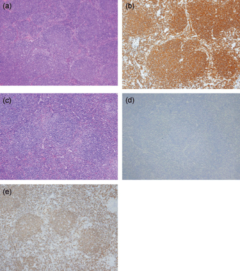
(a–e) A case of pattern 1 change in CD20‐positive follicular lymphoma (FL) to CD20‐negative FL. FL, grade 1, (a) in a lymph node (hematoxylin–eosin, ×40) and (b) with CD20‐positive phenotype, pre‐rituximab (×100). FL, grade 2, (c) in a lymph node (hematoxylin–eosin, ×40) with (d) CD20‐negative (×40) and (e) CD79a‐positive (×40) phenotypes, post‐rituximab.
Figure 2.
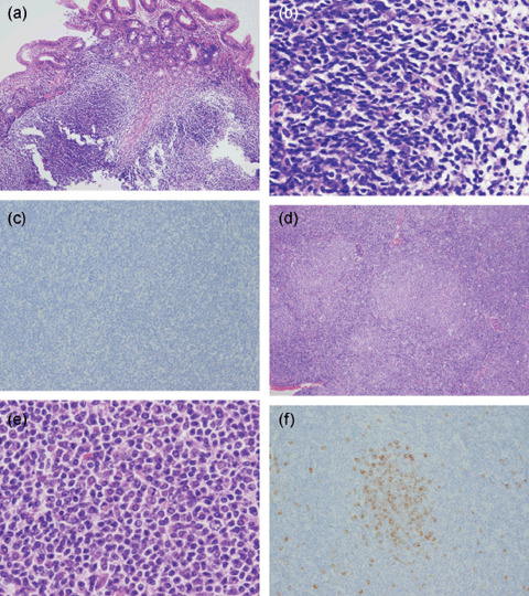
(a–f) A case of pattern 2 change of follicular lymphoma (FL) to FL with plasma cells. FL, grade 1 in duodenum (hematoxylin–eosin), (a) ×40, (b) ×400, with (c) IgM‐negative phenotype (×100), pre‐rituximab. FL, grade 1 with plasmacytoid differentiation in lymph node (hematoxylin–eosin), (d) ×40, (e) ×400, with (f) IgM‐positive phenotype (×100), post‐rituximab.
Figure 3.
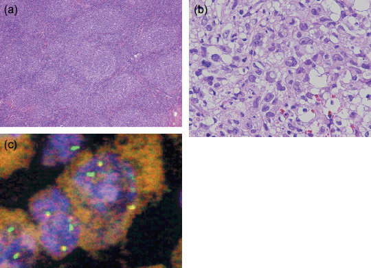
(a–c) A case of pattern 4 change of follicular lymphoma (FL) to anaplastic large‐cell lymphoma (ALCL)‐like undifferentiated lymphoma. (a) FL, grade 2 in lymph node, pre‐rituximab (hematoxylin–eosin, ×40). (b) ALCL‐like undifferentiated lymphoma in liver, post‐rituximab (hematoxylin–eosin, ×400). (c) The result of fluorescence in situ hybridization of ALCL‐like undifferentiated lymphoma. IGH and BCL2 fusion pattern. Two fusion IGHBCL2 signals were present.
Relationship between rituximab therapy and CD20 expression, and histological changes. The relationships between CD20 expression and interval after the last dose of rituximab, treatment regimens, clinical response, and frequency of rituximab administration were not detected. Among 16 cases showing loss of CD20, the clinical response to treatment was no change (NC) in three cases, and CR or partial response (PR) in the others.
Fourteen patients underwent rebiopsy within 1 month of the last dose of rituximab. Among them, only three cases (21%) were negative for CD20. Seven cases showing loss of CD20 expression after rituximab therapy were subsequently observed and rebiopsied and, among them, two cases (cases FL4‐2 and FL8) regained CD20 expression at 7 and 15 months after the last dose of rituximab. The other five cases were found not to have regained CD20 expression at 2, 7, 12, 28, and 44 months after the last dose of rituximab.
Nine patients with FL achieved CR or PR after treatment with a rituximab‐containing regimen, but their lymphomas showed early relapse (within 3 months to 1 year later). Among them, five cases (cases FL12, FL18, FL25, FL33, and FL34) relapsed as CD20‐positive DLBCL, two (cases FL16 and FL21) as CD20‐positive low‐grade FL, one (case FL3‐2) as CD20‐negative FL grade 2, and one (case FL8) as transformation to Hodgkin's lymphoma.
Discussion
Recent reports have indicated that the treatment of B‐NHL with rituximab may be associated with CD20‐negative lymphoma relapse,( 5 , 6 , 7 , 8 , 9 , 10 , 11 , 12 , 13 , 14 , 15 , 16 , 17 ) and the frequency of loss of CD20 after rituximab treatment varies widely (24, 56, 60, and 94%).( 9 , 11 , 13 , 14 ) In the present study, 16 of 59 B‐NHL (27%) showed loss of CD20 after rituximab‐containing therapy using a larger series than in previous reports. Our results also suggested that the frequency of CD20 loss was not largely affected by the period before rebiopsy. Although the site of sampling (bone marrow vs non‐bone marrow) might affect the observed degree of CD20 loss,( 9 ) this issue needs to be studied further using a larger number of cases. Tumors with loss of CD20 in the present study included FL, DLBCL, MZBCL, and CLL. Although Goteri et al. reported that MCL frequently showed loss of CD20 expression in bone marrow, none of our MCL cases showed CD20 loss.( 13 ) Because a previous report indicated that rituximab + CHOP combination therapy (R‐CHOP) had insufficient efficacy for MCL,( 30 ) it was considered that this regimen might not have been sufficiently potent to induce selection of a CD20‐negative clone.
Four histological patterns of CD20 loss were evident. The majority were patterns 1 or 2, whereas patterns 3 or 4 were rare. Recently, several reports have described relapse with pattern 1 or 2 histology after rituximab therapy.( 13 , 16 ) Using flow cytometry, Goteri et al. demonstrated that 26 cases of low‐grade B‐cell lymphoma showed CD20 loss in bone marrow aspirates, including cases with no histological change, and with residual plasmacytoid tumor cells of lymphoplasmacytic lymphoma.( 13 ) It has also been reported that mucosa‐associated lymphoid tissue lymphoma changes to a pure plasma‐cell neoplasm.( 16 )
Case FL8, which showed transformation to Hodgkin lymphoma (pattern 3), was one of the transformed FL that we reported previously.( 29 ) As composite Hodgkin lymphoma and FL is reported to be very rare,( 31 , 32 ) rituximab might have induced transformation to Hodgkin lymphoma. Recently, a case of Hodgkin lymphoma subsequent to FL in a patient receiving maintenance rituximab was reported.( 17 )
Transformation from FL to ALCL‐like undifferentiated lymphoma (pattern 4) has been reported previously as neither transformation of FL nor histological change after rituximab therapy. Although two cases of FL with relapse to peripheral T‐cell lymphoma after rituximab have been reported,( 33 , 34 ) it was suspected that the T‐cell lymphomas were another clone, thus differing from the present case. Cohen et al. reported large‐cell transformation of CLL and FL during or soon after treatment with a fludarabine‐ and rituximab‐containing regimen,( 35 ) thus resembling the present case treated with fludarabine and rituximab.
Several mechanisms of resistance to rituximab have been suggested, including selection of a CD20‐negative clone as a consequence of rituximab exposure, masking of CD20 epitopes by rituximab itself, or true loss of CD20 antigen due to genetic and epigenetic changes.( 12 , 13 , 15 , 19 , 20 , 21 , 22 , 23 , 24 ) Although the present study was not intended to address the mechanism of CD20 loss, several remarkable phenomena were evident. No relationships were detected between loss of CD20 and the interval between the last dose of rituximab and rebiopsy, frequency of rituximab administration, clinical responses, and treatment regimens. It is suspected that susceptibility to rituximab differs greatly among lymphomas. Our results also indicated that loss of CD20 immediately after rituximab therapy was not frequent. Although some cases do regain CD20 expression, loss of CD20 persisting for more than 6 months is not infrequent.
A previous case report has emphasized that early relapse of FL after rituximab therapy was related to CD20‐negative transformation to DLBCL.( 15 ) However, this was not confirmed in our study using a larger series: most of the relapses were CD20‐positive DLBCL, and only two were CD20‐negative FL or Hodgkin lymphoma. Our results suggested that CD20‐positive relapse with histological transformation occurred most frequently in cases of early relapse, and that CD20‐negative relapse was relatively rare.
In conclusion, our findings indicate that 27% of B‐NHL show loss of CD20 expression with four histological patterns after rituximab therapy. As the changes in morphology and CD20 expression after rituximab therapy vary widely, including not only loss of CD20 expression but also proliferation of plasmacytoid cells or transformation to special subtypes of lymphoma, and clinical outcomes are very confused, careful follow up and rebiopsy are recommended.
Acknowledgments
The authors are grateful to Ms S. Miura and Ms C. Kina for their excellent technical assistance.
References
- 1. Reff ME, Carner K, Chambers KS et al . Depletion of B cells in vivo by a chimeric mouse human monoclonal antibody to CD20. Blood 1994; 83: 435–45. [PubMed] [Google Scholar]
- 2. Demidem A, Lam T, Alas S et al . Chimeric anti‐CD20 (IDEC‐C2B8) monoclonal antibody sensitizes a B cell lymphoma cell line to cell killing by cytotoxic drugs. Cancer Biother Radiopharm 1997; 12: 177–86. [DOI] [PubMed] [Google Scholar]
- 3. Shan D, Ledbetter JA, Press OW. Apoptosis of malignant human B cells by ligation of CD20 with monoclonal antibodies. Blood 1998; 91: 1644–52. [PubMed] [Google Scholar]
- 4. Cardarelli PM, Quinn M, Buckman D et al . Binding to CD20 by anti‐B1 antibody or F(ab′)(2) is sufficient for induction of apoptosis in B‐cell lines. Cancer Immunol Immunother 2002; 51: 15–24. [DOI] [PMC free article] [PubMed] [Google Scholar]
- 5. Meeker T, Lewder J, Cteary ML et al . Emergence of idiotype variants during treatment of B‐cell lymphoma with anti‐idiotype antibodies. N Eng J Med 1985; 312: 1658–65. [DOI] [PubMed] [Google Scholar]
- 6. Kinoshita T, Nagai H, Murate T et al . CD20‐negative relapse in B‐cell lymphoma after treatment with rituximab. J Clin Oncol 1998; 16: 3916. [PubMed] [Google Scholar]
- 7. Davis TA, Czerwinski DK, Levy R. Therapy of B‐cell lymphoma with anti‐CD20 antibodies can result in the loss of CD20 antigen expression. Clin Cancer Res 1999; 5: 611–15. [PubMed] [Google Scholar]
- 8. Schmitz K, Brugger W, Weiss B et al . Clonal selection of CD20‐negative non‐Hodgkin's lymphoma cells after treatment with anti‐CD20 antibody rituximab. Br J Haematol 1999; 106: 571–2. [DOI] [PubMed] [Google Scholar]
- 9. Foran JM, Norton AJ, Micallef IN et al . Loss of CD20 expression following treatment with rituximab (chimaeric monoclonal anti‐CD20): a retrospective cohort analysis. Br J Haematol 2001; 114: 881–3. [DOI] [PubMed] [Google Scholar]
- 10. Chu PG, Chen YY, Molina A et al . Recurrent B‐cell neoplasms after Rituximab therapy: an immunophenotypic and genotypic study. Leuk Lymphoma 2002; 43: 2335–41. [DOI] [PubMed] [Google Scholar]
- 11. Kennedy GA, Tey SK, Cobcroft R et al . Incidence and nature of CD20‐negative relapses following rituximab therapy in aggressive B‐cell non‐Hodgkin's lymphoma: a retrospective review. Br J Haematol 2002; 119: 412–16. [DOI] [PubMed] [Google Scholar]
- 12. Jilani I, O’Brien S, Manshuri T et al . Transient down‐modulation of CD20 by rituximab in patients with chronic lymphocytic leukemia. Blood 2003; 102: 3514–20. [DOI] [PubMed] [Google Scholar]
- 13. Goteri G, Olivieri A, Ranaldi M et al . Bone marrow histopathological and molecular changes of small B‐cell lymphomas after rituximab therapy: comparison with clinical response and patients’ outcome. Int J Immunopathol Pharmacol 2006; 19: 421–31. [DOI] [PubMed] [Google Scholar]
- 14. Seliem RM, Freeman JK, Steingart RH et al . Immunophenotypic changes and clinical outcome in B‐cell lymphomas treated with rituximab. Appl Immunohistochem Mol Morphol 2006; 14: 18–23. [DOI] [PubMed] [Google Scholar]
- 15. Alovaro‐Naranjo TA, Jaen‐Martinez JJ, Guma‐Padro JG et al . CD20‐negative DLBCL transformation after rituximab treatment in follicular lymphoma: a new case report and review of the literature. Ann Hematol 2003; 82: 585–8. [DOI] [PubMed] [Google Scholar]
- 16. Woehrer S, Streubel B, Chott A et al . Transformation of MALT lymphoma to pure plasma cell histology following treatment with the anti‐CD20 antibody rituximab. Leuk Lymphoma 2005; 46: 1645–9. [DOI] [PubMed] [Google Scholar]
- 17. Scaramucci L, Perrotti A, Niscola P et al . Hodgkin disease subsequent to follicular lymphoma on maintenance rituximab. Leuk Lymphoma 2007; 48: 1878–80. [DOI] [PubMed] [Google Scholar]
- 18. Pijuan L, Vicioso L, Bellosillo B et al . CD20‐negative T‐cell‐rich B‐cell lymphoma as a progression of a nodular lymphocyte‐predominant Hodgkin's lymphoma treated with rituximab: a molecular analysis using laser capture microdissection. Am J Surg Pathol 2005; 29: 1399–403. [DOI] [PubMed] [Google Scholar]
- 19. Cragg MS, Bayne MC, Illidge TM et al . Apparent modulation of CD20 by rituximab: an alternative explanation. Blood 2004; 103: 3989–90. [DOI] [PubMed] [Google Scholar]
- 20. Rawal YB, Nuovo GJ, Frambach GE et al . The absence of CD20 messenger RNA in recurrent cutaneous B‐cell lymphoma following rituximab therapy. J Cutan Pathol 2005; 32: 616–21. [DOI] [PubMed] [Google Scholar]
- 21. Takei K, Yamazaki T, Sawada U et al . Analysis of changes in CD20, CD55, and CD59 expression on established rituximab‐resistant B‐lymphoma cell lines. Leuk Res 2006; 30: 625–31. [DOI] [PubMed] [Google Scholar]
- 22. Tomita A, Hiraga J, Kiyoi H et al . Epigenetic regulation of CD20 protein expression in a novel B‐cell lymphoma cell line, RRBL1, established from a patient treated repeatedly with rituximab‐containing chemotherapy. Int J Hematol 2007; 86: 49–57. [DOI] [PubMed] [Google Scholar]
- 23. Jazirehi AR, Vega MI, Bonavida B. Development of rituximab‐resistant lymphoma clones with altered cell signaling and cross‐resistance to chemotherapy. Cancer Res 2007; 67: 1270–81. [DOI] [PubMed] [Google Scholar]
- 24. Czuczman MS, Olejniczak S, Gowda A et al . Acquirement of rituximab resistance in lymphoma cell lines is associated with both global CD20 gene and protein down‐regulation regulated at the pretranscriptional and posttranscriptional levels. Clin Cancer Res 2008; 14: 1561–70. [DOI] [PubMed] [Google Scholar]
- 25. Jaffe ES, Harris NL, Stein H et al . World Health Organization Classification of Tumours Pathology & Genetics: Tumours of Haematopoietic and Lymphoid Tissues. Lyon: IARC Press, 2001. [Google Scholar]
- 26. Sekiguchi N, Kobayashi Y, Yokota Y et al . Follicular lymphoma subgrouping by fluorescence in situ hybridization analysis. Cancer Sci 2005; 96: 77–82. [DOI] [PMC free article] [PubMed] [Google Scholar]
- 27. Keith TA, Cousar JB, Glick AD et al . Plasmacytic differentiation in follicular center cell (FCC) lymphomas. Am J Clin Pathol 1985; 84: 283–90. [DOI] [PubMed] [Google Scholar]
- 28. Vago JF, Hurtubise PE, Redden‐Borowski MM et al . Follicular center‐cell lymphoma with plasmacytic differentiation, monoclonal paraprotein, and peripheral blood involvement: recapitulation of normal B‐cell development. Am J Surg Pathol 1985; 9: 764–70. [DOI] [PubMed] [Google Scholar]
- 29. Maeshima AM, Omatsu M, Nomoto J et al . Diffuse large B‐cell lymphoma after transformation from low‐grade follicular lymphoma: morphological, immunohistochemical and FISH analyses. Cancer Sci 2008; 99: 1760–8. [DOI] [PMC free article] [PubMed] [Google Scholar]
- 30. Lenz G, Dreyling M, Hoster E et al . Immunochemotherapy with rituximab and cyclophosphamide, doxorubicin, vincristine, and prednisone significantly improves response and time to treatment failure, but not long‐term outcome in patients with previously untreated mantle cell lymphoma: results of a prospective randomized trial of the German Low Grade Lymphoma Study Group (GLSG). J Clin Oncol 2005; 23: 1984–92. [DOI] [PubMed] [Google Scholar]
- 31. Brauninger A, Hansmann ML, Strickler JG et al . Identification of common germinal‐center B‐cell precursors in two patients with both Hodgkin's disease and non‐Hodgkin's lymphoma. N Eng J Med 1999; 340: 1239–47. [DOI] [PubMed] [Google Scholar]
- 32. Marafioti T, Hummel M, Anagnostopoulos I et al . Classical Hodgkin's disease and follicular lymphoma originating from the same germinal center B cell. J Clin Oncol 1999; 17: 3804–9. [DOI] [PubMed] [Google Scholar]
- 33. Micallef IN, Kirk A, Norton A et al . Peripheral T‐cell lymphoma following rituximab therapy for B‐cell lymphoma. Blood 1999; 93: 2427–8. [PubMed] [Google Scholar]
- 34. Tetreault S, Abler SL, Robbins B et al . Peripheral T‐cell lymphoma after anti‐CD20 antibody therapy. J Clin Oncol 1998; 16: 1635–7. [DOI] [PubMed] [Google Scholar]
- 35. Cohen Y, Da’as N, Libstet D et al . Large‐cell transformation of chronic lymphocytic leukemia and follicular lymphoma during or soon after treatment with fludarabine‐rituximab‐containing regimens: natural history‐ or therapy‐related complication? Eur J Haematol 2002; 68: 80–3. [DOI] [PubMed] [Google Scholar]