



Abstract
Prostate cancer often relapses during androgen‐depletion therapy, even under the castration condition in which circulating androgens are drastically reduced. High expressions of androgen receptor (AR) and genes involved in androgen metabolism indicate a continued role for AR in castration‐resistant prostate cancers (CRPCs). There is increasing evidence that some amounts of 5α‐dihydrotestosterone (DHT) and other androgens are present sufficiently to activate AR within CRPC tissues, and enzymes involved in the androgen and steroid metabolism, such as 5α‐steroid reductases, are activated in CRPCs. In this report, we screened eight natural 5αDH‐steroids to search for novel products of 5α‐steroid reductases, and identified 11‐deoxycorticosterone (DOC) as a novel substrate for 5α‐steroid reductases in CRPCs. 11‐Deoxycorticosterone (DOC) and 5α‐dihydro‐deoxycorticosterone (5αDH‐DOC) could promote prostate cancer cell proliferation through AR activation, and type 1 5α‐steroid reductase (SRD5A1) could convert from DOC to 5αDH‐DOC. Sensitive liquid chromatography‐tandem mass spectrometric analysis detected 5αDH‐DOC in some clinical CRPC tissues. These findings implicated that under an extremely low level of DHT, 5αDH‐DOC and other products of 5α‐steroid reductases within CRPC tissues might activate the AR pathway for prostate cancer cell proliferation and survival under castration. (Cancer Sci 2010)
Prostate cancer (PC) is the most common malignancy in men and the second‐leading cause of cancer‐related death in Western countries.( 1 ) The androgen–androgen receptor (AR) signaling pathway plays a central role in PC development and progression, and PC growth is androgen‐stimulated. Androgen depletion (castration) is usually effective for a limited duration and eventually PC evolves to regain the ability to grow despite low levels of circulating androgens.( 2 ) This more aggressive and castration‐resistant phenotype has been termed castration‐resistant prostate cancer (CRPC). Treatment options for CRPC are an unmet need with docetaxel being the only agent that has been shown to prolonged survival,( 3 , 4 ) but its survival benefit is limited. Hence, many groups are now attempting various approaches to identify novel molecular targets or signaling pathways that contribute to the CRPC phenotype and some strategies are currently being tested in clinical trials in CRPC.( 5 , 6 )
Several clinical observations have been offering clues that AR signaling is still active and required in most CRPC. PSA (prostate specific antigen) declines after the initiation of androgen‐depletion therapy (ADT), but a subsequent rise of PSA is commonly the first sign of disease progression. This indicates that reactivation of AR signaling accompanies the development of CRPC.( 2 , 5 , 6 ) Within several androgen‐target tissues such as the prostate, testosterone is converted to 5α‐dihydrotestosterone (DHT), which is the most potent natural androgen. Although ADT can lead to a drastic reduction of the serum circulating testosterone level (to <5%), the intraprostatic concentration of DHT remains at ∼40% in CRPCs,( 7 , 8 , 9 , 10 ) indicating that CRPC cells preserve their DHT level to maintain their AR signaling pathways and survive under the castrated condition. The conversion from testosterone to DHT is catalyzed by 5α‐steroid reductase enzymes, and three types of human 5α‐steroid reductase enzymes have been reported so far.( 11 ) In the normal prostate and benign prostate diseases, type 2 isozyme is expressed dominantly and responsible for DHT production. On the other hand, in prostate cancer tissues, the type 2 isozyme expression is dramatically down‐regulated and the expressions of types 1 and 3 isozymes are up‐regulated, indicating that type 1 and type 3 isozymes are mainly responsible for DHT production in PC tissues, especially in CRPCs under the castration condition.( 10 , 11 ) Although the biological significance and the mechanism of this switching toward types 1 and 3 isozymes in CRPCs remain completely unknown, the question then arises whether these 5α‐steroid reductase enzymes can produce other 5αDH steroids to activate AR or other steroid receptors as well as DHT and may provide some survival advantages to CRPC cells in the castration condition.
In this study, we screened eight natural 5αDH steroids for candidates that could be produced by 5α‐steroid reductases to activate the AR signaling pathway in CRPCs, and identified 11‐deoxycorticosterone (DOC) as a novel substrate for 5α‐steroid reductases in CRPCs. We demonstrated that DOC and 5α‐dihydro‐deoxycorticosterone (5αDH‐DOC) could stimulate AR activity in CRPC cells and type 1 5α‐steroid reductase could convert from DOC to 5αDH‐DOC in vitro and in vivo. Furthermore, we measured 5αDH‐DOC levels in clinical CRPC tissues, and detected 5αDH‐DOC in some clinical CRPC tissues. These findings implicated that under the castration condition, CRPC cells might take advantage of 5αDH‐DOC and other products of 5α‐steroid reductases to maintain their AR pathway for their survival, and also they provide new insights in molecular mechanisms of CRPC progression and some clues to develop new therapeutic strategies for CRPC.
Materials and Methods
Cell lines and clinical PC tissues. COS7 cell and four PC cell lines (LNCaP, 22Rv1, PC‐3, and DU‐145) were purchased from the American Type Culture Collection (ATCC, Rockville, MD, USA). They were grown in Delbecco’s modified Eagle’s medium with or without phenol red (Invitrogen, Carlsbad, CA, USA); these media were supplemented with 10% fetal bovine serum (FBS) or 10% charcoal/dextran‐treated FBS (Hyclone, Logan, UT, USA) and 1% antibiotic/antimycotic solution (Sigma‐Aldrich, St. Louis, MO, USA). Cells were maintained at 37°C in atmospheres of humidified air with 5% CO2. Clinical CRPC tissues were obtained by autopsy and transurethral resection of prostate (TURP) at Kochi University Medical School, Iwate Medical University, Kyoto Prefectural University of Medicine, and Okayama University Medical School with the appropriate informed consents, as described before,( 12 ) and the tissues included one liver metastasis, two lymphnode metastases, and two bone metastases from autopsies. All patients were undergoing the treatment of LH‐RH agonist even after the emergence of CRPC.
Steroid compounds. Steroid compounds used in this study were purchased from Sigma or Steraloids (Newport, RI, USA) and included: androstanedione (Sigma), cholestanone (Steraloids), 5α‐dihydrocorticosterone (Steraloids), 5α‐dihydrocortisone (Steraloids), 5α‐dihydroprogesterone (Sigama), 5α‐dihydroDOC (Steraloids), 5α‐dihydrocortisol (Steraloids), 5α‐dihydro‐17OH‐progesterone (Steraloids), corticosterone (Sigma), cortisone (Sigma), progesterone (Sigma), DOC (Sigma), cortisol (Sigma). All steroids were dissolved by pure ethanol.
MTT (3‐[4,5‐dimethylthiazol‐2‐yl]‐2,5‐diphenyltetrazolium bromide) assays. Prostate cancer (PC) cells were cultured in the medium containing 10% charcoal/dextran‐treated FBS. Cell proliferation assay was performed by plating 1.5 × 10⁵ of 22Rv1 cells, 2.0 × 10⁵ of LNCaP cells, 4.0 × 10⁴ of DU‐145 cells, or 5.0 × 10⁴ of PC‐3 cells on six‐well plates. Twenty‐four hours later, cells were treated with the indicated concentration of each steroid and cultured for additional 72 h. Cell viability was measured by MTT assay using Cell‐Counting kit‐8 (Dojindo, Kumamoto, Japan). Absorbance at 490 nm and at 630 nm as a reference was measured with a multi‐label counter ARVO MX (Perkin Elmer, Fremont, CA, USA). Each experiment was performed six times.
Dual luciferase assays. Twenty‐four hours after plating 3 × 10⁵ cells of 22Rv1, DU‐145, and PC‐3 or 5 × 10⁵ cells of LNCaP on six‐well plates in the media with 10% charcoal/dextran‐treated FBS, 22Rv1, DU‐145, or PC‐3 cells were co‐transfected by FuGENE6 (Roche, Basel, Switzerland) with 0.5 μg of pGL3‐PSA luciferase reporter plasmid and 0.1 μg of Renila luciferase expression plasmid serving as an internal control. As for LNCaP, Lipoectamine 2000 (Invitrogen) was used as a transfection reagent. Human AR expression plasmid pSG5‐AR (wild type), pSV‐ARmut T877S, or mock vector (provided by Dr. Chang at University of Rochester Medical Center) was co‐transfected to PC‐3 cells, and 24 h after transfection, cells were treated with the indicated concentration of each steroid. Cells were harvested 48 h after addition of the steroid and lysed by the lysis buffer (Dual‐Luciferase Reporter Assay System; Promega, Madison, WI, USA). The luciferase activity was quantified by a luminometer, and the results were normalized by Renila luciferase activity. To examine the specificity of AR activation by each steroid, 10 μM of bicalutamide (Sigma‐Aldrich) was added 30 min before the cells were treated with each steroid.
Small‐interfering RNA (siRNA) to SRD5A1. To inhibit SRD5A1 expression in PC cells efficiently and specifically, we synthesized RNA duplexes corresponding to the target sequence of SRD5A1 (siSRD5A1: 5′‐GAGCCAUUGUGCAGUGUAUTT‐3′ and 5′‐AUACACUGCACAAUGGCUCTT‐3′), as well as control RNA duplex (siEGFP: 5′‐GAAGCAGCACGACUUCUUCTT‐3′ and 5′‐GAAGAAGUCGUGCUGCUUCTT‐3′). 1.8 × 10⁶ of 22RV1 cells onto a 10‐cm dish were transfected with a final concentration of 100 nmol/L of RNA duplex using Lipofectamin RNAiMAX (Invitrogen) according to the manufacturer’s instructions. Forty‐eight hours after transfection, total RNAs were extracted from the transfected cells to evaluate the knockdown effects by semi‐quantitative RT‐PCR. The primer sequences were 5′‐TTGGCTTGACTCAGGATTTA‐3′ and 5′‐ATGCTATCACCTCCCCTGTG‐3′ for β‐actin (ACTB) as an internal control; 5′‐ CCTGTTTGTTCTTTGTTGATTGAA‐3′ and 5′‐CCAGATGAGATGATAAGGCAAAG‐3′ for SRD5A1; and 5′‐TTTAATCAGGCCCTGTCTGC‐3′ and 5′‐GGGGTATAGAAATGGAATGGAGA‐3′ for SRD5A3. Forty‐eight hours after transfection, MTT assays were performed as described above. For 5α‐steroid reductase assay in vitro, 48 h after transfection, the cells were treated with 10−6 M DOC, and after 1 h of incubation, their conditioned media were collected to measure their 5αDH‐DOC levels by liquid chromatography‐tandem mass spectrometry (LC‐MS/MS) as described below.
In‐vivo 5α‐steroid reductase reaction. To construct the expression vectors for SRD5A1 and SRD5A3, the entire coding sequences of SRD5A1 (GenBank accession no. NM_001047) and SRD5A3 (GenBank accession no. NM_024592) were amplified by PCR using Prime STAR DNA polymerase (Takara, Kyoto, Japan). COS7 cells were transfected with each of HA‐tagged expression vectors (SRD5A1, SRD5A3, and mock). The expressions of exogenous SRD5A1 and SRD5A3 were evaluated by western blot analysis using anti‐HA tag antibody (Sigma‐Aldrich). Forty‐eight hours after the transfection, cells were treated with 10−6 M DOC, and after 1 h of incubation, the conditioned media were harvested. 5α‐Dihydro‐deoxycorticosterone (5αDH‐DOC) levels were measured by LC‐MS/MS described below. To inhibit 5α‐steroid reductase activity, we added 1 μM dutasteride (provided by GlaxoSmithKline, Middlesex, UK) to the media 30 min before DOC treatment.
Quantitative analysis of 5αDH‐DOC and DHT by LC‐MS/MS. 5α‐Dihydrotestosterone (DHT) measurement by LC‐MS/MS was described previously.( 11 ) One ng of DHT‐d3 [17,16,16‐2H3]‐DHT and 1 ng of Corticosterone‐d8 as an internal standard was added to the individual homogenized cells or the conditioned media, which was extracted with ether. The organic layer was evaporated and the extracts were dissolved in 1 mL of 20% acetonitrile–H2O in an ultrasonic bath and applied to a 3‐mL Bond Elut C18 cartridge column (Varian, Harbor City, CA, USA). These columns were then washed successively with 1 mL water and 3 mL of 30% acetonitrile–H2O, and the steroidal fraction was eluted with 2.5 mL of 70% acetonitrile–H2O and dried using a centrifugal evaporator. To increase the sensitivity of MS analysis, the dried steroidal fraction was reacted with 50 μL reagent mixture (50 mg 2‐methyl‐6‐nitrobenzoic anhydride, 20 mg 4‐dimethyl‐aminopyridine, and 50 mg picolinic acid in 1 mL tetrahydrofuran) and 15 μL triethylamine for 60 min at room temperature. The reaction mixture diluted with 1% acetic acid was applied to a 3‐mL Bond Elut C18 cartridge column. The columns were washed with distilled water and 30% acetonitrile–H2O, and the steroidal fraction was eluted with 3 mL of 70% acetonitrile–H2O. The collected fraction was evaporated and dissolved in 100 μL of 40% acetonitrile–H2O. Ten millilitres was applied to the LC‐MS/MS instrument: API4000 QTRAP (Applied Biosystems, Foster City, CA, USA) equipped with an ESI ion source and a Shimadzu high‐performance liquid chromatography (HPLC) system (Shimazu, Kyoto, Japan). The HPLC column was a Cadenza CD‐C18 (150 × 2 mm, inner diameter 3 μm; Imtakt, Kyoto, Japan). The mobile phase consisting of acetonitrile–methanol (50:50 v/v, solvent A) and 0.1% formic acid (solvent B) was used with a gradient elution of A:B = 60:40–100:0) at a flow rate of 0.4 mL/min. The electrospray (ESI)/MS conditions were as follows: spray voltage, 3300V; Collison gas, 1.5 psi (gas pressure) nitrogen; curtain gas nitrogen, 11 psi (gas pressure); ion source temperature, 600°C; and ion polarity, positive. For 5αDH‐DOC determination, m/z 333.3 was activated as a precursor ion, and the product m/z 279.2 ions were monitored. The produced ion mass monitored for the internal standards was m/z 125.1. The assay was validated to ensure that the result was within the 20% range of accuracy and precision. The lower limit value for 5αDH‐DOC was 1 pg.
Results
5αDH Steroids stimulated PC cell growth. For this analysis, we selected eight 5α‐DH‐oxosteroids (5αDH steroids) that were reported to be present naturally in the human body (Table 1). To investigate whether they could stimulate PC cell proliferation, we treated four sets of PC cell lines (22Rv1, LNCaP, DU‐145, and PC‐3) with serial concentration of eight 5α‐DH‐oxosteroids. Simultaneously, we treated the four PC cell lines with their precursors 4‐ene‐3‐oxosteroids, which were expected to be converted to 5α‐DH‐oxosteroids by endogenous 5α‐steroid reductases in PC cells, and examined their growth‐promoting effect in the same way. As shown in Figure 1a,c treatment of 5αDH‐DOC and 5αDH‐progesterone at 10−7 M or lower concentration showed some growth‐promoting effect on both 22Rv1 and LNCaP cells which expressed AR, but not on DU‐145 and PC‐3 cells which did not express AR. Their precursor steroids (DOC and progesterone) also showed significant growth‐promoting effect on AR‐positive 22Rv1 and LNCaP cells, but not on AR‐negative PC‐3 and DU‐145 cells (Fig. 1b,d). These findings indicate that 5αDH‐DOC and 5αDH‐progesterone could be good candidates to stimulate PC proliferation through AR activation. On the other hand, corticosterone and cortisol had a positive effect in proliferating cells of AR‐negative DU‐145, as well as LNCaP or 22RV1 cells (Fig. S1a,c), and this effect on AR‐negative DU‐145 cells might be mediated by other steroid receptors, rather than AR. Their 5αDH steroids did not show any significant growth‐promoting effect on PC cell lines (Fig. S1b,d). These data are summarized in Table 1.
Table 1.
Summary of the growth‐promoting effects of eight 5αDH steroids and their precursors

Figure 1.
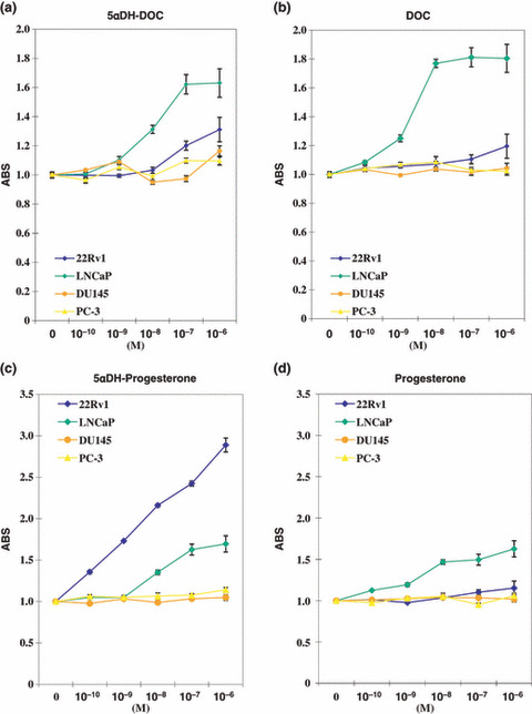
Cell growth assays of 5αDH steroids and their precursors. 5α‐Dihydro‐deoxycorticosterone (5αDH‐DOC) (a) and 5αDH‐progesterone (c) at 10−7 M or lower concentration showed growth‐promoting effect on both androgen receptor (AR)‐positive prostate cancer (PC) cell lines 22Rv1 and LNCaP, but did not on AR‐negative PC cells DU‐145 and PC‐3. (b,d) Their 4‐ene‐3‐oxosteroids (DOC and progesterone) had also some ability to stimulate cell proliferation. Prostate cancer (PC) cells were treated with the indicated concentration of each steroid (x‐axis, 10−10–10−6 M) and 72 h later, MTT (3‐[4,5‐dimethylthiazol‐2‐yl]‐2,5‐diphenyltetrazolium bromide) assay was performed. Y‐axis: absorbance (ABS) at 490 nm (MTT assay), and at 630 nm as a reference, measured with a microplate reader. Each assay was tested six times and the means ± SD were plotted.
5α‐Dihydro‐deoxycorticosterone (5αDH‐DOC) activated wild‐type AR more efficiently than DOC. Focusing on 5αDH‐DOC and 5αDH‐progesterone, to evaluate their direct ability to stimulate AR transactivation activity, we transfected LNCaP cells and 22Rv1 cells with a luciferase plasmid (pGL3‐PSA) driven by the PSA enhancer which included androgen response elements, and compared the luciferase activity reflecting AR transactivation activity by the treatment of these 5αDH‐steroids and their putative precursors. LNCaP cells and 22Rv1 cells expressed mutant AR (T877A and H874Y, respectively) and mutant AR has been reported to respond to steroids differently from wild‐type AR.( 14 , 15 ) Then, to evaluate their effects on wild‐type AR, we also co‐transfected AR‐null PC‐3 cells with wild‐type AR or mutant AR (T877S) and examined the transactivation activity of AR by each of the steroids. As shown in Figure 2(a), both 5αDH‐DOC and DOC at 10−7 M or lower concentration showed some level of AR transactivation in LNCaP cells (mutant AR: T877A) and 22Rv1 cells (mutant AR: H874Y). 5α‐Dihydro‐deoxycorticosterone (5αDH‐DOC) and DOC treatment showed transactivation of wild‐type AR (Fig. 2b, left) and mutant AR (Fig. 2b, right) which was introduced in AR‐null PC‐3 cells, although they did not show any transactivation activity in mock‐transfected PC‐3 cells (Fig. 2b, lower). These transactivation activities were blocked by the AR antagonist bicalutamide (BCL), confirming that they were caused via AR, regardless of its mutation status. Interestingly, 5αDH‐DOC showed more impact on the transactivation activity of wild‐type AR than DOC (Fig. 2b, left), while 5αDH‐DOC did not show any advantageous impact on the transactivation activity of mutant AR (T877S) over DOC (Fig. 2b, right). This was consistent with the findings that 5αDH‐DOC showed less effect on the transactivation of mutant ARs in LNCaP cells and 22Rv1 cells than DOC. On the other hand, 5αDH‐progesterone and other 5α‐DH‐oxosteroids did not show such a characteristic feature to induce AR transactivation (Fig. 2c,d for 5αDH‐progesterone, and data are not shown as for other 5αDH‐steorids), except for DHT. These findings suggest that the conversion from DOC to 5αDH‐DOC by 5α‐steroid reductases could provide some advantageous effects on AR transactivation to PC cells with wild‐type AR, as well as the conversion from testosterone to DHT. Most of CRPC cells showed overexpression of wild‐type AR, not mutant AR,( 13 ) and these effects of 5αDH‐DOC on wild‐type AR might be beneficial for CRPC cells. Thus we focused on 5αDH‐DOC and DOC in further experiments.
Figure 2.
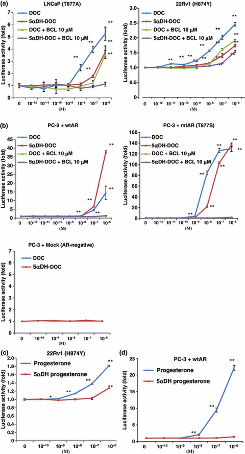
Luciferase assays of 5αDH steroids and their precursors for androgen receptor (AR) transactivation activity. (a) LNCaP cells and 22Rv1 cells were transfected with pGL3‐PSA and the luciferase activity in the presence of indicated concentration (X‐axis) of 5α‐dihydro‐deoxycorti‐costerone (5αDH‐DOC) and 11‐deoxycorticosterone (DOC) was compared. LNCaP cells and 22Rv1 cells expressed mutant AR, T877A, and H874Y, respectively. (b) Androgen receptor (AR)‐null PC‐3 cells were co‐transfected with wild‐type AR (left), mutant AR‐T877A (right), or mock (lower) and pGL3‐PSA and the luciferase activity in the presence of indicated concentration (x‐axis) of 5αDH‐DOC and DOC was compared. These luciferase activities were blocked by antiandrogen bicalutamide (BCL). Each assay was tested six times and the means ± SD were plotted. *P < 0.05, **P < 0.01 by Student’s t‐test. (c) 22Rv1 cells were transfected with pGL3‐PSA and the luciferase activity in the presence of indicated concentration (x‐axis) of 5αDH‐progesterone and progesterone was compared. (d) Androgen receptor (AR)‐null PC‐3 cells were co‐transfected with wild‐type AR and pGL3‐PSA and the luciferase activity in the presence of indicated concentration (x‐axis) of 5αDH‐progesterone and progesterone was compared. Each assay was tested six times and the means ± SD were plotted. *P < 0.05, **P < 0.01 by Student’s t‐test.
Type 1 5α‐steroid reductase (SRD5A1) was responsible for 5αDH‐DOC production in PC cells. In PC tissues, especially CRPC tissues, type 1 (SRD5A1) and type 3 (SRD5A3) isozymes are up‐regulated and dominant( 11 , 14 ) and also their expressions were observed in all available PC cell lines (Fig. S2). They are likely to be responsible for DHT and other 5αDH‐steroid production in PC tissues. To investigate which of type 1 and/or type 3 is responsible for 5αDH‐DOC production in PC cells, COS7 cells were transfected with each of HA‐tagged expression vectors (SRD5A1, SRD5A3, and mock, Fig. 3a) and the cells were treated with 10−6 M DOC. One hour after incubation under 10−6 M DOC, the conditioned media were harvested to measure the amounts of 5αDH‐DOC by sensitive LC‐MS/MS analysis. As a result, a much higher amount of 5αDH‐DOC was observed in the cells that overexpressed SRD5A1 than in the SRD5A3 or mock‐transfected (Fig. 3b) cells. The production of 5αDH‐DOC was inhibited by pre‐treatment of 1 μM dutasteride which could inhibit the activity of SRD5A1 (Fig. 3c). These findings indicated that SRD5A1 could be responsible for 5αDH‐DOC production in vitro.
Figure 3.
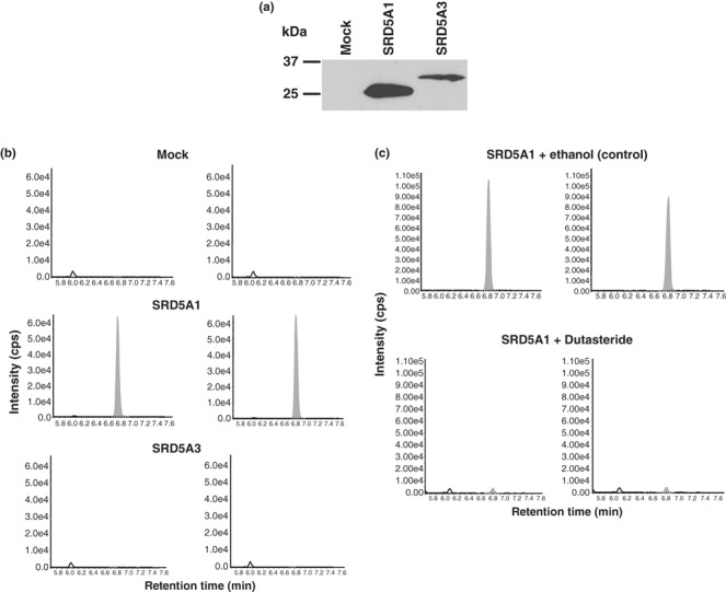
Type 1 5α‐steroid reductase (SRD5A1) was responsible for the production of 5α‐dihydro‐deoxycorticosterone (5αDH‐DOC). (a) COS7 cells were transfected with SRD5A1, SRD5A3, or mock vector and the expression of exogenous SRD5A1 and SRD5A3 were evaluated by western blot analysis using anti‐HA tag antibody. (b) The transfected cells were treated with 10−6 M 11‐deoxycorticosterone (DOC), and after 1 h of incubation, the conditioned media were harvested. Liquid chromatography‐tandem mass spectrometry (LC‐MS/MS) analysis of the media specifically detected the production of 5αDH‐DOC which was converted from DOC in COS7 cells overexpressing SRD5A1 (middle panel), but not in COS7 cells overexpressing SRD5A3 (lower panel) or mock cells (upper panel). These experiments were performed in duplicate (right and left panels). (c) 1‐μM Dutasteride treatment inhibited 5αDH‐DOC production in COS7 cells transfected with SRD5A1 vector. 5α‐Dihydro‐deoxycorticosterone (5αDH‐DOC)was detected by LC‐MS/MS and these experiments were performed in duplicate (right and left panels).
Subsequently, to evaluate the endogenous activity of SRD5A1 for 5αDH‐DOC, 22Rv1 cells were treated with siRNA duplex specific to SRD5A1 (siSRD5A1) or the control siRNA duplex (siEGFP). Reverse transcription (RT)‐PCR validated the knockdown effect by siSRD5A1 (Fig. 4a), and siSRD5A1 treatment suppressed the cell viability of 22Rv1 cells, compared with the control siEGFP (Fig. 4b, P < 0.01). The amount of 5αDH‐DOC production was also significantly decreased in the conditioned media of 22Rv1 of which SRD5A1 was knocked down (Fig. 4c, P < 0.01). These findings suggest that SRD5A1 is likely to play some important roles in converting DOC to 5αDH‐DOC in PC cells, as well as PC cell viability.
Figure 4.
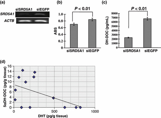
(a) Reverse transcription (RT)‐PCR confirmed knockdown effect on type 1 5α‐steroid reductase (SRD5A1) expression by siSRD5A1 in 22Rv1 cells. β‐Actin (ACTB) was used to quantify the input RNAs. (b) Knockdown of SRD5A1 expression by siSRD5A1 suppressed the proliferation of 22Rv1 cells, compared with the control RNA duplex siEGFP (P < 0.01, Student’s t‐test). Y‐axis, absorbance (ABS) at 490 nm (MTT assay), and at 630 nm as a reference, measured with a microplate reader. (c) Suppression of SRD5A1 expression by siSRD5A1 reduced 5α‐dihydro‐deoxycorticosterone (5αDH‐DOC) production in 22Rv1 cells (P < 0.01, Student’s t‐test). 5α‐Dihydro‐deoxycorticosterone (5αDH‐DOC) in the media was measured by liquid chromatography‐tandem mass spectrometry (LC‐MS/MS) analysis. (d) Detection of 5αDH‐DOC and DHT in 13 clinical CRPC tissues. 5α‐Dihydro‐deoxycorticosterone (5αDH‐DOC) was detected by sensitive LC‐MS/MS in clinical CRPC tissues. 5α‐Dihydrotestosterone (DHT) was also measured by sensitive LC‐MS/MS in the same tissues, and the level of 5αDH‐DOC was inversely correlated with the low level of DHT concentration (Pearson r = −0.5727, P < 0.05).
5α‐Dihydro‐deoxycorticosterone (5αDH‐DOC) was detected in clinical CRPC tissues by LC‐MS/MS. To prove the presence of 5αDH‐DOC in clinical CRPC tissues, we extracted steroid fractions from 13 fresh frozen CRPC tissues and measured the intratumoral 5αDH‐DOC and DHT levels by the sensitive LC‐MS/MS analysis we established here. As expected, the intratumoral DHT level in all CRPC samples was below 1000 pg/g tissues, and 5αDH‐DOC was detected in eight out of 13 CRPC tissues (Fig. 4d). We did not find any clinical features of these 5αDH‐DOC‐positive CRPCs. Interestingly, 5αDH‐DOC‐positive CRPCs had a comparatively lower level of DHT, and the level of 5αDH‐DOC was significantly inversely correlated to the DHT level within CRPC tissues (Pearson r = −0.5727, P < 0.05).
Discussion
Several molecular events that drive the progression to the castration‐resistant state have been proposed and extensively reviewed.( 5 , 6 , 15 , 16 , 17 ) One of the key and main mechanisms is overexpression of wild‐type AR with or without amplification of the AR gene, which is commonly observed in clinical CRPCs.( 6 , 12 , 18 , 19 ) On the other hand, AR mutations allow AR activation by low androgen levels or by other endogenous steroids such as corticosteroids and antiandrogens,( 20 , 21 , 22 , 23 ) but the incidence of AR mutation is <20% in clinical CRPCs.( 13 ) In this study, we searched for novel 5αDH steroids that could have any potential to stimulate PC cell growth as well as AR activity, and among them, we here focused on 5αDH‐DOC and demonstrated that it could stimulate wild‐type AR preferentially rather than mutant AR, and type 1 5α‐steroid reductase overexpressed in CRPC cells could be responsible for 5αDH‐DOC production. These findings indicate that 5αDH‐DOC and other unknown products of 5α‐steroid reductases (type 1 and type 3) could activate AR under extremely low levels of DHT in some CRPC cases and provide some survival advantages to PC cells, although the concentration of 5αDH‐DOC we here showed was relatively lower than that of DHT. In situ steroidogenesis including this 5α‐steroid reduction is likely to be one of the key mechanisms of CRPC development and a good molecular target for CRPC treatment.
In the standard steroidogenesis pathway, the testes provide the major source of androgens, particularly testosterone. Alternatively, prostate cells can convert adrenal‐derived steroids, such as androstenediol and DHEA (dehydroepiandrosterone), to testosterone by 17β‐hydroxysteroid dehydrogenase and 3β‐hydroxysteroid dehydrogenase.( 17 , 24 ) Currently, there is increasing evidence that PC cells can synthesize androgens de novo instead of receiving them through the bloodstream.( 17 , 25 ) This concept is supported by the fact that expressions of several enzymes responsible for steroid synthesis were up‐regulated in CRPCs( 26 , 27 , 28 ) and they are likely to play some critical roles in the CRPC phenotype. Among them, cytochrome P17 (CYP17) catalyzes the key reactions to androgen and estrogen biosynthesis, and its selective inhibitor, abiraterone, is now in clinical trials for CRPCs, showing significant antitumor activity in CRPCs,( 29 ) indicating that CRPCs remain dependent on ligand‐activated AR signaling, and also these findings suggest that such steroids or androgen metabolism enzymes could be potential molecular therapeutic targets for CRPCs. Among several steroid metabolism enzymes altered in CRPCs, 5α‐steroid reductases mostly contribute to both the standard steroidogenesis pathway (testosterone to DHT), and the alternative steroidogenesis pathway which is likely to be activated when testosterone are not available from the blood circulation.( 16 , 24 ) In addition to DHT, other various types of steroids can be subject to the reduction of 5α‐steroid reductases to acquire more potentials as functional steroids or modify/change their functions.( 30 ) Neuroendocrine cells in the brain highly express type 1 5α‐steroid reductase (SRD5A1) and produce several neurosteroids such as 5αDH‐progesterone, allopregnolone (ALLO), and tetrahydrodeoxycorticosterone (THDOC), which modulate GABA action at GAGAA receptors to contribute to neuroendocrine transmission.( 31 ) In this point, 5αDH‐DOC production by SRD5A1 in CRPCs may reflect the neuroendocrine‐like phenotype of CRPCs,( 32 , 33 ) and in addition to AR activation, it may regulate or stimulate other signaling pathways similar to how other 5αDH neurosteroids to contribute to the CRPC phenotype.
The source of DHT in prostatic tissue after ADT is likely to be intracrine production within the prostate, which converts adrenal androgens to DHT.( 9 ) Interestingly, we observed that the level of 5αDH‐DOC was significantly inversely correlated to the DHT level within CRPC tissues, although the concentration of 5αDH‐DOC was quite lower than that of DHT. This might implicate that when DHT levels were declined within CRPC tissues, there might still be compensatory increases in 5α‐DH‐DOC and other unknown 5αDH steroids to activate AR by taking advantage of overexpressing SRD5A1 or SRD5A3. There is no evidence obtained so far by clinical trials to support the sole treatment with dutastride; dual inhibitor of type 1 and 2 5α‐steroid reductases could effectively suppress CRPC growth, although the DHT level within PC tissues was not assessed.( 34 , 35 ) Several other ligands can activate the AR pathway or other pathways to support the survival of PC cells under the castrated and 5α‐steroid reductase‐free environment, and the combination hormonal or AR‐targeting therapy with dutastride might be required to deplete intratumoral active steroids in CRPCs and suppress CRPC growth effectively.
Acknowledgments
We thank Ms U. Mami for her technical assistance. This work was supported in part by research grants (#18590323 to H. Nakagawa) and (#00L01402 to Y. Nakamura) from the Japan Society for the Promotion of Science.
Supporting information
Fig. S1. Cell growth assays of 5αDH steroids and their precursors. Corticosterone (a) and cortisol (c) had positive effect on the proliferation of androgen receptor (AR)‐negative DU‐145 cells, as well as LNCaP or 22RV1 cells. 5αDH‐Corticosterone (b) and 5αDH‐cortisol (d) did not show any significant growth‐promoting effect on prostate cancer (PC) cell lines. Prostate cancer (PC) cells were treated with indicated concentration of each steroid (x‐axis, 10−10–10−6 M) and 72 h later, MTT assay was performed. Y‐axis: absorbance (ABS) at 490 nm (MTT assay), and at 630 nm as a reference, measured with a microplate reader. Each assay was tested six times and the means ± SD. were plotted.
Fig. S2. Semi‐quantitative RT‐PCR showed that all of the prostate cancer (PC) cell lines we used expressed type 3 5α‐steroid reductase (SRD5A1) and SRD5A1. Expression of β‐actin (ACTB) served as the quantitative control.
Please note: Wiley‐Blackwell are not responsible for the content or functionality of any supporting materials supplied by the authors. Any queries (other than missing material) should be directed to the corresponding author for the article.
Supporting info item

Supporting info item

References
- 1. Gronberg H. Prostate cancer epidemiology. Lancet 2003; 361: 859–64. [DOI] [PubMed] [Google Scholar]
- 2. Smaletz O, Scher HI. Outcome predictions for patients with metastatic prostate cancer. Semin Urol Oncol 2002; 20: 155–63. [DOI] [PubMed] [Google Scholar]
- 3. Petrylak DP, Tangen CM, Hussain MHA et al. Docetaxel and estramustine compared with mitoxantrone and prednisone for advanced refractory prostate cancer. N Engl J Med 2004; 351: 1513–20. [DOI] [PubMed] [Google Scholar]
- 4. Tannock IF, De Wit R, Berry WR et al. Docetaxel plus prednisone or mitoxantrone plus prednisone for advanced prostate cancer. N Engl J Med 2004; 351: 1502–12. [DOI] [PubMed] [Google Scholar]
- 5. Scher HI, Sawyers CL. Biology of progressive, castration‐resistant prostate cancer: directed therapies targeting the androgen‐receptor signaling axis. J Clin Oncol 2005; 23: 8253–61. [DOI] [PubMed] [Google Scholar]
- 6. Chen Y, Sawyers CL, Scher HI. Targeting the androgen receptor pathway in prostate cancer. Curr Opin Pharmacol 2008; 8: 440–8. [DOI] [PMC free article] [PubMed] [Google Scholar]
- 7. Thigpen AE, Silver RI, Guileyardo JM, Casey ML, McConnell JD, Russell DW. Tissue distribution and ontogeny of steroid 5 alpha‐reductase isozyme expression. J Clin Invest 1993; 92: 903–10. [DOI] [PMC free article] [PubMed] [Google Scholar]
- 8. Mohler JL, Gregory CW, Ford OH et al. The androgen axis in recurrent prostate cancer. Clin Cancer Res 2004; 10: 440–8. [DOI] [PubMed] [Google Scholar]
- 9. Nishiyama T, Hashimoto Y, Takahashi K. The influence of androgen deprivation therapy on dihydrotestosterone levels in the prostatic tissue of patients with prostate cancer. Clin Cancer Res 2004; 10: 7121–6. [DOI] [PubMed] [Google Scholar]
- 10. Titus MA, Schell MJ, Lih FB, Tomer KB, Mohler JL. Testosterone and dihydrotestosterone tissue levels in recurrent prostate cancer. Clin Cancer Res 2005; 11: 4653–7. [DOI] [PubMed] [Google Scholar]
- 11. Uemura M, Tamura K, Chung S et al. Novel 5α‐steroid reductase (SRD5A3, type‐3) is overexpressed in hormone‐refractory prostate cancer. Cancer Sci 2008; 99: 81–6. [DOI] [PMC free article] [PubMed] [Google Scholar]
- 12. Tamura K, Furihata M, Tsunoda T et al. Molecular features of hormone‐refractory prostate cancer cells by genome‐wide gene expression profiles. Cancer Res 2007; 67: 5117–25. [DOI] [PubMed] [Google Scholar]
- 13. Taplin M‐E, Rajeshkumar B, Halabi S et al. Androgen receptor mutations in androgen‐independent prostate cancer: Cancer and Leukemia Group B Study 9663. J Clin Oncol 2003; 21: 2673–8. [DOI] [PubMed] [Google Scholar]
- 14. Titus MA, Gregory CW, Ford OH, Schell MJ, Maygarden SJ, Mohler JL. Steroid 5α‐reductase isozymes I and II in recurrent prostate cancer. Clin Cancer Res 2005; 11: 4365–71. [DOI] [PubMed] [Google Scholar]
- 15. Feldman BJ, Feldman D. The development of androgen‐independent prostate cancer. Nat Rev Cancer 2001; 1: 34–45. [DOI] [PubMed] [Google Scholar]
- 16. Vis AN, Schroder FH. Key targets of hormonal treatment of prostate cancer. Part 1: the androgen receptor and steroidogenic pathways. BJU Int 2009; 104: 438–48. [DOI] [PubMed] [Google Scholar]
- 17. Attar RM, Takimoto CH, Gottardis MM. Castration‐resistant prostate cancer: locking up the molecular escape routes. Clin Cancer Res 2009; 15: 3251–5. [DOI] [PubMed] [Google Scholar]
- 18. Linja MJ, Savinainen KJ, Saramaki OR, Tammela TLJ, Vessella RL, Visakorpi T. Amplification and overexpression of androgen receptor gene in hormone‐refractory prostate cancer. Cancer Res 2001; 61: 3550–5. [PubMed] [Google Scholar]
- 19. Ford OH, Gregory CW, Kim D, Smitherman AB, Mohler JL. Androgen receptor gene amplification and protein expression in recurrent prostate cancer. J Urol 2003; 170: 1817–21. [DOI] [PubMed] [Google Scholar]
- 20. Culig Z, Hobisch A, Cronauer MV et al. Mutant androgen receptor detected in an advanced‐stage prostatic carcinoma is activated by adrenal androgens and progesterone. Mol Endocrinol 1993; 7: 1541–50. [DOI] [PubMed] [Google Scholar]
- 21. Taplin M‐E, Bubley GJ, Shuster TD et al. Mutation of the androgen‐receptor gene in metastatic androgen‐independent prostate cancer. N Engl J Med 1995; 332: 1393–8. [DOI] [PubMed] [Google Scholar]
- 22. Zhao XY, Malloy PJ, Krishnan AV et al. Glucocorticoids can promote androgen‐independent growth of prostate cancer cells through a mutated androgen receptor. Nat Med 2000; 6: 703–6. [DOI] [PubMed] [Google Scholar]
- 23. Mizokami A, Koh E, Fujita H et al. The adrenal androgen androstenediol is present in prostate cancer tissue after androgen deprivation therapy and activates mutated androgen receptor. Cancer Res 2004; 64: 765–71. [DOI] [PubMed] [Google Scholar]
- 24. Ghayee HK, Auchus RJ. Clinical implications of androgen synthesis via 5α‐reduced precursors. Endocr Dev 2008; 13: 55–66. [DOI] [PubMed] [Google Scholar]
- 25. Attard G, Reid AHM, Olmos D, De Bono JS. Antitumor activity with CYP17 blockade indicates that castration‐resistant prostate cancer frequently remains hormone driven. Cancer Res 2009; 69: 4937–40. [DOI] [PubMed] [Google Scholar]
- 26. Stanbrough M, Bubley GJ, Ross K et al. Increased expression of genes converting adrenal androgens to testosterone in androgen‐independent prostate cancer. Cancer Res 2006; 66: 2815–25. [DOI] [PubMed] [Google Scholar]
- 27. Locke JA, Guns ES, Lubik AA et al. Androgen levels increase by intratumoral de novo steroidogenesis during progression of castration‐resistant prostate cancer. Cancer Res 2008; 68: 6407–15. [DOI] [PubMed] [Google Scholar]
- 28. Montgomery RB, Mostaghel EA, Vessella R et al. Maintenance of intratumoral androgens in metastatic prostate cancer: a mechanism for castration‐resistant tumor growth. Cancer Res 2008; 68: 4447–54. [DOI] [PMC free article] [PubMed] [Google Scholar]
- 29. Attard G, Reid AHM, Yap TA et al. Phase I clinical trial of a selective inhibitor of CYP17, abiraterone acetate, confirms that castration‐resistant prostate cancer commonly remains hormone driven. J Clin Oncol 2008; 26: 4563–71. [DOI] [PubMed] [Google Scholar]
- 30. Wilson JD. The role of 5α‐reduction in steroid hormone physiology. Reprod Fertil Dev 2001; 13: 673–8. [DOI] [PubMed] [Google Scholar]
- 31. Agis‐Balboa RC, Pinna G, Zhubi A et al. Characterization of brain neurons that express enzymes mediating neurosteroid biosynthesis. Proc Natl Acad Sci USA 2006; 103: 14602–7. [DOI] [PMC free article] [PubMed] [Google Scholar]
- 32. Hirano D, Okada Y, Minei S, Takimoto Y, Nemoto N. Neuroendocrine differentiation in hormone refractory prostate cancer following androgen deprivation therapy. Eur Urol 2004; 45: 586–92. [DOI] [PubMed] [Google Scholar]
- 33. Yuan T‐C, Veeramani S, Lin M‐F. Neuroendocrine‐like prostate cancer cells: neuroendocrine transdifferentiation of prostate adenocarcinoma cells. Endocr Relat Cancer 2007; 14: 531–47. [DOI] [PubMed] [Google Scholar]
- 34. Shah SK, Trump DL, Sartor O, Tan W, Wilding GE, Mohler JL. Phase II study of dutasteride for recurrent prostate cancer during androgen deprivation therapy. J Urol 2009; 181: 621–6. [DOI] [PMC free article] [PubMed] [Google Scholar]
- 35. Taplin M‐E, Regan MM, Ko Y‐J et al. Phase II study of androgen synthesis inhibition with ketoconazole, hydrocortisone, and dutasteride in asymptomatic castration‐resistant prostate cancer. Clin Cancer Res 2009; 15: 7099–105. [DOI] [PMC free article] [PubMed] [Google Scholar]
Associated Data
This section collects any data citations, data availability statements, or supplementary materials included in this article.
Supplementary Materials
Fig. S1. Cell growth assays of 5αDH steroids and their precursors. Corticosterone (a) and cortisol (c) had positive effect on the proliferation of androgen receptor (AR)‐negative DU‐145 cells, as well as LNCaP or 22RV1 cells. 5αDH‐Corticosterone (b) and 5αDH‐cortisol (d) did not show any significant growth‐promoting effect on prostate cancer (PC) cell lines. Prostate cancer (PC) cells were treated with indicated concentration of each steroid (x‐axis, 10−10–10−6 M) and 72 h later, MTT assay was performed. Y‐axis: absorbance (ABS) at 490 nm (MTT assay), and at 630 nm as a reference, measured with a microplate reader. Each assay was tested six times and the means ± SD. were plotted.
Fig. S2. Semi‐quantitative RT‐PCR showed that all of the prostate cancer (PC) cell lines we used expressed type 3 5α‐steroid reductase (SRD5A1) and SRD5A1. Expression of β‐actin (ACTB) served as the quantitative control.
Please note: Wiley‐Blackwell are not responsible for the content or functionality of any supporting materials supplied by the authors. Any queries (other than missing material) should be directed to the corresponding author for the article.
Supporting info item
Supporting info item