Intensive care has been defined as “a service for patients with potentially recoverable conditions who can benefit from more detailed observation and invasive treatment than can safely be provided in general wards or high dependency areas.” It is usually reserved for patients with potential or established organ failure. The most commonly supported organ is the lung, but facilities should also exist for the diagnosis, prevention, and treatment of other organ dysfunction.
Who to admit
Intensive care is appropriate for patients requiring or likely to require advanced respiratory support, patients requiring support of two or more organ systems, and patients with chronic impairment of one or more organ systems who also require support for an acute reversible failure of another organ. Early referral is particularly important. If referral is delayed until the patient’s life is clearly at risk, the chances of full recovery are jeopardised.
Categories of organ system monitoring and support
(Adapted from Guidelines on admission to and discharge from intensive care and high dependency units. London: Department of Health, 1996.)
| Advanced respiratory support | Circulatory support |
| • Mechanical ventilatory support (excluding mask continuous positive airway pressure (CPAP) or non-invasive (eg, mask) ventilation) | • Need for vasoactive drugs to support arterial pressure or cardiac output |
| • Possibility of a sudden, precipitous deterioration in respiratory function requiring immediate endotracheal intubation and mechanical ventilation | • Support for circulatory instability due to hypovolaemia from any cause which is unresponsive to modest volume replacement (including post-surgical or gastrointestinal haemorrhage or haemorrhage related to a coagulopathy) |
| Basic respiratory monitoring and support | • Patients resuscitated after cardiac arrest where intensive or high dependency care is considered clinically appropriate |
| • Need for more than 50% oxygen | • Intra-aortic balloon pumping |
| • Possibility of progressive deterioration to needing advanced respiratory support | Neurological monitoring and support |
| • Need for physiotherapy to clear secretions at least two hourly | • Central nervous system depression, from whatever cause, sufficient to prejudice the airway and protective reflexes |
| • Patients recently extubated after prolonged intubation and | • Invasive neurological monitoring |
| mechanical ventilation | Renal support |
| • Need for mask continuous positive airway pressure or non-invasive | • Need for acute renal replacement therapy (haemodialysis, haemofiltration, or haemodiafiltration) |
| ventilation | |
| • Patients who are intubated to protect the airway but require no | |
| ventilatory support and who are otherwise stable | |
Factors to be considered when assessing suitability for admission to intensive care
Diagnosis
Severity of illness
Age
Coexisting disease
Physiological reserve
Prognosis
Availability of suitable treatment
Response to treatment to date
Recent cardiopulmonary arrest
Anticipated quality of life
The patient’s wishes
As with any other treatment, the decision to admit a patient to an intensive care unit should be based on the concept of potential benefit. Patients who are too well to benefit or those with no hope of recovering to an acceptable quality of life should not be admitted. Age by itself should not be a barrier to admission to intensive care, but doctors should recognise that increasing age is associated with diminishing physiological reserve and an increasing chance of serious coexisting disease. It is important to respect patient autonomy, and patients should not be admitted to intensive care if they have a stated or written desire not to receive intensive care—for example, in an advanced directive.
Severity of illness scoring systems such as the acute physiology and chronic health evaluation (APACHE) and simplified acute physiology score (SAPS) estimate hospital mortality for groups of patients. They cannot be used to predict which patients will benefit from intensive care as they are not sufficiently accurate and have not been validated for use before admission.
When to admit
Patients should be admitted to intensive care before their condition reaches a point from which recovery is impossible. Clear criteria may help to identify those at risk and to trigger a call for help from intensive care staff. Early referral improves the chances of recovery, reduces the potential for organ dysfunction (both extent and number), may reduce length of stay in intensive care and hospital, and may reduce the costs of intensive care. Patients should be referred by the most senior member of staff responsible for the patient—that is, a consultant. The decision should be delegated to trainee doctors only if clear guidelines exist on admission. Once patients are stabilised they should be transferred to the intensive care unit by experienced intensive care staff with appropriate transfer equipment.
Criteria for calling intensive care staff to adult patients
(Adapted from McQuillan et al BMJ 1998;316:1853-8.)
Threatened airway
All respiratory arrests
Respiratory rate ⩾40 or ⩽8 breaths/min
Oxygen saturation <90% on ⩾50% oxygen
All cardiac arrests
Pulse rate <40 or >140 beats/min
Systolic blood pressure <90 mm Hg
Sudden fall in level of consciousness (fall in Glasgow coma score >2 points)
Repeated or prolonged seizures
Rising arterial carbon dioxide tension with respiratory acidosis
Any patient giving cause for concern
Initial treatment
In critical illness the need to support the patient’s vital functions may, at least initially, take priority over establishing a precise diagnosis. For example, patients with life threatening shock need immediate treatment rather than diagnosis of the cause as the principles of management are the same whether shock results from a massive myocardial infarction or a gastrointestinal bleed. Similarly, although the actual management may differ, the principles of treating other life threatening organ failures—for example, respiratory failure or coma—do not depend on precise diagnosis.
Basic monitoring requirements for seriously ill patients
Heart rate
Blood pressure
Respiratory rate
Pulse oximetry
Hourly urine output
Temperature
Blood gases
Respiratory support
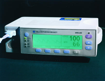
All seriously ill patients without pre-existing lung disease should receive supplementary oxygen at sufficient concentration to maintain arterial oxygen tension ⩾8 kPa or oxygen saturation of at least 90%. In patients with depressed ventilation (type II respiratory failure) oxygen will correct the hypoxaemia but not the hypercapnia. Care is required when monitoring such patients by pulse oximetry as it does not detect hypercapnia.
A few patients with severe chronic lung disease are dependent on hypoxic respiratory drive, and oxygen may depress ventilation. Nevertheless, life threatening hypoxaemia must be avoided, and if this requires concentrations of oxygen that exacerbate hypercapnia the patient will probably need mechanical ventilation.
Any patient who requires an inspired oxygen concentration of 50% or more should ideally be managed at least on a high dependency unit. Referral to intensive care should not be based solely on the need for endotracheal intubation or mechanical ventilation as early and aggressive intervention, high intensity nursing, and careful monitoring may prevent further deterioration. Endotracheal intubation can maintain a patent airway and protect it from contamination by foreign material such as regurgitated or vomited gastric contents or blood. Putting the patient in the recovery position with the head down helps protect the airway while awaiting the necessary expertise for intubation. Similarly, simple adjuncts such as an oropharyngeal airway may help to maintain airway patency, although it does not give the protection of an endotracheal tube.
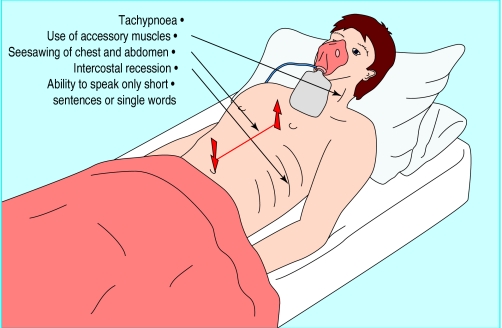
Breathlessness and respiratory difficulty are common in acutely ill patients. Most will not need mechanical ventilation, but those that do require ventilation need to be identified as early as possible and certainly before they deteriorate to the point of respiratory arrest. The results of blood gas analysis alone are rarely sufficient to determine the need for mechanical ventilation. Several other factors have to be taken into consideration:
Degree of respiratory work—A patient with normal blood gas tensions who is working to the point of exhaustion is more likely to need ventilating than one with abnormal tensions who is alert, oriented, talking in full sentences, and not working excessively.
Likely normal blood gas tensions for that patient—Some patients with severe chronic lung disease will lead surprisingly normal lives with blood gas tensions which would suggest the need for ventilation in someone previously fit.
Likely course of disease—If imminent improvement is likely ventilation can be deferred, although such patients need close observation and frequent blood gas analysis.
Adequacy of circulation—A patient with established or threatened circulatory failure as well as respiratory failure should be ventilated early in order to gain control of at least one major determinant of tissue oxygen delivery.
Circulatory support
Shock represents a failure of tissue perfusion. As such, it is primarily a failure of blood flow and not blood pressure. Nevertheless, an adequate arterial pressure is essential for perfusion of major organs and glomerular filtration, particularly in elderly or hypertensive patients, and for sustaining flow through any areas of critical narrowing in the coronary and cerebral vessels. A normal blood pressure does not exclude shock since pressure may be maintained at the expense of flow by vasoconstriction. Conversely, a high cardiac output (for example, in sepsis) does not preclude regional hypoperfusion associated with systemic vasodilatation, hypotension, and maldistribution.
Signs suggestive of failing tissue perfusion
Tachycardia
Confusion or diminished conscious level
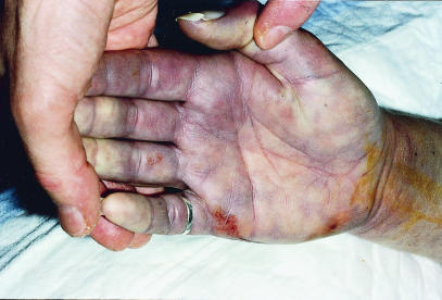
Poor peripheral perfusion (cool, cyanosed extremities, poor capillary refill, poor peripheral pulses)
Poor urine output (<0.5 ml/kg/h)
Metabolic acidosis
Increased blood lactate concentration
Normal blood pressure does not exclude shock
Shock may be caused by hypovolaemia (relative or actual), myocardial dysfunction, microcirculatory abnormalities, or a combination of these factors. To identify shock it is important to recognise the signs of failing tissue perfusion.
Neurological considerations in referral to intensive care
Airway obstruction
Absent gag or cough reflex
Measurement of intracranial pressure and cerebral perfusion pressure
Raised intracranial pressure requiring treatment
Prolonged or recurrent seizures which are resistant to conventional anticonvulsants
Hypoxaemia
Hypercapnia or hypocapnia
All shocked patients should receive supplementary oxygen. Thereafter, the principles of management are to ensure an adequate circulating volume and then, if necessary, to give vasoactive drugs (for example, inotropes, vasopressors, vasodilators) to optimise cardiac output (and hence tissue oxygen delivery) and correct hypotension. Most patients will need intravenous fluid whatever the underlying disease. Central venous pressure may guide volume replacement and should be considered in patients who fail to improve despite an initial litre of intravenous fluid or sooner in patients with known or suspected myocardial dysfunction. Any patients needing more than modest fluid replacement or who require vasoactive drugs to support arterial pressure or cardiac output should be referred for high dependency or intensive care.
Neurological support
Neurological failure may occur after head injury, poisoning, cerebral vascular accident, infections of the nervous system (meningitis or encephalitis), cardiac arrest, or as a feature of metabolic encephalopathy (such as liver failure). The sequelae of neurological impairment may lead to the patient requiring intensive care. For instance, loss of consciousness may lead to obstruction of airways, loss of protective airway reflexes, and disordered ventilation that requires intubation or tracheostomy and mechanical ventilation.
Neurological disease may also cause prolonged or recurrent seizures or a rise in intracranial pressure. Patients who need potent anaesthetic drugs such as thiopentone or propofol to treat seizures that are resistant to conventional anticonvulsants, or monitoring of intracranial pressure and cerebral perfusion pressure must be referred to a high dependency or intensive care unit. Patients with neuromuscular disease (for example,
Guillain-Barré syndrome, myasthenia gravis) may require admission to intensive care for intubation or ventilation because of respiratory failure, loss of airway reflexes, or aspiration.
Renal support
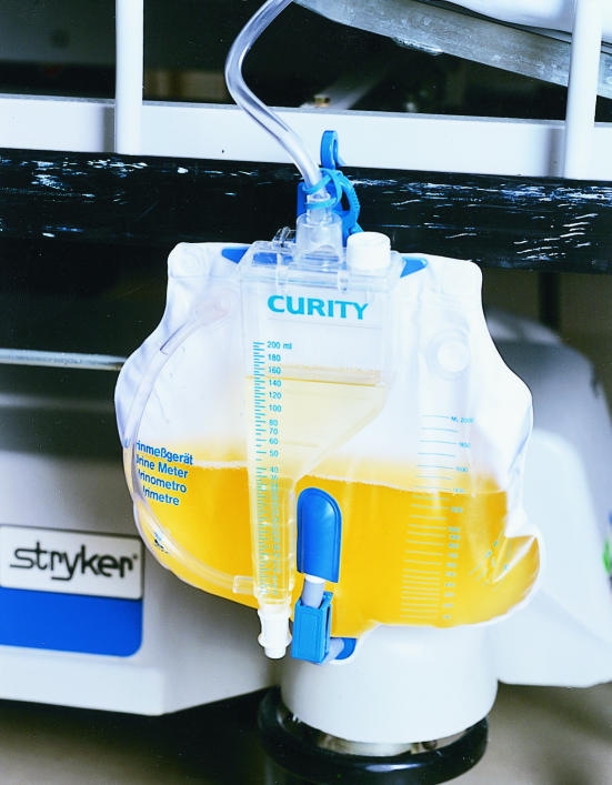
Renal failure is a common complication of acute illness or trauma and the need for renal replacement therapy (haemofiltration, haemodialysis, or their variants) may be a factor when considering referral to intensive or high dependency care. The need for renal replacement therapy is determined by assessment of urine volume, fluid balance, renal concentrating power (for example, urine:plasma osmolality ratio and urinary sodium concentration), acid-base balance, and the rate of rise of plasma urea, creatinine, and potassium concentrations. In ill patients hourly recording of urine output on the ward may give an early indication of a developing renal problem; prompt treatment, including aggressive circulatory resuscitation, may prevent this from progressing to established renal failure.
Indications for considering renal replacement therapy
Oliguria (<0.5ml/kg/h)
Life threatening hyperkalaemia (>6 mmol/l) resistant to drug treatment
Rising plasma concentrations of urea or creatinine, or both
Severe metabolic acidosis
Symptoms related to uraemia (for example, pericarditis, encephalopathy)
Figure.
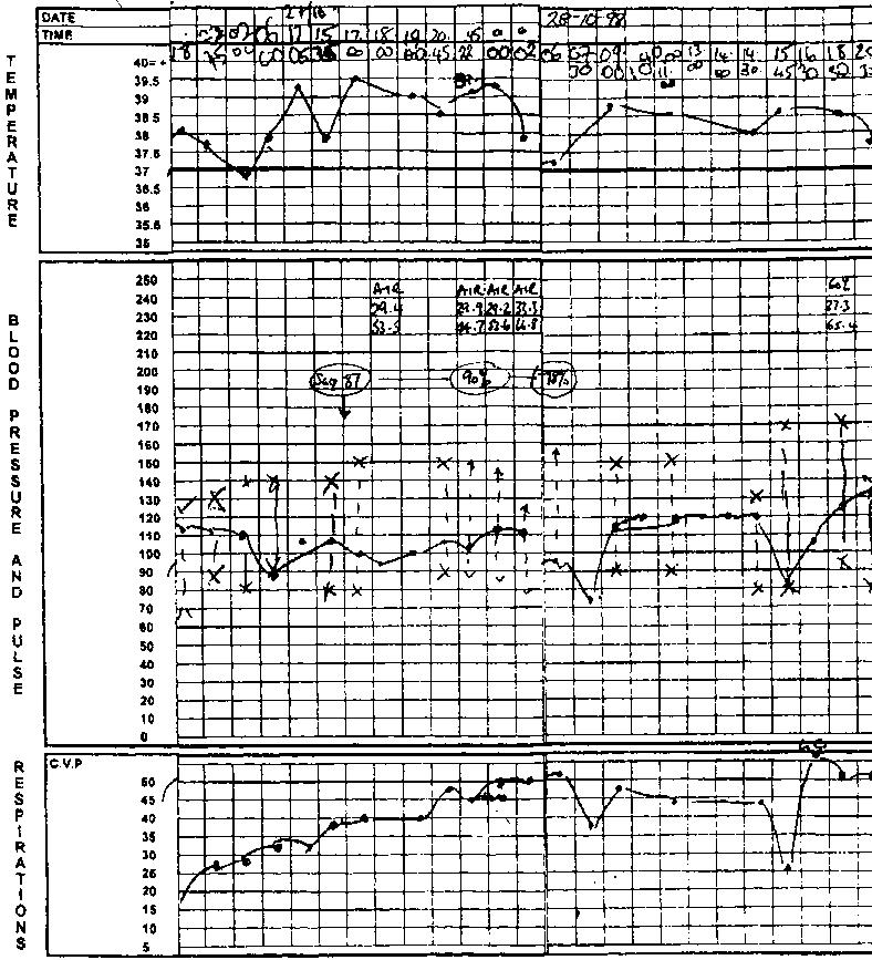
Ward observation chart showing serious physiological deterioration
Figure.
Pulse oximeters give no information about presence or absence of hypercapnia
Figure.
Signs of excessive respiratory work
Figure.
Peripheral cyanosis and poor capillary refill indicate failing circulation
Figure.
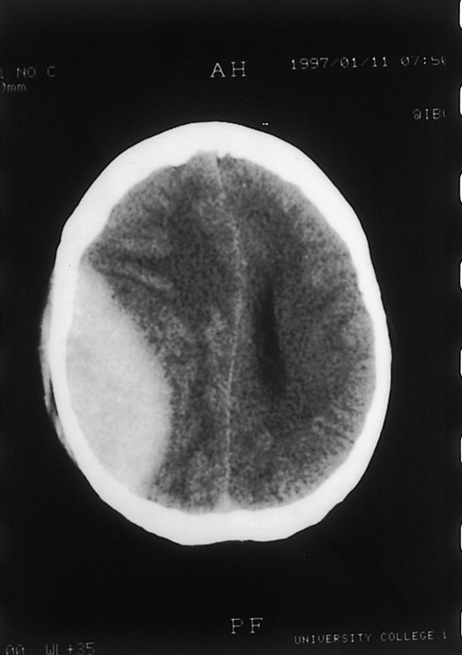
Extradural haematoma
Figure.
Measurement of urine output is important to detect renal problems promptly
Footnotes
Gary Smith is director of intensive care medicine, Queen Alexandra Hospital, Portsmouth, and Mick Nielsen is director of the general intensive care unit, Southampton General Hospital, Southampton.
The ABC of intensive care is edited by Mervyn Singer, reader in intensicve care medicine, Bloomsbury Institute of Intensive Care Medicine, University College London and Ian Grant, director of intensive care, Western General Hospital, Edinburgh. The series was conceived and planned by the Intensive Care Society’s council and research subcommittee.


