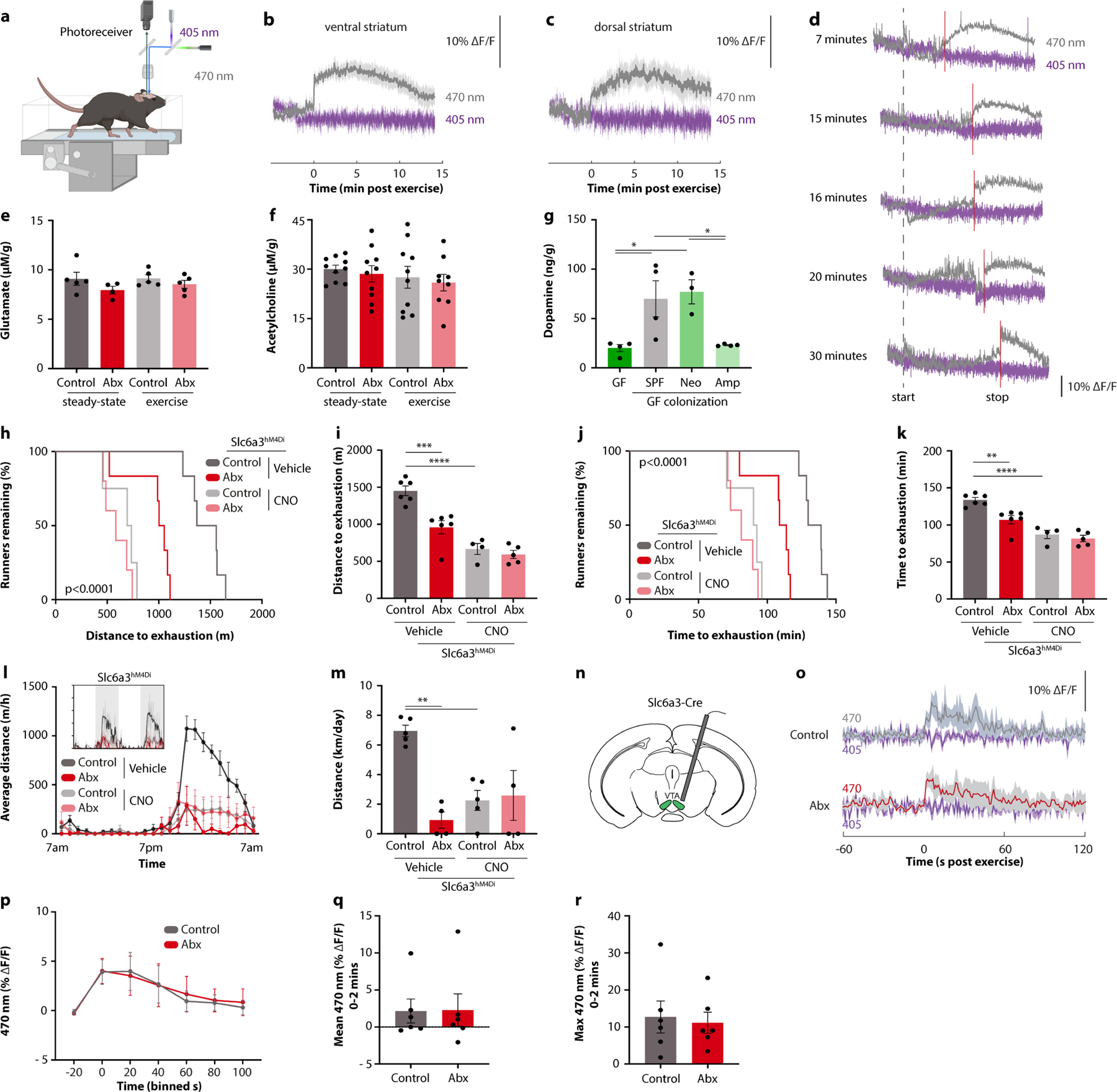
Extended Data Fig. 6 |. The role of microbiome-mediated dopamine responses in exercise performance.

a, Schematic depicting in vivo fibre photometry of dopamine sensor fluorescence in the nucleus accumbens of mice during treadmill running. b-d, Fibre photometry recording of dopamine dynamics at the end of exercise in the ventral striatum (b), the dorsal striatum (c), and the ventral striatum after different durations of exercise (d). e, f, Glutamate (e) and acetylcholine (f) levels in the brain of Abx-treated mice and controls, at steady-state and after endurance exercise. g, Post-exercise dopamine levels in brain tissue of GF mice, and GF mice colonized with stool from SPF, neomycin- or ampicillin-treated mice. h-k, Kaplan-Meier plots (h, j) and quantifications (i, k) of distance (h, i) and time ( j, k) on treadmill of Abx-treated Slc6a3hM4Di mice, with or without CNO treatment. l, m, Averaged hourly distance (l) and quantification (m) of voluntary wheel activity of Abx-treated Slc6a3hM4Di mice, with or without CNO treatment. Inset shows representative recording traces. n-r, Schematic (n), recording traces (o), quantification (p), mean signals (q), and maximum signals (r) of fibre photometry recording from the VTA of Slc6a3-Cre mice injected with a GCamp6-expressing virus. Mice were recorded at the end of the exercise protocol. Error bars indicate means ± SEM. * p < 0.05, ** p < 0.01, *** p < 0.001, **** p<0.0001. Exact n and p-values are presented in Supplementary Table 2.
