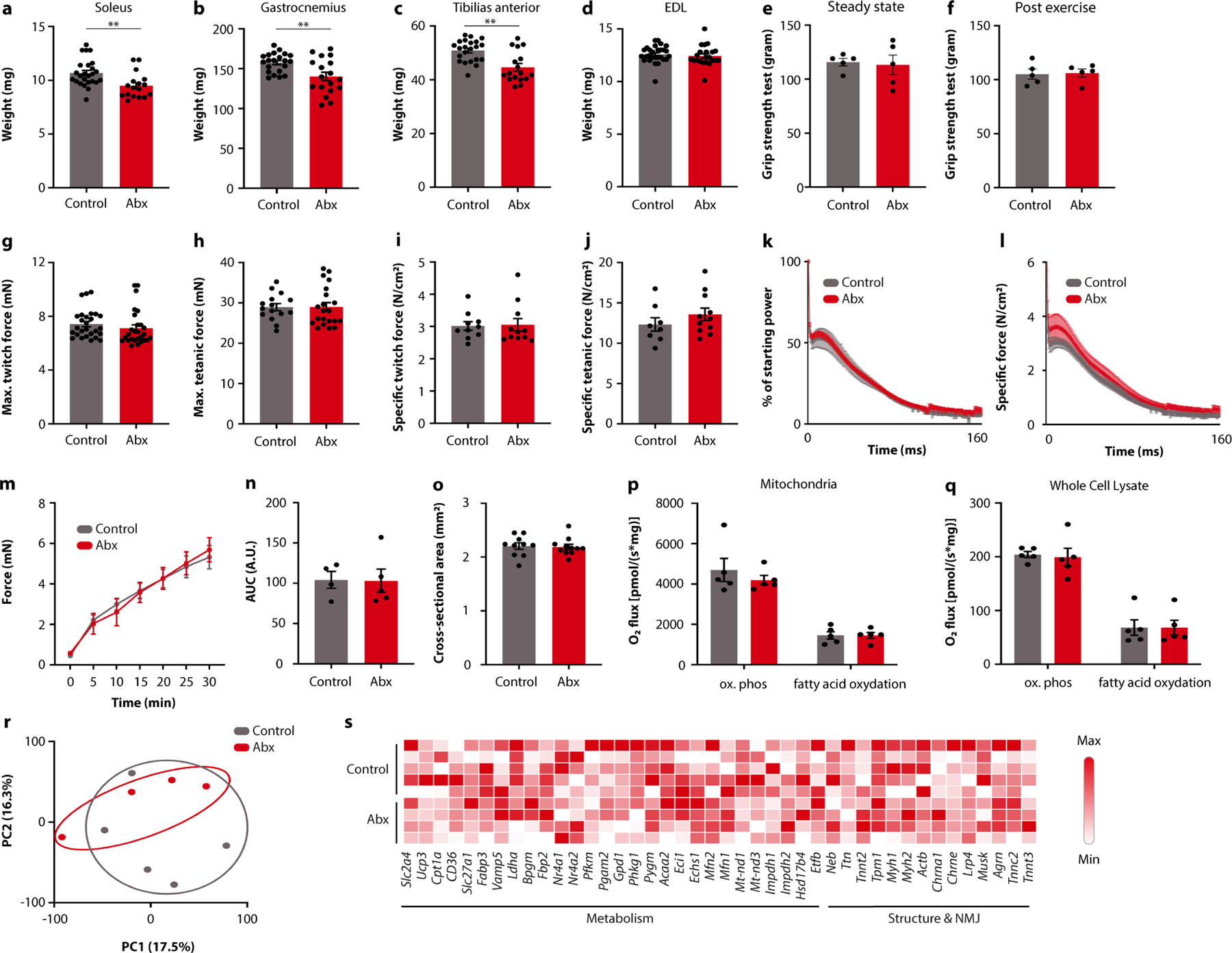
Extended Data Fig. 4 |. The impact of the microbiome on muscle physiology.

a-d, Weight of soleus (a), gastrocnemius (b), tibilias anterior (c), and extensor digitorum longus (EDL) muscles in Abx-treated mice and controls. e, f, Grip strength of Abx-treated mice and controls before (e) and after (f) exercise. g-j, Maximum twitch (g), tetanic force (h), specific twitch force (i), and specific tetanic force ( j) of EDL muscle from Abx-treated mice and controls. k-n, Decrease in power (k) and specific muscle force (l), force recovery over time (m) and force recovery quantification (n) of EDL muscle from Abx-treated mice and controls. o, EDL muscle cross-sectional area in Abx-treated mice and controls. p, q, Quantification of oxidative phosphorylation and fatty acid oxidation of isolated mitochondria (p) and whole cell lysates (q) from EDL muscle obtained from either Abx-treated mice or controls. r, s, PCA plot (r) and heatmap of selected genes (s) from EDL transcriptomes obtained from Abx-treated mice and controls. NMJ, neuro-muscular junction. Error bars indicate means ± SEM. ** p < 0.01. Exact n and p-values are presented in Supplementary Table 2.
