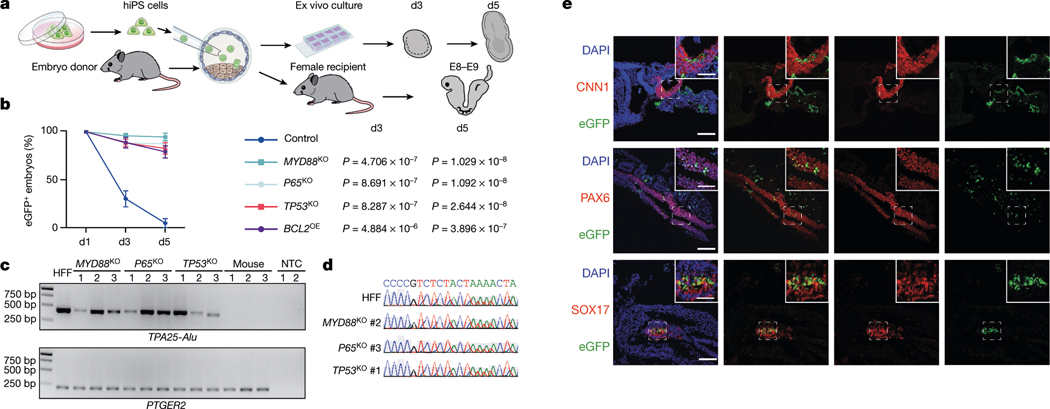
Fig. 3 |. Overcoming interspecies PSC competition enhances human primed PSCs survival and chimerism in early mouse embryos.
a, Schematic showing the generation of ex vivo and in vivo human–mouse chimeric embryos. b, Line graphs showing the percentages of eGFP+ mouse embryos at indicated time points during ex vivo culture after blastocyst injection of wild-type (control), MYD88KO, P65KO, TP53KO and BCL2OE hiPS cells. n = 3 (control), n = 4 (MYD88KO), n = 6 (P65KO), n = 6 (TP53KO) and n = 3 (BCL2OE) independent injection experiments. Data are mean ± s.e.m. P values (versus control) determined by one-way ANOVA with least significant difference (LSD) multiple comparison. c, Genomic PCR analysis of E8–E9 mouse embryos derived from blastocyst injection of MYD88KO, P65KO and TP53KO hiPS cells, and non-injected control blastocysts. TPA25-Alu denotes a human-specific primer; PTGER2 was used as a loading control. HFF, HFF-hiPS cells. NTC, non-template control. This experiment was repeated independently three times with similar results. For gel source data, see Supplementary Fig. 1. d, Sanger sequencing results of representative PCR products generated by human-specific TPA25-Alu primers. A stretch of TPA25-Alu DNA sequences derived from HFF, MYD88KO (#2), P65KO (#3) and TP53KO (#1) from c are shown. e, Representative immunofluorescence images showing contribution and differentiation of eGFP-labelled MYD88KO hiPS cells in E8–E9 mouse embryos. Embryo sections were stained with antibodies against eGFP and lineage markers including CNN1 (mesoderm, top), PAX6 (ectoderm, middle) and SOX17 (endoderm, bottom). Scale bars, 100 μm and 50 μm (insets). Images are representative of three independent experiments.

