



Abstract
Among molecular imaging modalities that can monitor enzyme activity in vivo, optical imaging provides sensitive, molecular-level information at low cost using safe and non-ionizing wavelengths of light. Yet, obtaining quantifiable optical signals in vivo poses significant challenges. Benchmarking using ratiometric signals can overcome dependence on dosing, illumination variability, and pharmacokinetics to provide quantitative in vivo optical data. This review highlights recent advances using fluorescent probes that are processed by enzymes to induce photophysical changes that can be monitored by ratiometric imaging. These diverse strategies include caged fluorophores that change photophysical properties upon enzymatic cleavage, as well as multi-fluorophore systems that are triggered by enzymatic cleavage to alter optical outputs in one or more fluorescent channels. The strategies discussed here have great potential for further development as well as potential broad applications for targeting diverse enzymes important for a wide range of human diseases.
Keywords: Optical imaging, Fluorescent probes, Enzyme probes
Introduction
Functional imaging of enzyme activity in living systems provides valuable insights into biological processes and disease states [1]. While in vitro experiments can be used to make fundamental observations using simplified models, in vivo imaging provides information regarding the regulation of enzyme activity in more complex and therapeutically relevant models of human health and disease [2]. In vivo imaging experiments can provide detailed biological information about enzyme function at various levels, including tissues, organs, systems, and whole organisms. Additionally, in vivo imaging methods developed for clinical use can provide activity-based measurements of disease state and progression that can be used for diagnostics, therapy monitoring and surgical guidance.[3]
Molecular imaging tools for a variety of imaging modalities have enabled non-invasive analysis of enzyme activity in vivo. In particular, enzyme probes suitable for use in magnetic resonance imaging (MRI) [4,5] have high spatial resolution and unrestricted depth penetration in tissues [6]. However, due to the low sensitivity of MRI imaging, enzymes of interest must have fast kinetics and/or high local concentrations to be detected by probes [7]. Enzyme responsive positron emission tomography (PET) and single-photon emission computed tomography (SPECT) probes also are highly sensitive and provide imaging through any tissue depth [8]. Yet, the time and resources required for isotope production and/or on demand probe synthesis limits their use outside of specialized facilities. Further, radiation exposure can limit applications in longitudinal imaging. Recent advances in ultrasound probes that respond to enzyme activity show promise for diverse imaging applications in deep tissues within the vascular and gastrointestinal systems [9].
Optical imaging agents can provide sensitive, molecular-level information about enzyme activity at low cost, using safe and non-ionizing wavelengths of light [10]. There have been a wide range of fluorescent tools, including “always-on” targeted probes, and “turn-on” probes, developed for monitoring enzyme activity [11]. However, in vivo imaging requires measurements of signals within deep tissues, posing challenges for optical imaging agents which have limited penetration depth [12]. These challenges include variable probe distribution among organs and tissues, differences in cellular uptake and permeability, inconsistent incident illumination, and impact of biological environments on overall signal intensity. Optical tomography methods rely on estimations of scattering and absorption to provide quantitative volumetric measurements but cannot exclude signal from probes taken up in healthy tissues. Thus, obtaining quantitative data for both biological and clinical applications is more challenging in vivo compared to live cell or ex vivo imaging methods [13].
Ratiometric imaging is a benchmarking strategy that involves recording and analyzing two linked signals as a ratio. Ratiometric imaging can overcome challenges associated with quantifying optical probe signals in vivo, as ratiometric signals can be normalized to eliminate effects from dosing, illumination variability, and pharmacokinetics. In this review, we explore recent strategies and applications of in vivo ratiometric imaging probes. These strategies include using caging groups which change the photophysical properties of a single chromophore upon enzymatic cleavage (Figure 1a), using two fluorophores that interact photophysically, such as through Förster Resonance Energy Transfer (FRET)), that respond to enzymatic processing (Figure 1b) and using a single fluorophore that can be benchmarked to a separate static fluorescent signal (Figure 1c). Here, we describe probes with a change in either excitation or emission wavelength upon enzyme processing that enable a ratiometric readout. However, fluorescence lifetime is another parameter that can be modulated by enzyme activity [14]. Recently, fluorescent lifetime-based ratiometric imaging methods such as, Macroscopic Fluorescence Lifetime Imaging (MFLI) have been developed for applications including drug-target engagement studies [15].
Figure 1.
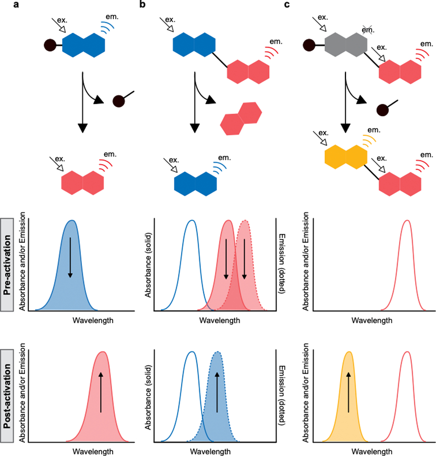
Representative strategies for ratiometric optical imaging. Schematic of enzymatic processing and resulting spectral changes by a) release of a caging group that induces photophysical changes in a single fluorophore, b) release of a photophysically interacting second fluorophore, and c) release of a caging group that unquenches a fluorophore in the presence of a second, spectrally separated fluorophore. Shaded spectra indicate change upon enzymatic reaction in the direction of the appended arrow. Unshaded spectra indicate spectra remain constant upon enzymatic reaction.
The studies highlighted here provide insights for the development of robust and widely applicable probes to enable longitudinal tracking of enzyme activity in vivo. We focus on recent examples of fluorescent, ratiometric optical imaging probes developed and applied in preclinical in vivo models. Readers are encouraged to refer to existing work, particularly reviews on chemical probes for ratiometric fluorescence imaging [16,17] and optical imaging of enzyme activity [18,19,20] for more comprehensive overviews of the surrounding fields.
Strategies to image enzyme activity in vivo: Single fluorophore methods
Caged single fluorophores in the far-red and near-infrared (NIR)
Perhaps the most common strategy used in the development of enzyme-responsive optical probes is to employ a fluorophore whose fluorescent signal is quenched by FRET using a second non-fluorescent chromophore. Enzyme processing leads to unquenching of the dye and turn on of a signal. This same general strategy can be used for ratiometric imaging by replacing the quencher with a fluorophore, resulting in a FRET signal. As an alternative strategy, it is possible to use a single dye appended to a cleavable caging group, whose photochemical properties change upon processing by an enzyme. This simplifies synthesis and reduces the overall size of the probe. Several examples of this strategy in cellular microscopy experiments include boron dipyrromethene (BODIPY) probes that respond to esterase activity [21] and aminocoumarin probes that respond to processing by acyl-protein thioesterases [22]. In vivo imaging probes using this strategy often rely on red-shifted chromophores such that the excitation and emission wavelengths are within the far-red (600 – 700 nm) or near-infrared (NIR; 700 – 1000 nm) regions to increase depth penetration of optical signals through tissue and reduce background autofluorescence. Early efforts in the development of caged ratiometric probes with far-red emission include styryl push-pull chromophores caged with galactose that respond to β-galactosidase (β-gal) activity [23,24]. Ratiometric measurements in vitro and in live cells enabled quantitation of β-gal activity, however this approach has not yet been translated to in vivo imaging.
More recently, Xiang et al. developed a NIR ratiometric probe selective for butyrylcholinesterase (BChE), an enzyme that plays roles in metabolic functions in the liver [25]. BChE activity has been recognized as a general indicator of healthy liver function and can be used to monitor progression of chronic liver diseases, such as non-alcoholic fatty liver disease (NAFLD) [26]. Previously, in vitro ratiometric diagnostic methods using carbon dots [27] coupled with point-of-care microfluidic devices [28] provided sensitive detection of BChE and acetylcholinesterase (AChE) activities. These methods improved sensitivity over traditional colorimetric or non-ratiometric strategies, thus allowing detection of acetylcholinesterase activity in complex samples such as whole blood. However, the need for exogenous reagents (i.e. metal ions, substrates, selective inhibitors) prevents their use for in vivo imaging. Furthermore, a general lack of selectivity between BChE and AChE and interference from glutathione (GSH) make their in vivo use challenging.
To address the challenges with existing probes, Xiang et al. converted a tricyanofuranyl iminosalicylaldehyde (TCFIS) chromophore (2) [29] previously used to sense HNO [30] and hydrogen polysulfide [31] into a BChE responsive probe by modification with a cyclopropionyl ester (1, Figure 2a) [25]. The presence of the electron-donating ester weakens the intramolecular charge transfer (ICT) of TCFIS, resulting in a chromophore that displays two weak emission peaks at 626 nm and 760 nm when excited at 575 nm. Upon cleavage of the ester to regenerate the free phenol, ICT is restored, and an aggregation induced emission (AIE) response is observed as a large increase in fluorescence at 626 nm relative to 730 nm. When the probe, TB-BChE (1), was applied in vivo and excited at 570 nm, a ratiometric response was measured by collecting emission at 600–650 nm (red) and 700–780 nm (NIR) and calculating the Ired/INIR ratio (Figure 2b). The probe has a limit of detection (LOD) of 39 ng/mL and has high selectivity for BChE over AChE. Application of the probe to an in vivo model of NAFLD demonstrated that increased BChE activity is correlated with markers of disease progression such as high levels of liver steatosis and fat deposition. A similar probe that employs the TCIFS chromophore was developed [32] using an ester that is selectively processed by AChE.[33] However, the overall low fluorescence of the caged chromophore likely precludes its application for ratiometric imaging applications.
Figure 2.
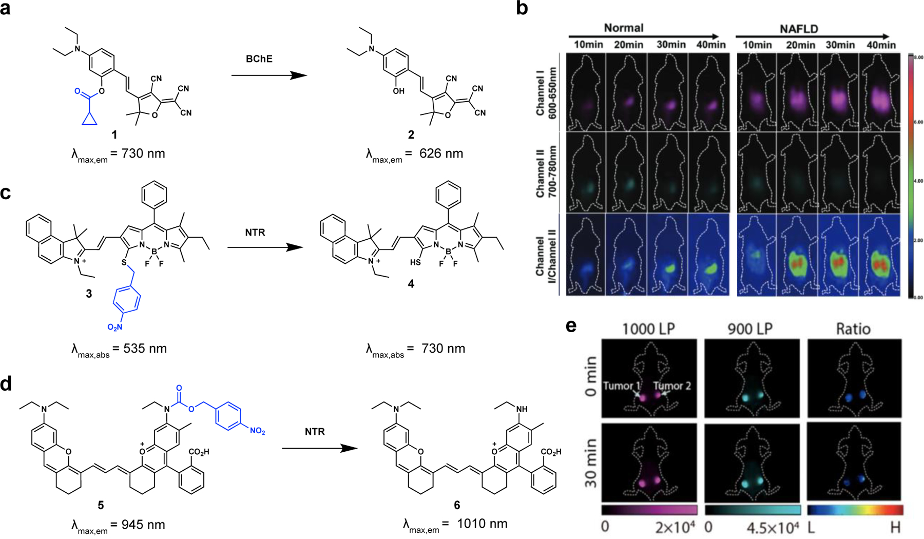
Single fluorophore-based ratiometric fluorescent probes for in vivo imaging. a) Ester-capped TCFIS chromophore 1 is cleaved by BChE, resulting in 2, with a blue shifted emission wavelength. b) Ratiometric in vivo imaging of a lung cancer model with 1 (ex. 570 nm). c) NTR-InD (3) is converted to 4 with a red-shifted absorption wavelength by treatment with NTR and reduction and elimination of the nitrobenzyl group. d) Rap-N (5) is converted to red-shifted 6 upon treatment with NTR and reduction and elimination of the nitrobenzylcarbamate. e) Ratiometric in vivo imaging of a breast tumor model with 5 (ex. 808 nm).
Caged single fluorophores in the SWIR
Recently, there has been increasing interest in using probes with excitation and/or emission wavelengths beyond the NIR region for optical imaging. Shortwave infrared (SWIR; 1000 – 1700 nm, also called NIR-II) detection has the benefit of increased tissue penetration as well as improved resolution and contrast compared to NIR wavelengths.[34] Several fluorophore classes have been developed for SWIR imaging, including, Donor-Acceptor-Donor (DAD) [35], polymethine [36], and aza-boron dipyrromethene (aza-BODIPY) dyes [37]. Furthermore, diverse probes that report in this wavelength region have been developed for tracking metabolites, pH, viscosity, and enzyme activity [38].
In a recent example, Wang et al. developed enzyme responsive probes using red-shifted BODIPY chromophores caged with a self-immolative benzyl thioether linker attached to substrates that can be processed by enzymes [39]. Using this approach, it was possible to generate probes that change their excitation and emission properties upon processing by nitroreductase (NTR), NAD(P)H quinone oxidoreductase isozyme 1 (NQO), or alkaline phosphatase (ALP). For the NTR probe, NTR-InD (3), reduction of the nitro group by NTR and subsequent 1,6-elimination results in conversion to 4, and a concomitant increase in SWIR emission (~850 – 1150 nm) with 730 nm excitation and a decrease in NIR emission (~575 – 725 nm) with 535 nm excitation (Figure 2c). Thus, a ratiometric change can be calculated for Iex730/Iex535. A similar probe that responds to β-Gal activity was developed to image ovarian tumors [40]. In vivo imaging experiments, however, have yet to capitalize on the ratiometric response of these probes.
NTR responsive probes have also been generated by Lan et al. using SWIR emissive polymethine dyes [41]. Capping a polymethine similar to CX-2 [42] with a para-nitro carbamate produces probe Rap-N (5) (λmax,abs = 795 nm, λmax,em = 945 nm). Reduction of the nitro group on Rap-N (5) by NTR produces an aniline which subsequently undergoes 1,6-elimination and carbamic acid hydrolysis to release the free polymethine dye Py-2 (6) (λmax,abs = 965 nm, λmax,em = 1010 nm; Figure 2d). In vivo imaging with excitation at 808 nm and collection of signals with long pass filtering at 900 nm or 1000 nm, enables calculation of the ratio of ILP1000/ILP900 in mouse models of cancer (Figure 2e). In a 4T1 model of breast cancer and a CT6 model of colon cancer, NTR activity was found to correlate with tumor hypoxia, in agreement with prior studies [43].
Two-photon imaging of caged single fluorophores
Two-photon excitation offers an alternate strategy to using red-shifted fluorophores for increasing tissue penetrance of excitation light. The longer wavelength excitation provides higher signal to background ratios due to reduced interference from endogenous biomolecules compared to one photon imaging with probes that absorb at similar wavelengths. However, imaging with two-photon excitation involves laser-scanning illumination, often resulting in long acquisition times and limiting the focal volume which can be observed. Thus, two-photon strategies are often limited to in vivo applications with small fields of view and using optical windows.[44] Further, the ratiometric imaging using two-photon excitation has been so far limited to ex vivo applications, but the studies highlighted here provide a starting point for translating these technologies in vivo.
Several probes using various chromophores linked to a L-glutamate substrate have been developed to image γ‑glutamyltransferase (GGT) activity and applied to two-photon ratiometric imaging in cancer models. GGT is a commonly used imaging target due to its overexpression on the cell surface of many human tumor types [45]. Park et al. used a cresyl violet chromophore to perform 2-photon microscopy on excised mouse colons using 1000 nm excitation and 550–590 nm (green) and 610–650 nm (red) detection. Notably, the ratiometric response, Ired/Igreen was time, but not depth dependent and could be detected at up to ~30 µm below the tissue surface [46]. A different strategy using a naphthalene-benzothiazole donor-acceptor chromophore (7) was developed for GGT by Kim et al. [47] (Figure 3a). Ratiometric imaging in human colon tissues for the detection of cancerous lesions was performed using 750 nm excitation and emission windows of 400−450 nm (blue) and 500−600 nm (green), to obtain the Igreen/Iblue ratio to distinguish 7 and 8. Using a similar strategy, Reo et al. used an L-glutamate caged amino-benzocoumarin fluorophore for two-photon microscopy in a CT-26 mouse cancer model [48]. These methods for ratiometric imaging of GGT activity were all applied to excised tissues, but have yet to be used for in vivo imaging, likely due to the relatively blue-shifted detection channels required.
Figure 3.
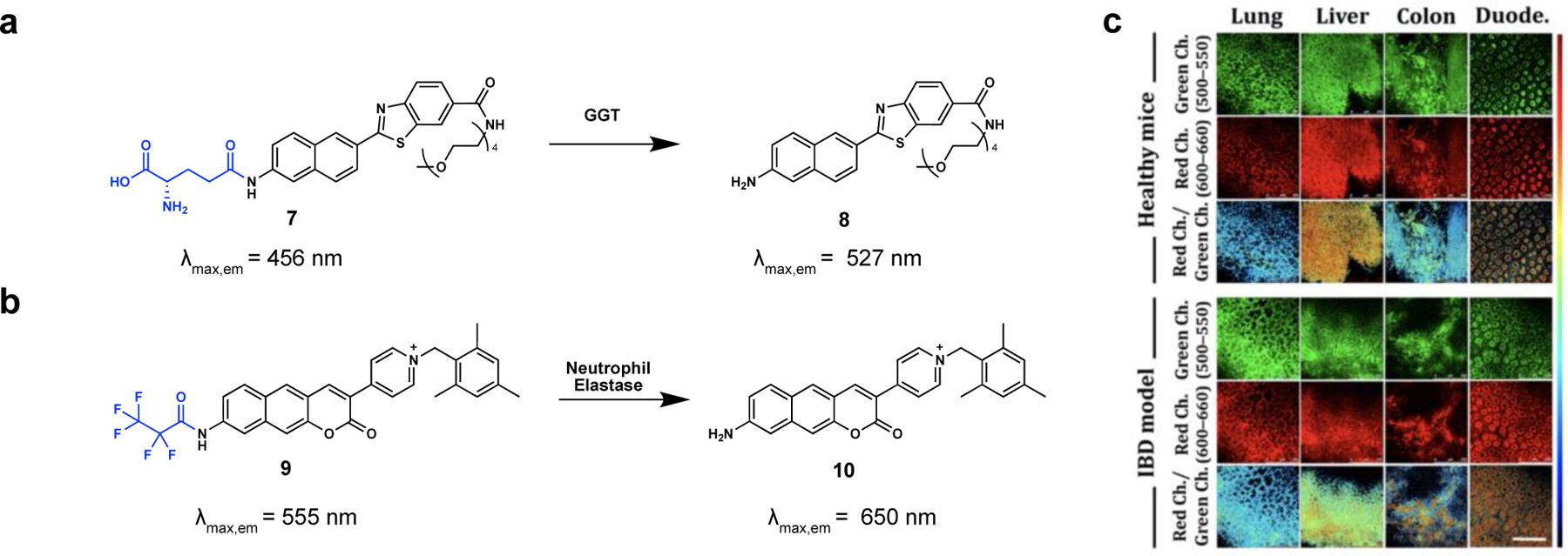
Caged single-fluorophores imaged with two-photon ratiometric imaging. a) L-glutamate caging group on naphthalene-benzothiazole chromophore 7 is cleaved by GGT, resulting in chromophore 8, with a red-shifted emission wavelength. b) Neutrophil elastase cleaves the pentafluoropropanamide group on rTP-BC3 (9) resulting in 10, with a red-shifted emission wavelength. c) Ratiometric imaging of collected tissues of an IBD mouse model, treated with 9 (2-photon ex. 800 nm).
Probes composed of amino-benzocoumarin fluorophores have recently been developed to monitor the activity of the serine protease, neutrophil elastase [49]. Early work produced turn-on fluorescent probes using coumarin, [50] polymethine, [51] and rhodamine [52] dyes linked to neutrophil elastase-targeting molecules through a pentafluoropropionamide capping group. A turn-on response was observed when the capping group was cleaved by the protease. More recently, Sarkar et al. expanded on this work to enable ratiometric detection by increasing the conjugation length of the chromophore and adding an electron acceptor to induce changes in fluorescence wavelength upon cleavage of the pentafluoropropionamide capping group [49]. Using 4-pyridinium as an electron acceptor reduced solvent and pH sensitivity, and a N-methylmesitylene substituent on the pyridinium increased the brightness of the probe. The emission peak of optimized rTP-BC3 probe (9) changes from 555 nm to 650 nm (Δλ = 95 nm) after incubation with neutrophil elastase (10) (Figure 3b). By using 2-photon excitation at 800 nm, detection at 500–550 nm (green), 600–650 nm (red), and ratiometric analysis (Ired/Igreen), it was possible to image neutrophil migration and changes in activity in ex vivo tissues in a mouse model of inflammatory bowel disease (IBD) (Figure 3c).
Strategies to image enzyme activity in vivo: Multi fluorophore methods
Ratiometric FRET probes
In contrast to changing the photophysical properties of a single fluorophore by enzymatic reaction, another commonly employed strategy for producing ratiometric fluorescence employs two fluorophores whose Forester Resonance Energy Transfer (FRET) interaction is changed by reaction with an enzyme. Seminal work in this area by Tsein, Nguyen and colleagues focused on an enzyme cleavable sequence flanked by poly(arginine) and poly(glutamic acid) and Cy5 and Cy7, respectively (Figure 4a, 11) [53]. The intact probes place the Cy5 and Cy7 within close proximity (<10 nm) enabling FRET between the two dyes. Using 620 nm excitation and collection at 670 nm (Cy5 emission) and 780 nm (Cy7 emission) the intact probe produces a low ratio of I670/I780 as the Cy5 is quenched by Cy7. Upon cleavage of the substrate by a protease, Cy5 and Cy7 diffuse apart, disrupting the FRET interaction between the two fluorophores and increasing the ratio signal I670/I780, resulting in a ~40-fold increase in dynamic range [53]. Additionally, the release of the polycationic poly(arginine) sequence which acts as a cell penetrating peptide, drives intracellular accumulation of the Cy5 fluorophore labeled peptide fragment leading to prolonged retention at the tumor site [54]. This strategy has been primarily developed with peptide cleavage sequences targeting MMP-2/MMP-9 and elastases, with applications in metastatic lymph node detection [53] pancreatic cancer [55], head and neck cancers[56,57], and breast cancer. An optimized probe, AVB-620 (Figure 4b) [58] has recently completed a phase II clinical trial for detecting primary tumors, tumor margins and malignant lymph nodes in breast cancer surgeries, including for ductal carcinoma in situ (DCIS) and primary invasive carcinoma of the breast (stages I-III) [59]. The same approach has also been applied to image inflammation by targeting thrombin [60].
Figure 4.
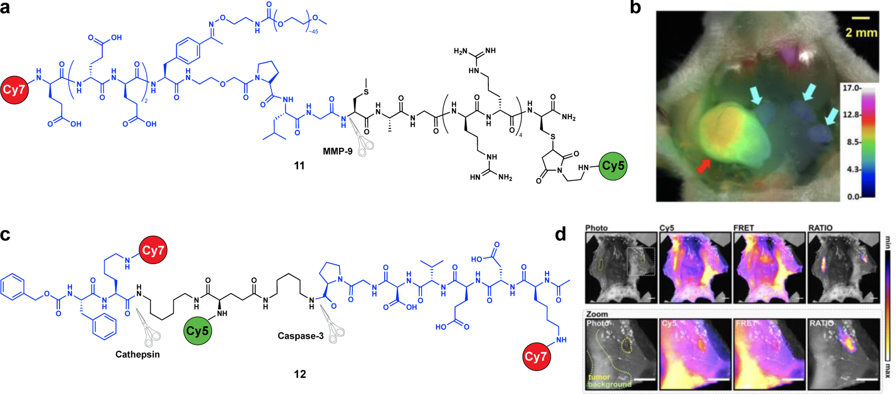
Imaging of enzyme activity with ratiometric FRET probes. a) Structure of ratiometric FRET probe AVB-620 (7) that responds to MMP-9 protease activity. b) Ratio image (ICy5/ICy7) of cervical lymph nodes following dosing with AVB-620 (11). Cyan and red arrows designate cancer negative and positive lymph nodes, respectively. c) Structure of Death-Cat-RATIO (12), a dual substrate “AND-gated” ratiometric FRET probe with response to cathepsins and caspase-3. d) Ex-vivo fluorescent images of metastases-like breast tumors after injection with Death-Cat-RATIO 24 h prior to imaging. Ratio channel is (ICy5/IFRET). Dashed lines indicate the tumor boundaries and areas of background tissue.
In recent work, FRET ratiometric imaging was used to image tumor-associated cathepsin and caspase proteases [61]. Faucher et al. adapted both single substrate [62] and dual substrate logic gated probes [63] that were previously developed as quenched turn-on probes in response to cathepsin and caspase-3 activities. Cathepsins are cysteine proteases secreted by tumor-associated macrophages (TAMs) while caspase-3 is active in the cytosol of apoptotic cells. The use of a dual substrate “AND-gate” probe for two complementary enzymes expressed in the tumor with different substrate specificities reduced off-target activation and increased the specificity of the signal for the disease area of interest. By maintaining the donor fluorophore (Cy5) and converting the FRET-acceptor from a fluorescence quencher (QSY21) to a fluorophore (Cy7), tumor-to-background ratios were improved from ~1.2 to ~4.2 for the single substrate probe (6QC-RATIO) and from ~2.2 to ~7.2 for the dual substrate probe (Death-Cat-RATIO, 12) at a 24 h time point after probe injection (Figure 4c–d). These improved tumor-to-background ratios in the ratiometric imaging experiments enabled visualization of small metatheses-like tumors (~4.6 mm in diameter) as well as distal tumors not directly in line with the illumination source. Additionally, quenched turn-on probes often produce nonspecific signal in metabolic and excretory organs, such as the liver and kidneys, respectively, due to high levels of enzymatic activity and/or probe accumulation. Imaging with the ratiometric probe Death-Cat-RATIO (8) demonstrated drastically reduced signal in the liver and kidneys, compared to the turn-on probe with analogous substrates. The ratiometric probe provided significantly higher tumor signal compared to liver and kidney signals, opening up opportunities to distinguish malignant tissue within these clearance organs in future studies. Overall, this strategy of reconfiguring existing turn-on probes into ratiometric versions offers a relatively simple approach that could be broadly applied to improve signal to background and sensitivity as well as mitigate challenges with uneven illumination that occur across heterogenous biological tissues.
Benchmarking a turn on dye with a linked chromophore:
Ratiometric imaging can be achieved by relying on two fluorophores which are not interacting photophysically with each other. In this case, one chromophore signal can be benchmarked to a static signal from the other. Early work in this area employed a mixture of dye-iron oxide nanoparticle conjugates with two different color fluorophores, wherein one probe contained a cathepsin B-cleavable sequence between the dye and nanoparticle and the other contained a non-cleavable linkage [64]. Because the iron oxide nanoparticles serve as a fluorescent quencher, the activity of cathepsin B could be monitored by observing Cy5.5 signal (λmax,abs = 630 nm, λmax,em = 700 nm) and benchmarking it against Cy3.5 fluorescence (λmax,abs = 520 nm, λmax,em = 600 nm), which is expected to remain constant. As the overall structure of the nanoparticles is similar, any differences in biodistribution between the particle are estimated to be negligible. Recently, Huang et al. developed a ratiometric probe for GGT with an activatable polymethine dye (λmax,abs = 660 nm, λmax,em = 735 nm) conjugated to an always-on BODIPY dye (λmax,abs = 480 nm, λmax,em = 517 nm; Figure 5a, 13) [65]. The probe showed sensitive and selective detection of GGT activity after intratumoral injection of mice bearing subcutaneous HeLa tumors.
Figure 5.
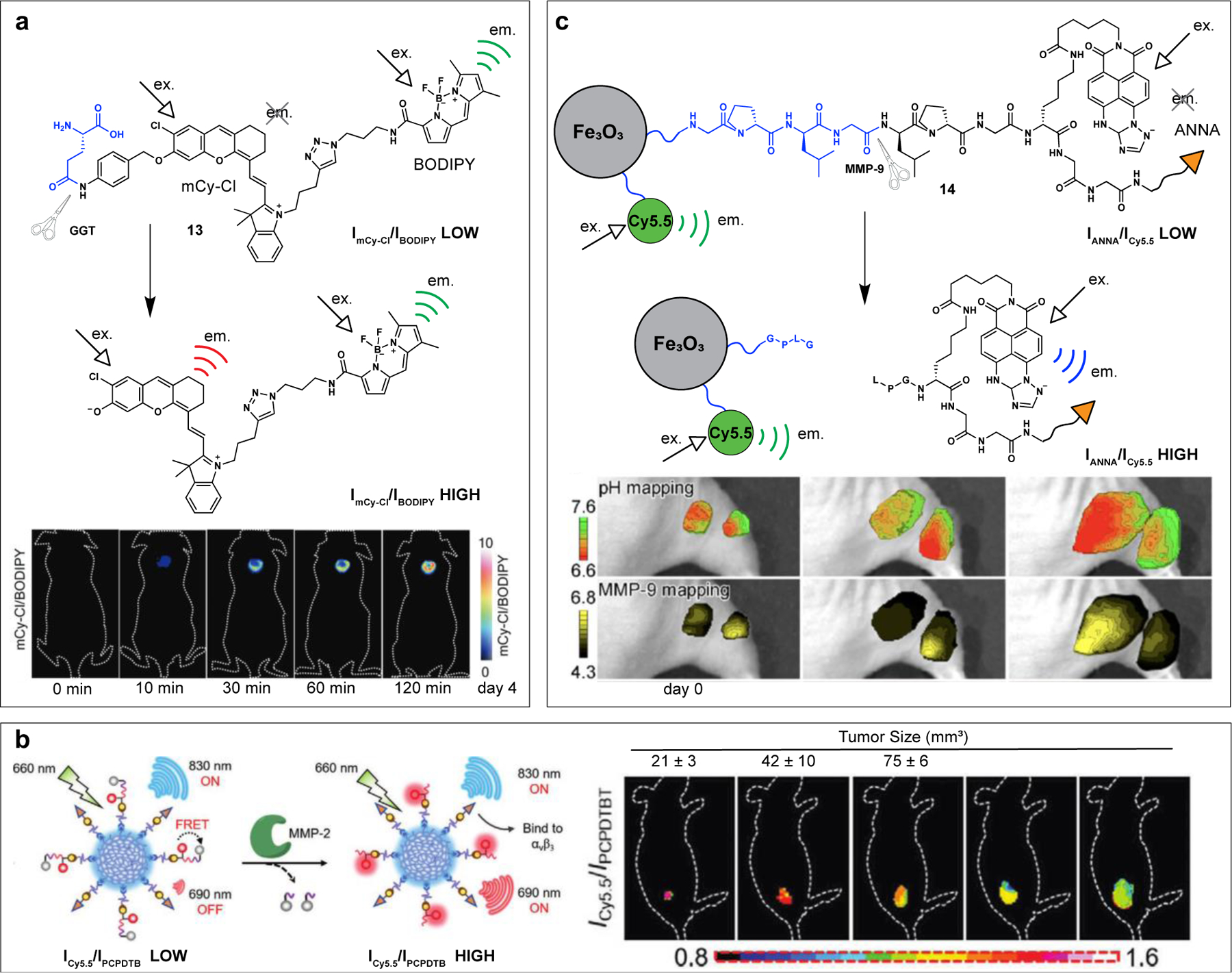
Ratiometric imaging with an “always on” and a turn-on” fluorophore. a) Ratiometric probe for GGT (9) before and after enzymatic cleavage. Ratio images of HeLa tumor mouse models after treatment with 13 (mCy-Cl channel: λex = 660 nm, λem = 710 ± 20 nm); BODIPY channel: λex = 480 nm, λem 520 ± 20 nm). b) Conjugated polymer (PCPDTBT) nanoparticles appended with MMP-2 cleavable peptide flanked with Cy5.5 and a quencher. Ratio images of primary gastric tumors in mice after treatment with polymer agent (Cy5.5 channel: λex = 660 nm, λem = 690 nm; PCPDTB channel: λex = 660 nm, λem 830 nm;). c) Ratiometric probe for MMP-9 (14) before and after enzymatic cleavage. Ratio images of LS180 tumors after treatment with 14 to observe pH mapping (ANNA channel 1: λex = 465–500 nm, λem 500 nm; ANNA channel 2: λex = 465–500 nm, λem 540 nm) and MMP-9 activity (ANNA channel: λex = 465–500 nm, λem 500 nm LP; Cy5.5 channel: λex = 640–675 nm, λem 680 nm LP).
In recent work by Zeng et al., the authors describe a system for imaging MMP-2 activity by using two chromophores with similar excitation wavelength but distinct stokes shifts, and thus spectrally separated emission wavelengths [66]. The use of a single excitation wavelength for both chromophores ensures consistent illumination and accurate overlay of the two emission channels. Specifically, nanoparticles composed of a conjugated polymer, poly[2,6-(4,4-bis-(2-ethylhexyl)-4H-cyclopenta[2,1-b;3,4-b′]dithiophene)-alt-4,7)(2,1,3-benzothiadiazole)] (PCPDTBT), were decorated with Cy5.5 that is linked to a quencher through an MMP-2-selective peptide substrate (QSY21-GGPLGVRGK-Cy5.5; Figure 5b). Before activation, under 660 nm excitation light, Cy5.5 is nonfluorescent, while PCPDTBT displays fluorescence at 830 nm. Upon treatment with MMP-2, the substrate linking Cy5.5 and the quencher is cleaved, QSY21-GGPLG diffuses away, and the fluorescence of Cy5.5 at 690 nm increases. The turn-on signal of Cy5.5 can be benchmarked against the constant fluorescence of PCPDTBT at 830 nm by calculating the ratiometric signal of I690/I830 (ex. 660 nm). The authors observe a 176-fold enhancement in I690/I830 upon treatment with MMP-2 in vitro, and apply the probe in vivo to track MMP-2 activity and identify metastases in mice with primary gastric tumors (Figure 5b).
Using a similar strategy, Ma et al. labelled iron oxide nanoparticles with both an “always on” Cy5.5 chromophore and an N-carboxyhexyl derivative of 3-amino-1,2,4-triazole fused 1,8-naphthalimide (ANNA) chromophore with an MMP-9-cleavable linker (Figure 5c, 14) [67]. Before cleavage by MMP-9, the fluorescence of the ANNA chromophore is quenched by FRET interaction with the iron oxide nanoparticles. Upon cleavage, ANNA is released from the nanoparticle, and fluorescence is activated. Thus, MMP-9 activity within the tumor can be monitored using the integrated IANNA/ICy5.5 intensity. Furthermore, due to the pH sensitivity of the ANNA fluorophore, both MMP-9 activity and pH could be mapped longitudinally with tumor progression (Figure 5c).
Finally, methods have been developed to used biological autofluorescence itself as a benchmark for fluorescence intensity of an exogenous fluorophore. Hwang et al. demonstrated this approach using ratiometric spectral imaging with a relatively blue-shifted chromophore, HerGa [68]. By creating a spectral signature of the tissue autofluorescence before injection and recording the spectral signature of both the autofluorescence and probe after injection, the background could be treated as a constant benchmark to provide quantitative data on accumulation of the tumor targeting probe. Applying spectral methods such as these could be beneficial in improving quantitation of enzyme activity using existing one-channel probes, as well as for the establishment of clinical thresholds and standards [69].
Conclusion
Using small molecule fluorophores from the far red to the SWIR, it is possible to apply diverse ratiometric strategies to facilitate activity-based imaging of a variety of enzyme classes. It is important to note that optical properties of tissue vary greatly over the visible, NIR and SWIR regions [70]. Thus, changes in excitation wavelength and emission wavelength can change the absorption, scattering, and autofluorescence observed in imaging experiments. Depending on the spectral region, these changes can impact image resolution and/or depth [71] and should be characterized and accounted for when deemed to be significant.
Quantitative measurements are essential in both small animal imaging experiments as well as for diagnostics and surgical guidance in human patients. The incorporation of ratiometric measurements into fluorescence imaging workflows offers improved biological quantitation compared to single channel measurements. Yet it has not yet been widely adopted in clinical applications. This is likely due to the relatively small number of ratiometric probes developed compared to “always on” or a single-channel “turn-on” probes, as well as the lack of commercially available instruments designed for ratiometric imaging, for both preclinical and clinical applications. However, commonly used commercial preclinical optical imaging systems can be employed for a wide range of ratiometric imaging experiments using post-processing to obtain the ratiometric results. Dedicated real-time ratiometric fluorescence imaging instruments have also been developed as companions to specific probes and to enable clinical translation [59,61,72]. Advances in both ratiometric probes and instrumentation are poised to improve quantitation of enzyme activity in vivo across a wide range of applications from biological discovery research to surgical guidance in human patients.
Acknowledgements
This work was supported by the National Institutes of Health [R01 EB028628 to M.B., T32CA11868 to E.D.C.]
Footnotes
Publisher's Disclaimer: This is a PDF file of an unedited manuscript that has been accepted for publication. As a service to our customers we are providing this early version of the manuscript. The manuscript will undergo copyediting, typesetting, and review of the resulting proof before it is published in its final form. Please note that during the production process errors may be discovered which could affect the content, and all legal disclaimers that apply to the journal pertain.
Declaration of Interest: None.
Declaration of interests
The authors declare that they have no known competing financial interests or personal relationships that could have appeared to influence the work reported in this paper.
References
*of special interest
- [1].Zmudzinski M, Malon O, Poręba M, Drąg M, Imaging of proteases using activity-based probes, Curr. Opin. Chem. Biol 74 (2023) 102299. [DOI] [PubMed] [Google Scholar]
- [2].Yang C, Wang Q, Ding W, Recent progress in the imaging detection of enzyme activities in vivo, RSC Adv 9 (2019) 25285–25302. [DOI] [PMC free article] [PubMed] [Google Scholar]
- [3].James ML, Gambhir SS, A molecular imaging primer: modalities, imaging agents, and applications. Physiol. Rev 92 (2012), 897–965. [DOI] [PubMed] [Google Scholar]
- [4].Louie AY, Hüber MM, Ahrens ET, Rothbächer U, Moats R, Jacobs RE, Fraser SE, Meade TJ, In vivo visualization of gene expression using magnetic resonance imaging, Nat. Biotechnol 18 (2000) 321–325. [DOI] [PubMed] [Google Scholar]
- [5].Ye D, Shuhendler AJ, Pandit P, Brewer KD, Tee SS, Cui L, Tikhomirov G, Rutt B, Rao J, Caspase-responsive smart gadolinium-based contrast agent for magnetic resonance imaging of drug-induced apoptosis, Chem. Sci 5 (2014) 3845–3852. [DOI] [PMC free article] [PubMed] [Google Scholar]
- [6].Li H, Meade TJ, Molecular magnetic resonance imaging with Gd(III)-based contrast agents: challenges and key advances, J. Am. Chem. Soc 141 (2019) 17025–17041. [DOI] [PMC free article] [PubMed] [Google Scholar]
- [7].Angelovski G, What we can really do with bioresponsive MRI contrast agents, Angew. Chem. Int. Ed 55 (2016) 7038–7046. [DOI] [PubMed] [Google Scholar]
- [8].Rempel BP, Price EW, Phenix CP, Molecular imaging of hydrolytic enzymes using PET and SPECT, Mol. Imaging 16 (2017) 1536012117717852. [DOI] [PMC free article] [PubMed] [Google Scholar]
- [9].Lakshmanan A, Jin Z, Nety SP, Sawyer DP, Lee-Gosselin A, Malounda D, Swift MB, Maresca D, Shapiro MG, Acoustic biosensors for ultrasound imaging of enzyme activity, Nat. Chem. Biol 16 (2020) 988–996. [DOI] [PMC free article] [PubMed] [Google Scholar]
- [10].Shah K, Weissleder R, Molecular optical imaging: Applications leading to the development of present day therapeutics, NeuroRX 2 (2005) 215–225. [DOI] [PMC free article] [PubMed] [Google Scholar]
- [11].Garland M, Yim JJ, Bogyo M, A bright future for precision medicine: advances in fluorescent chemical probe design and their clinical application, Cell Chem. Biol 23 (2016),122–136. [DOI] [PMC free article] [PubMed] [Google Scholar]
- [12].Bashkatov AN, Genina EA, Kochubey VI, Tuchi VV, Optical properties of human skin, subcutaneous and mucous tissues in the wavelength range from 400 to 2000 nm, J. Phys. D: Appl. Phys 38 (2005) 2543. [Google Scholar]
- [13].Bagheri N, Carpenter AE, Lundberg E, Plant AL, Horwitz R, The new era of quantitative cell imaging-challenges and opportunities, Mol. Cell 82 (2022) 241–247. [DOI] [PMC free article] [PubMed] [Google Scholar]
- [14].Terai T, Ito H, Hanaoka K, Komatsu T, Ueno T, Nagano T, Urano Y, Detection of NAD(P)H-dependent enzyme activity by time-domain ratiometry of terbium luminescence, Bioorganic Medicinal Chem. Lett 26 (2016) 2314–2317. [DOI] [PubMed] [Google Scholar]
- [15].Ochoa M, Rudkouskava A, Smith JT, Intes X, Barroso M, Macroscopic fluorescence lifetime imaging for monitoring of drug–target engagement, in: Rasooly A, Baker H, Ossandon MR (Eds.), Biomedical Engineering Technologies. Methods in Molecular Biology, vol 2394, Humana, New York, New York, 2022. pp. 837–856. [DOI] [PMC free article] [PubMed] [Google Scholar]
- [16].Lee MH, Kim JS, Sessler JL, Small molecule-based ratiometric fluorescence probes for cations, anions, and biomolecules, Chem. Soc. Rev 44 (2014) 4185–4191. [DOI] [PMC free article] [PubMed] [Google Scholar]
- [17].Huang X, Song J, Yung BC, Huang X, Xiong Y, Chen X, Ratiometric optical nanoprobes enable accurate molecular detection and imaging, Chem. Soc. Rev 47 (2018) 2873–2920. [DOI] [PMC free article] [PubMed] [Google Scholar]
- [18].Ou Y, Wilson RE, Weber SG, Methods of measuring enzyme activity ex vivo and in vivo, Annu. Rev. Anal. Chem 11 (2018) 1–25. [DOI] [PMC free article] [PubMed] [Google Scholar]
- [19].Yang C, Wang Q, Ding W, Recent progress in the imaging detection of enzyme activities in vivo, RSC Adv 9 (2019) 25285–25302. [DOI] [PMC free article] [PubMed] [Google Scholar]
- [20].Chyan W, Raines RT, Enzyme-activated fluorogenic probes for live-cell and in vivo imaging, ACS Chem. Biol 13 (2018) 1810–1823. [DOI] [PMC free article] [PubMed] [Google Scholar]
- [21].Komatsu T, Urano Y, Fujikawa Y, Kobayashi T, Kojima H, Terai T, Hanaoka K, Nagano T, Development of 2,6-carboxy-substituted boron dipyrromethene (BODIPY) as a novel scaffold of ratiometric fluorescent probes for live cell imaging, Chem. Commun (2009) 7015–7017. [DOI] [PubMed]
- [22].Beck MW, Kathayat RS, Cham CM, Chang EB, Dickinson BC, Michael addition-based probes for ratiometric fluorescence imaging of protein S-depalmitoylases in live cells and tissues, Chem. Sci 8 (2017), 7588–7592. [DOI] [PMC free article] [PubMed] [Google Scholar]
- [23].Gu K, Xu Y, Li H, Guo Z, Zhu S, Zhu S, Shi P, James TD, Tian H, Zhu W-H, Real-time tracking and in vivo visualization of β-galactosidase activity in colorectal tumor with a ratiometric near-infrared fluorescent probe, J. Am. Chem. Soc 138 (2016) 5334–5340. [DOI] [PubMed] [Google Scholar]
- [24].Kim E-J, Kumar R, Sharma A, Yoon B, Kim HM, Lee H, S Hong K, Kim JS, In vivo imaging of β-galactosidase stimulated activity in hepatocellularcarcinoma using ligand-targeted fluorescent probe, Biomaterials 122 (2017) 83–90. [DOI] [PubMed] [Google Scholar]
- [25]. * Xiang C, Xiang J, Yang X, Li C, Zhou L, Jiang D, Peng Y, Xu Z, Deng G, Zhu B, et al. , Ratiometric imaging of butyrylcholinesterase activity in mice with nonalcoholic fatty liver using an AIE-based fluorescent probe, J. Mater. Chem. B 10 (2022), 4254–4260. The authors report a ratiometric probe selective for BChE over AChE and demonstrate its utility in an in vivo model of NAFLD. The report enables the translation of BChE reporting probes to in vivo experiments.
- [26].Turecky L, Kupcova V, Durfinova M, Uhlikova E, Serum butyrylcholinesterase activities in patients with non-alcoholic fatty liver disease. Comparison with liver proteosynthetic function and liver fibrosis, Bratisl. Lek. Listy 122 (2021) 689–694. [DOI] [PubMed] [Google Scholar]
- [27].Xu X, Cen Y, Xu G, Wei F, Shi M, Hu Q, A ratiometric fluorescence probe based on carbon dots for discriminative and highly sensitive detection of acetylcholinesterase and butyrylcholinesterase in human whole blood, Biosens. Bioelectron 131 (2019) 232–236. [DOI] [PubMed] [Google Scholar]
- [28].Zhu Y, Tong X, Wei Q, Cai G, Cao Y, Tong C, Shi S, Wang F, 3D origami paper-based ratiometric fluorescent microfluidic device for visual point-of-care detection of alkaline phosphatase and butyrylcholinesterase, Biosens. Bioelectron 196 (2022) 113691. [DOI] [PubMed] [Google Scholar]
- [29].Gainsford GJ, Ashraf M, Ka AJ, 2-{3-Cyano-4-[2-(4-diethylamino-2-hydroxyphenyl)ethenyl]-5,5-dimethyl-2,5-dihydrofuran-2-ylidene}malononitrile acetone 0.25-solvate, Acta Crystallogr. Sect. E Struct. Reports Online 68 (2012) o2991–o2992. [DOI] [PMC free article] [PubMed] [Google Scholar]
- [30].Li C, Jiang G, Liu X, Lai Q, Kang M, Wang D, Zhang P, Wang J, Tang BZ, An easily available ratiometric AIE probe for nitroxyl visualization in vitro and in vivo, Mater. Chem. Frontiers 5 (2021) 1817–1823. [Google Scholar]
- [31].Xiang C, Li C, Xiang J, Luo Y, Peng J, Deng G, Wang J, Kolemen S, Li H, Zhang P, et al. , An easily available lysosomal-targeted ratiometric fluorescent probe with aggregation induced emission characteristics for hydrogen polysulfide visualization in acute ulcerative colitis, Mater. Chem. Frontiers 5 (2021) 7638–7644. [Google Scholar]
- [32].Xiang C, Dirak M, Luo Y, Peng Y, Cai L, Gong P, Zhang P, Kolemen S, A responsive AIE-active fluorescent probe for visualization of acetylcholinesterase activity in vitro and in vivo, Mater. Chem. Frontiers 6 (2022) 1515–1521. [Google Scholar]
- [33].Wu X, An JM, Shang J, Huh E, Qi S, Lee E, Li H, Kim G, Ma H, Oh MS, et al. , A molecular approach to rationally constructing specific fluorogenic substrates for the detection of acetylcholinesterase activity in live cells, mice brains and tissues, Chem. Sci 11 (2020) 111285–11292. [DOI] [PMC free article] [PubMed] [Google Scholar]
- [34].Elijah T, Bryce S, Berezin MY, Shortwave-infrared (SWIR) emitters for biological imaging: a review of challenges and opportunities, Nanophotonics 6 (2017) 1043–1054. [Google Scholar]
- [35].Antaris AL, Hao C, Kai C, Yao S, Guosong H, Chunrong Q, Shuo D, Zixin D, Xianming H, Bo Z, et al. , A small-molecule dye for NIR-II imaging, Nat. Mater 15 (2016), 235–242. [DOI] [PubMed] [Google Scholar]
- [36].Cosco ED, Caram JR, Bruns OT, Franke D, Day RA, Farr EP, Bawendi MG, Sletten EM, Flavylium polymethine fluorophores for near‐ and shortwave infrared imaging, Angew. Chem. Int. Ed 56 (2017) 13126–13129. [DOI] [PubMed] [Google Scholar]
- [37].Godard A, Kalot G, Pliquett J, Busser B, Guével XL, Wegner KD, Resch-Genger U, Rousselin Y, Coll J-L, Denat F, et al. , Water-soluble aza-BODIPYs: biocompatible organic dyes for high contrast in vivo NIR-II imaging, Bioconjugate Chem 31 (2020) 1088–1092. [DOI] [PubMed] [Google Scholar]
- [38].Wong KCY, Sletten EM, Extending optical chemical tools and technologies to mice by shifting to the shortwave infrared region, Curr. Opin. Chem. Biol 68 (2022) 102131. [DOI] [PMC free article] [PubMed] [Google Scholar]
- [39].Wang R, Chen J, Gao J, Chen J-A, Xu G, Zhu T, Gu X, Guo Z, Zhu W—H, Zhao C, A molecular design strategy toward enzyme-activated probes with near-infrared I and II fluorescence for targeted cancer imaging, Chem. Sci 10 (2019) 7222–7227. [DOI] [PMC free article] [PubMed] [Google Scholar]
- [40].Chen J-A, Pan H, Wang Z, Gao J, Tan J, Ouyang Z, Guo W, Gu X, Imaging of ovarian cancers using enzyme activatable probes with second near-infrared window emission, Chem. Commun 56 (2020) 2731–2734. [DOI] [PubMed] [Google Scholar]
- [41]. * Lan Q, Yu P, Yan K, Li X, Zhang F, Lei Z, Polymethine molecular platform for ratiometric fluorescent probes in the second near-infrared window, J. Am. Chem. Soc 144 (2022) 21010–21015. SWIR emissive probes for several triggers, including activity of NTR are developed and applied to ratiometric in vivo imaging. The report represents the first use of ratiometric imaging in the SWIR region for in vivo studies.
- [42].Lei Z, Sun C, Pei P, Wang S, Li D, Zhang X, Zhang F, Stable, wavelength‐tunable fluorescent dyes in the NIR‐II region for in vivo high‐contrast bioimaging and multiplexed biosensing, Angewandte Chemie Int. Ed 58 (2019) 8166–8171. [DOI] [PubMed] [Google Scholar]
- [43].Chen Q, Xu L, Chen J, Yang Z, Liang C, Yang Y, Liu Z, Tumor vasculature normalization by orally fed erlotinib to modulate the tumor microenvironment for enhanced cancer nanomedicine and immunotherapy, Biomaterials 148 (2017) 69–80. [DOI] [PubMed] [Google Scholar]
- [44].Helmchen F, Denk W, Deep tissue two-photon microdcopy, Nat. Methods 2 (2005) 932–940. [DOI] [PubMed] [Google Scholar]
- [45].Urano Y, Sakabe M, Kosaka N, Ogawa M, Mitsunaga M, Asanuma D, Kamiya M, Young MR, Nagano T, Choyke PL, et al. , Rapid cancer detection by topically spraying a g-glutamyltranspeptidase–activated fluorescent probe, Sci. Transl. Med 3 (2011) 110ra119. [DOI] [PMC free article] [PubMed] [Google Scholar]
- [46].Park Y, Ryu Y, Wang T, Jung Y, Kim S, Hwang S, Park J, Bae D, Kim J, Moon H, et al. , Colorectal cancer diagnosis using enzyme‐sensitive ratiometric fluorescence dye and antibody–quantum dot conjugates for multiplexed detection, Adv. Funct. Mater 28 (2018) 1703450. [Google Scholar]
- [47].Kim YJ, Park SJ, Lim CS, Lee DJ, Noh C-K, Lee K, Shin SJ, Kim HM, Ratiometric detection of γ-glutamyltransferase in human colon cancer tissues using a two-photon probe, Anal. Chem 91 (2019) 9246–9250. [DOI] [PubMed] [Google Scholar]
- [48].Reo YJ, Jun YW, Sarkar S, Dai M, Ahn KH, Ratiometric imaging of γ-glutamyl transpeptidase unperturbed by pH, polarity, and viscosity changes: a benzocoumarin-based two-photon fluorescent probe, Anal. Chem 91 (2019) 14101–14108. [DOI] [PubMed] [Google Scholar]
- [49]. *Sarkar S, Shil A, Nandy M, Singha S, Reo YJ, Yang YJ, Ahn KH, Rationally designed two-photon ratiometric elastase probe for investigating inflammatory bowel disease, Anal. Chem 94 (2022) 1373–1381. The report provides a ratiometric probe for neutrophil elastase that is optimized for two-photon imaging. The work demonstrates the development of ratiometric probes with improved photophysical properties compared to turn-on probes for the same enzyme.
- [50].Sun Q, Li J, Liu W-N, Dong Q-J, Yang W-C, Yang G-F, Non-peptide-based fluorogenic small-molecule probe for elastase, Anal. Chem 85 (2013) 11304–11311. [DOI] [PubMed] [Google Scholar]
- [51].Liu S-Y, Xiong H, Li R-R, Yang W-C, Yang G-F, Activity-based near-infrared fluorogenic probe for enabling in vitro and in vivo profiling of neutrophil elastase, Anal. Chem, 91 (2019) 3877–3884. [DOI] [PubMed] [Google Scholar]
- [52].Jia Z, Han H-H, Sedgwick AC, Williams GT, Gwynne L, Brewster JT, Bull SD, Jenkins ATA, He X-P, Schönherr H, et al. , Protein encapsulation: a nanocarrier approach to the fluorescence Imaging of an enzyme-based biomarker, Front. Chem 8 (2020) 389. [DOI] [PMC free article] [PubMed] [Google Scholar]
- [53].Savariar EN, Felsen CN, Nashi N, Jiang T, Ellies LG, Steinbach P, Tsien RY, Nguyen QT, Real-time in vivo molecular detection of primary tumors and metastases with ratiometric activatable cell-penetrating peptides, Cancer. Res 73 (2013) 855–864. [DOI] [PMC free article] [PubMed] [Google Scholar]
- [54].Nguyen QT, Olson ES, Aguilera TA, Jiang T, Scadeng M, Ellies LG, Tsien RY, Surgery with molecular fluorescence imaging using activatable cellpenetrating peptides decreases residual cancer and improves survival, Proc. Natl. Acad. Sci 107 (2010) 4317–4322. [DOI] [PMC free article] [PubMed] [Google Scholar]
- [55].Metildi CA, Felsen CN, Savariar EN, Nguyen QT, Kaushal S, Hoffman RM, Tsien RY, Bouvet M, Ratiometric activatable cell-penetrating peptides label pancreatic cancer, enabling fluorescence-guided surgery, which reduces metastases and recurrence in orthotopic mouse models, Ann. Surg. Oncol 22 (2015) 2082–2087. [DOI] [PMC free article] [PubMed] [Google Scholar]
- [56].Hussain T, Savariar EN, Diaz–Perez JA, Messer K, Pu M, Tsien RY, Nguyen QT, Surgical molecular navigation with ratiometric activatable cell penetrating peptide for intraoperative identification and resection of small salivary gland cancers, Head Neck 38 (2016) 715–723. [DOI] [PMC free article] [PubMed] [Google Scholar]
- [57].Hingorani DV, Lemieux AJ, Acevedo JR, Glasgow HL, Kedarisetty S, Whitney MA, Molinolo AA, Tsien RY, Nguyen QT, Early detection of squamous cell carcinoma in carcinogen induced oral cancer rodent model by ratiometric activatable cell penetrating peptides, Oral Oncol 71 (2017) 156–162. [DOI] [PMC free article] [PubMed] [Google Scholar]
- [58].Miampamba M, Liu J, Harootunian A, Gale AJ, Baird S, Chen SL, Nguyen QT, Tsien RY, González JE, Sensitive in vivo visualization of breast cancer using ratiometric protease-activatable fluorescent imaging agent, AVB-620, Theranostics 7 (2017) 3369–3386. [DOI] [PMC free article] [PubMed] [Google Scholar]
- [59].FDA. Study of AVB-620 in Women With Primary, Nonrecurrent Breast Cancer Undergoing Surgery, https://clinicaltrials.gov/show/NCT03113825
- [60].Whitney M, Savariar EN, Friedman B, Levin RA, Crisp JL, Glasgow HL, Lefkowitz R, Adams SR, Steinbach P, Nashi N, et al. , Ratiometric activatable cell‐penetrating peptides provide rapid in vivo readout of thrombin activation, Angew. Chem. Int. Ed 52 (2013) 325–330. [DOI] [PMC free article] [PubMed] [Google Scholar]
- [61]. * Faucher FF, Liu KJ, Cosco ED, Widen JC, Sorger J, Guerra M, Bogyo M, Protease activated probes for real-time ratiometric imaging of solid tumors, ACS Central. Sci 9 (2023) 1059–1069. The report demonstrates conversion of protease-cleavable turn on probes into to ratiometric probes for imaging of solid tumors. The ratiometric probes provide higher tumor to background ratios and enable visualization of smaller tumor metastases compared to the analogous single color turn-on probes.
- [62].Ofori LO, Withana NP, Prestwood TR, Verdoes M, Brady JJ, Winslow MM, Sorger J, Bogyo M, Design of protease activated optical contrast agents that exploit a latent lysosomotropic effect for use in fluorescence guided surgery, ACS Chem. Biol 10 (2015) 1977–1988. [DOI] [PMC free article] [PubMed] [Google Scholar]
- [63].Widen JC, Tholen M, Yim JJ, Antaris A, Casey KM, Rogalla S, Klaasse A, Sorger J, Bogyo M, AND-gate contrast agents for enhanced fluorescence-guided surgery, Nat. Biomed. Eng 5 (2021) 264–277. [DOI] [PMC free article] [PubMed] [Google Scholar]
- [64].Kircher MF, Josephson L, Weissleder R, Ratio imaging of enzyme activity using dual wavelength optical reporters, Mol. Imaging 1 (2002) 89–95. [DOI] [PubMed] [Google Scholar]
- [65].Huang Z, An R, Wei S, Wang J, Ye D, Noninvasive ratiometric fluorescence imaging of γ-glutamyltransferase activity using an activatable probe, Analyst 146 (2021) 1865–1871. [DOI] [PubMed] [Google Scholar]
- [66]. * Zeng W, Wu L, Sun Y, Wang Y, Wang J, Ye D, Ratiometric imaging of MMP‐2 activity facilitates tumor detection using activatable near‐infrared fluorescent semiconducting polymer nanoparticles, Small 17 (2021) 2101924. The authors develop a nanoparticle platform for ratiometric imaging with distinct signals from the conjugated polymer composing the nanoparticle and appended dyes, for which fluorescence can be modulated with a quencher. The report demonstrates a distinct benchmarking strategy for ratiometric imaging.
- [67].Ma T, Hou Y, Zeng J, C,. Liu, P. Zhang, L. Jing, D. Shangguan, M. Gao, Dual-ratiometric target-triggered fluorescent probe for simultaneous quantitative visualization of tumor microenvironment protease activity and pH in vivo, J. Am. Chem. Soc 140 (2018) 211–218. [DOI] [PubMed] [Google Scholar]
- [68].Hwang JY, Gross Z, Gray HB,Medina-Kauwe LK, Farkas DL, Ratiometric spectral imaging for fast tumor detection and chemotherapy monitoring in vivo, J. Biomed. Opt 16 (2011) 066007. [DOI] [PMC free article] [PubMed] [Google Scholar]
- [69].Warram JM, Boer E, Moore LS, Schmalbach CE, Withrow KP, Carroll WR, Richman JS, Morlandt AB, Brandwein‐Gensler M, Rosenthal EL, A ratiometric threshold for determining presence of cancer during fluorescence‐guided surgery, J. Surg. Oncol 112 (2015) 2–8. [DOI] [PMC free article] [PubMed] [Google Scholar]
- [70].Bashkatov AN, Genina EA, Kochubey VI, Tuchin VV, Optical properties of human skin, subcutaneous and mucous tissues in the wavelength range from 400 to 2000 nm. J. Phys. D: Appl. Phys 38 (2005) 2543. [Google Scholar]
- [71].Cosco ED, Spearman AL, Ramakrishnan S, Lingg JPG, Saccomano M, Pengshung M, Arús BA, Wong KCY, Glasl S, Ntziachristos V, et al. , Shortwave infrared polymethine fluorophores matched to excitation lasers enable non-invasive, multicolour in vivo imaging in real time, Nat. Chem 12 (2020) 1123–1130. [DOI] [PMC free article] [PubMed] [Google Scholar]
- [72].Moriyama EH, Kim A, Bogaards A, Lilge L, Wilson BC, A ratiometric fluorescence imaging system for surgical guidance. Adv. Opt. Technol (2008) 532368.