

After amputation many people experience vivid sensations of the body part they have lost.1 These “sensory ghosts” can arise within hours of the loss of the limb and are often painful. Such phenomena have been a mysterious part of medical lore for over a century, but recent research suggests that phantoms can teach us substantial lessons about the organisation and plasticity of the brain.
We stand to learn most from phantoms if we attend closely to patients’ subjective reports. One innovative study, for example, has made use of digital photography to depict how amputees perceive their phantoms (fig 1) (A Wright et al, Wellcome Trust Sci Art Project, 1997). By remaining true to patients’ own experiences, the researchers found it possible to document several neglected features. Patients reported a wide range of phantom sensations and many described striking changes in the phantom over time. Similar phantom sensations have been reported to occur after mastectomy2 and after stroke.3
Figure 1.
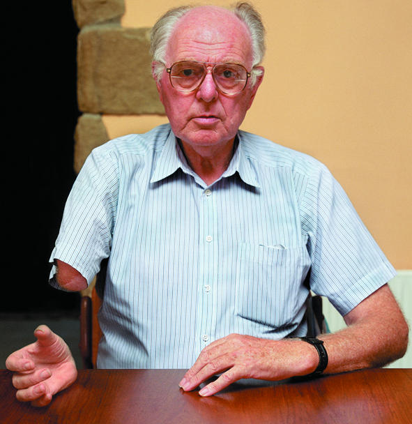
ALEXA WRIGHT
ALEXA WRIGHT
How amputees perceive their phantoms
Similar work by Aglioti with women undergoing mastectomy found that 25% of patients experienced a phantom breast when the pinna region of the ear lobe (on the same side as the mastectomy) was stimulated.2 This surprising finding suggests that these two regions might share neighbouring neural representations on the sensory homunculus in the brain.
Several recent brain imaging experiments also lend support to the functional remapping hypothesis. Studies of arm amputation using magnetoencephalography—a technique which allows high fidelity recordings of the electrical activity of small regions of the cortex—have shown that brain areas which ordinarily represent the hand were activated when either the lower face or upper arm was touched.6,7 Functional imaging techniques, such as positron emission tomography, provide another means of establishing which brain areas are activated by specific stimuli and tasks. One recent study demonstrated that stimulation of the trunk with touch reliably activated the representation of the hand on the sensory cortex, showing that sensory input to the trunk had gained access to the “homuncular hand.”8
Given the relatively short delay between amputation and phantom experience—usually less than 24 hours—this “invasion” of neuronal connections is unlikely to result from the sprouting of new connections. A more plausible explanation is that existing dormant synapses between neighbouring cortical areas are unmasked after large scale sensory loss. However, both processes could be at work, and indeed this remapping may involve neuronal changes at both cortical and thalamic levels.9
All of these findings question the widely held assumption within neuroscience that the primary sensory areas of the adult brain are hard wired. There is now growing evidence that complex brain adaptation can occur after stroke, and functional remapping may explain the striking degree of recovery seen in some patients. It has also been shown that extensive training can produce physical changes in specific parts of the brain: Braille readers and trained musicians show highly developed brain areas responsible for touch and musical appreciation.10
Understanding how the brain reorganises itself may eventually facilitate the treatment of patients with intractable phantom pain. Ramachandran, for example, has proposed a novel technique for the relief of the cramp-like pain caused by a clenched phantom fist. By observing the reflection of their free moving normal hand in a mirror, so that it appears in the position of the phantom, some patients report an immediate and compelling illusion whereby they are able to generate voluntary movements in the phantom hand. In some cases, the sensation of unclenching the phantom hand relieves the pain.5 The efficacy of this method will require a prospective controlled trial, but it offers the prospect of a real clinical benefit from an exciting insight into how the brain organises itself.
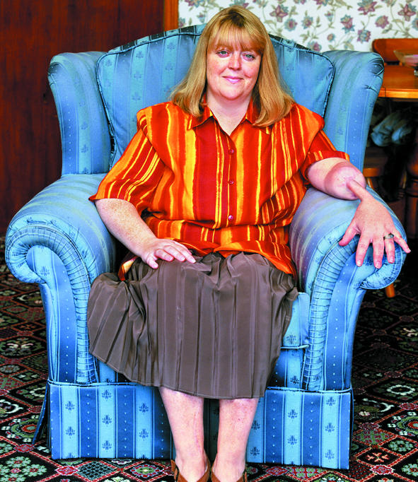
Figure 2.

The areas depicted on the right side of the face in this patient elicited the precisely localised referred sensations in the phantom hand, shown with supine position
References
- 1.Mitchell SW. Injuries of nerves and their consequences. Philadephia: Lippincott; 1872. p. 348. [Google Scholar]
- 2.Aglioti S, Cortese F, Franchini C. Rapid sensory remapping in the adult brain as inferred from phantom breast perception. Neuroreport. 1994;5:473–476. doi: 10.1097/00001756-199401120-00026. [DOI] [PubMed] [Google Scholar]
- 3.Halligan PW, Marshall JC, Wade DT. Three arms: a case study of supernumerary phantom limb after right hemisphere stroke. J Neurol Neurosurg Psych. 1993;56:159–166. doi: 10.1136/jnnp.56.2.159. [DOI] [PMC free article] [PubMed] [Google Scholar]
- 4.Weinstein S. Neuropsychological studies of the phantom. In: Benton AL, editor. Contributions to clinical neuropsychology. Chicago: Aldine; 1969. pp. 73–107. [Google Scholar]
- 5.Ramachandran VS, Hirstein W. The perception of phantom limbs. Brain. 1998;121:1603–1630. doi: 10.1093/brain/121.9.1603. [DOI] [PubMed] [Google Scholar]
- 6.Ramachandran VS, Stewart M, Rogers-Ramachandran DC. Perceptual correlates of massive cortical reorganization. Neuroreport. 1992;3:583–586. doi: 10.1097/00001756-199207000-00009. [DOI] [PubMed] [Google Scholar]
- 7.Halligan PM, Marshall LC, Wade DT, Davey J, Morrison D. Thumb in cheek? Sensory reorganization and perceptual plasticity after limb amputation. Neuroreport. 1993;4:233–236. doi: 10.1097/00001756-199303000-00001. [DOI] [PubMed] [Google Scholar]
- 8.Kew JJM, Halligan PW, Marshall JC, Rothwell JC, Ridding MC, Sooriakumaran S, et al. Abnormal access of axial vibrotactile input to deafferented somatosensory cortex in human upper limb amputees. J Neurophysiol. 1997;77:2753–2764. doi: 10.1152/jn.1997.77.5.2753. [DOI] [PubMed] [Google Scholar]
- 9.Jones EG, Pons TP. Thalamic and brainstem contributions to large scale plasticity of primate somatosensory cortex. Science. 1998;282:1121–1125. doi: 10.1126/science.282.5391.1121. [DOI] [PubMed] [Google Scholar]
- 10.Sadato N, Pascual-Leone A, Grafman J, Ibanez V, Deiber MP, Dold G, et al. Activation of the primary visual cortex by Braille reading in blind subjects. Nature. 1996;380:526–528. doi: 10.1038/380526a0. [DOI] [PubMed] [Google Scholar]