Virtual colonoscopy is a new procedure that fuses computed tomography of the large bowel with advanced techniques for rendering three dimensional images to produce views of the colonic mucosa similar to those obtained during “real” colonoscopy. Preliminary results suggest that it surpasses barium enema and approaches the sensitivity of conventional colonoscopy. This review summarises the technical developments that have made virtual colonoscopy possible and describes the expected advantages of this exciting technique over conventional endoscopy and more traditional imaging methods. We describe its possible applications and outline further technical developments that can be expected in the near future.
Background
In 1994 the Royal Mail issued a series of four stamps celebrating major medical advances of the 20th century; three of these were radiological (ultrasound scanning, computed tomography, and magnetic resonance imaging). Radiology is a technology based discipline and is one of the most rapidly developing medical specialties, not least because advances in computer technology are almost instantly incorporated into the various imaging modalities. Image acquisition and display are constantly improving, and image processing that required until recently a dedicated workstation is now possible with a personal computer. The possibilities for diagnostic imaging that are available today would have been unimaginable only 30 years ago, and computed tomography has played a central role in this development.
Predicted developments
New multi-slice spiral computed tomography will increase the sensitivity of virtual colonoscopy for detecting colorectal polyps
Faster and cheaper computer power will translate into faster and more complex image reconstruction, such as “virtual pathology”
Image analysis and polyp detection are likely to become automated
Oral labelling agents will eliminate the need for bowel cleansing
Magnetic resonance imaging will be increasingly used for virtual examinations
Because virtual colonoscopy is safe, easy, complete, and, once bowel cleansing is eliminated, non-invasive, it will assume a prominent role in screening for colorectal cancer
Conventional and spiral computed tomography
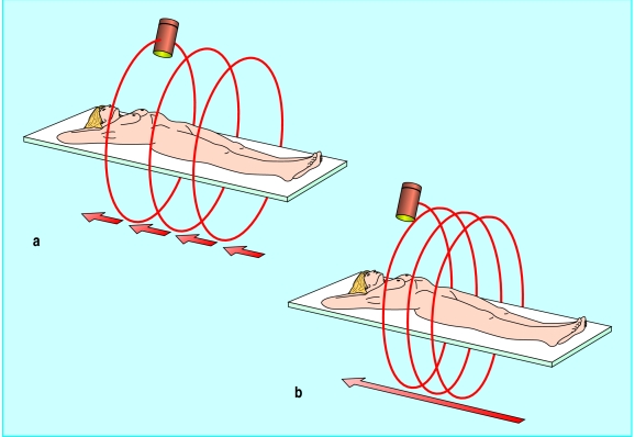
Invented by Godfrey Hounsfield in 1973,1 computed tomography involves rotating an x ray tube around a subject, the tube taking one dimensional views as it travels. These individual projections are digitally summated and reconstructed by means of mathematical algorithms to give a two dimensional “slice” through the body. Until recently, mechanical constraints limited tube rotation to 360°: a slice was taken, and the x ray tube would return to its original position while the table supporting the subject moved to the next acquisition position, after which the whole process started again (fig 1a). This process, known as dynamic incremental computed tomography, meant that studies comprised a series of consecutive slices of variable thickness and separation, and they were also relatively time consuming.
Figure 1.
Conventional computed tomography comprises a series of incremental 360° degree slices through the subject (a), whereas in spiral computed tomography the x ray tube continuously rotates around the subject, who passes through the scanner in a continuous movement (b)
The introduction of complex slip ring technology has made it possible to continuously rotate the x ray tube during image acquisition, and higher tube loading has also enabled prolonged exposure. It seems sensible to combine these developments with continuous table movement rather than time consuming, stepped increments, so that a subject can be examined in one, rapid sequence.2 Because the tube is continuously rotating while the subject moves smoothly through the scanner, the x ray beam describes a spiral pathway through the body (fig 1b)—hence the term spiral computed tomography.
There are several advantages with spiral computed tomography. Obviously it is faster, so that an entire study can be completed in a single breath-hold. This has eliminated misregistration artefacts, which were previously caused by uneven inspiration between individual slices. The reduced scanning time also means that, when intravenous iodine compounds are needed to enhance the contrast of some organs, the entire body part being studied can now be imaged at peak contrast. Most importantly, whereas the images produced by conventional computed tomography are slices through the patient, mirroring exactly how they are acquired, data acquisition during spiral computed tomography is continuous in three dimensions and therefore volumetric. Although images are conventionally presented as single slices for convenience, it should be borne in mind that these are two dimensional reconstructions from a truly three dimensional dataset.
Bowel imaging and computed tomography
These technical developments have been paralleled by changing attitudes towards luminal bowel imaging with computed tomography. It was initially thought that computed tomography of the abdomen and pelvis was best suited to solid organs, and the bowel was simply labelled with orally or rectally applied contrast medium so that it could be easily identified and distinguished from any disease. However, the concept of using computed tomography as the primary tool for colonic investigation was precipitated by investigators searching for an alternative to the barium enema in radiological investigations of frail, elderly patients.3
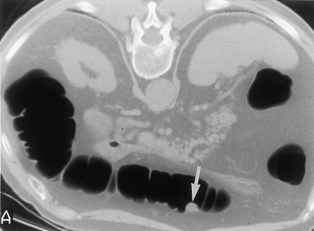
Indeed, it soon became apparent that the colon could be visualised in exquisite detail with spiral computed tomography if the principles underlying barium radiology were similarly applied—namely, distension of the colon, adequate tissue contrast, and temporary paralysis by means of intravenous smooth muscle relaxants—a procedure that has been termed “spiral CT pneumocolon.”4 Patients undergo full bowel preparation, an intravenous smooth muscle relaxant is administered, and the colon then insufflated with room air until fully distended. Once satisfactory distension has been achieved, spiral computed tomography is performed to encompass the entire colon, possibly with simultaneous intravenous injection of contrast medium. This approach allows detection of small polyps (fig 2), unlike examination without preparation, which is used to detect large cancers in frail elderly patients.
Figure 2.
Spiral CT pneumocolon of a patient lying prone. The large bowel is distended with air, and its lumen contrasts sharply with surrounding soft tissues, revealing a 10 mm polyp in the transverse colon (arrow)
Virtual colonoscopy
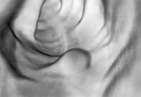
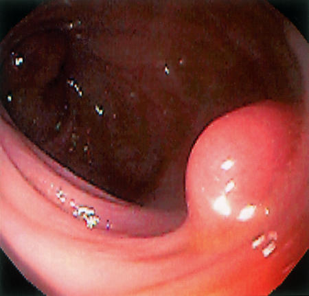
The combination of fast volumetric imaging, the application of barium techniques to computed tomography, and formidable developments in rendering three dimensional images by computer graphics packages has enabled virtual colonoscopy (computed tomography colography). First described by Vining and coworkers in 1994,5 virtual colonoscopy applies techniques for rendering complex images to spiral CT pneumocolon. The considerable difference in image contrast between intraluminal gas and the surrounding colonic wall and soft tissues makes this interface an easy target for the rendering of three dimensional images, allowing creation of a graphical representation of the colon through which the viewer can “navigate” (fig 3).
Figure 3.
Virtual colonoscopic view of a 10 mm transverse colon polyp (top) and same polyp imaged during subsequent conventional colonoscopy (bottom). (Same patient as in fig 2)
A spiral CT pneumocolon of a patient lying supine is performed in a single acquisition, with the patient holding a breath for the first 15-20 seconds (covering the upper abdomen) while the rest can be acquired during gentle respiration. The study is generally repeated with the patient lying prone to ensure that all colonic segments have been imaged distended. Reconstruction parameters vary but in a typical study would be at 2 mm intervals, with a 3 mm overlap of slices. The data are then downloaded to an independent workstation equipped with appropriate software for rendering perspective and volume. The computer generates a retrograde intraluminal “fly through” navigation from rectum to caecum and repeats it in the opposite direction.
Both antegrade and retrograde virtual colonoscopies are stored in a “cine loop” format and can be viewed directly from the workstation monitor. Image analysis is interactive, and the radiologist can choose to view the rendered mucosa from any angle, pass through the tightest stricture, and even cross the colonic wall into adjacent structures. Virtual studies are usually examined in combination with the standard axial computed tomograms. The computer can also generate two dimensional images at cross sectional and orthogonal angles to the long axis of the colon to aid interpretation.
Preliminary results
Preliminary results indicate that the accuracy of virtual colonoscopy for detecting colon polyps may exceed that of barium enema examination and approach that of conventional colonoscopy. In a study of 70 consecutive patients known to have polyps or being followed up one to five years after polypectomy the sensitivity and specificity of virtual colonoscopy were 75% and 90% respectively for adenomatous polyps >10 mm in diameter, 66% and 63% for adenomas >5 mm, and 45% and 80% for polyps <5 mm.6 Comparison with single contrast barium enema examination, still widely practised in the United States, suggests that virtual colonoscopy has a higher detection rate with fewer false positive results.7 At the time of writing, no study has compared virtual colonoscopy with double contrast barium enema examination. In studies of asymptomatic patients with no prior neoplasia virtual colonoscopy had a sensitivity of 75% for adenomas >10 mm in diameter,8 and other studies have reported sensitivities of between 91% and 100% for polyps >10 mm9,10 and sensitivity approaching 100% for carcinomas.11
Conventional colonoscopy is a difficult and technically demanding procedure, and is frequently incomplete for these reasons. A major attraction of virtual colonoscopy is the greater certainty of a complete examination, even when a carcinoma precludes proximal passage of a real colonoscope: computed tomography of 29 patients with occlusive cancers in whom colonoscopy and barium enema had failed to pass the stricture found all known tumours and revealed two further cancers and 24 polyps proximal to the site of obstruction.12 Furthermore, localisation of lesions is likely to be more accurate with computed tomography: in one study 38 cancers were correctly localised by means of computed tomography compared with 32 by conventional colonoscopy.13
Colorectal cancer screening
Almost simultaneous with the birth of virtual colonoscopy were suggestions that it could be used for screening for colorectal cancer, and there are several arguments in its favour. Virtual colonoscopy seems able to reliably detect adenomatous polyps, the precursor of cancer, with high accuracy. Furthermore, the entire colon is easily examined, in contrast to colonoscopy. It is quicker and less technically demanding than either colonoscopy or even barium enema examination. It is safe, although there is a theoretical risk from radiation exposure, roughly equal to that from barium enema radiography (higher if both supine and prone studies are performed). Other organs are also imaged and could be screened simultaneously. The skills needed to interpret virtual studies are different from those required for barium radiology and might be less demanding because of the wealth of imaging information provided. Lastly, virtual colonoscopy is perceived as a new and exciting development by radiologists, and this enthusiasm may help staff recruitment to any future screening programme.
Future developments
Virtual colonoscopy is in the early stages of its development. Its feasibility has clearly been demonstrated, and larger, multicentre studies are needed to determine its sensitivity and specificity outside specialist units. Multiple technical parameters can be altered during image acquisition, and a coherent technique has yet to emerge. Multi-slice spiral computed tomography has just been introduced and will reduce examination time even further. Furthermore, the principles underlying virtual colonoscopy by spiral computed tomography can be just as easily applied to magnetic resonance imaging, which carries no radiation penalty.14
Presently, most limitations apply to the processing and analysis of images after their acquisition—analysis of a single study takes about 30-60 minutes. However, the current constraints imposed by available processing power and digital storage are likely to diminish. Also, it seems that diagnosis can usually be made from the two dimensional images, and generating a three dimensional image, the most demanding application in terms of computer power, may be necessary only for problem solving, such as differentiating between a polyp and a haustral fold.15
Other rendering techniques are also being developed. It is possible to generate three dimensional images that resemble conventional fluoroscopic double contrast enema studies or to artificially straighten the colon, open it, and view the rendered, flattened surface en face, imitating the pathologist's view. Navigation software is also progressing; luminal “fly through” navigation can be automated and collision avoidance techniques used, with simultaneous antegrade and retrograde viewing.
At present, full colonic cleansing is mandatory for an acceptable examination, but workers are already investigating the possibility of using oral contrast medium with a view to tagging faecal residue so that it can be differentiated from pathological features and possibly digitally excluded from the image. Furthermore, because virtual colonoscopy generates images of the colonic wall, it is possible to apply algorithms that automatically detect and label regions where it is thickened, alerting the radiologist to the possible presence of polyps. Perhaps flat adenomas can be detected this way.
The development and refinement of all of these techniques are likely to have tremendous impact on the speed and accuracy of virtual colonoscopy and its interpretation in the near future. The performance characteristics of virtual colonoscopy are certain to improve with improvements in hardware and software innovation and radiologists' experience. With regard to its potential as a screening tool, its performance in real populations and its cost, patient acceptability, and availability will need to be determined.
Editorial by Atkin
Footnotes
Competing interests: None declared.
References
- 1.Hounsfield GN. Computerized transverse axial scanning (tomography): Part 1. Description of the system. Br J Radiol. 1973;46:1016–1022. doi: 10.1259/0007-1285-46-552-1016. [DOI] [PubMed] [Google Scholar]
- 2.Kalender WA, Seissler W, Klotz E, Vock P. Spiral volumetric CT with single breath-hold technique, continuous transport and continuous scanner rotation. Radiology. 1990;176:181–183. doi: 10.1148/radiology.176.1.2353088. [DOI] [PubMed] [Google Scholar]
- 3.Fink M, Freeman AH, Dixon AK, Coni NK. Computed tomography of the colon in elderly people. BMJ. 1994;308:101. doi: 10.1136/bmj.308.6935.1018. [DOI] [PMC free article] [PubMed] [Google Scholar]
- 4.Amin Z, Poulos PB, Lees WR. Spiral CT pneumocolon for suspected colonic neoplasms. Clin Radiol. 1996;51:56–61. doi: 10.1016/s0009-9260(96)80221-2. [DOI] [PubMed] [Google Scholar]
- 5.Vining DJ, Gelfand DW, Bechtold RE, Scharding ES, Grishaw EK, Shifrin RY, et al. Technical feasibility of colon imaging with helical CT and virtual reality [abstract] AJR Am J Roentgenol. 1994;162(S):104. [Google Scholar]
- 6.Hara AK, Johnson CD, Reed JE, Ahlquist DA, Nelson H, MacCarty RL, et al. Detection of colorectal polyps with CT colography: initial assessment of sensitivity and specificity. Radiology. 1997;205:59–65. doi: 10.1148/radiology.205.1.9314963. [DOI] [PubMed] [Google Scholar]
- 7.Hara AK, Johnson CD, Reed JE, Ahlquist DA, Nelson H, Harmsen WS, et al. Computed tomographic colography for polyp detection: early comparison against barium enema. Gastroenterology. 1997;112:A575. [Google Scholar]
- 8.Rex DK, Vining D, Kopecky K. Screening for polyps using spiral CT with and without virtual colonoscopy [abstract] Gastrointest Endosc. 1997;45:AB116. doi: 10.1053/ge.1999.v50.97776. [DOI] [PubMed] [Google Scholar]
- 9.Kay CL, Kulling D, Evandelou H, Hawes RH, Young JW, Cotton PB. CT-based virtual colonoscopy—correlation with colonoscopy in the detection of space-occupying lesions of the colon [abstract] Radiology. 1997;205:195. [Google Scholar]
- 10.Dachman AH, Kuniyoshi JK, Boyle CM, Samara Y, Hoffmann KR, Rubin DT, et al. CT colonography with three-dimensional problem solving for detection of colonic polyps. AJR Am J Roentgenol. 1998;171:989–995. doi: 10.2214/ajr.171.4.9762982. [DOI] [PubMed] [Google Scholar]
- 11.Royster AP, Fenlon HM, Clarke PD, Nunes DP, Ferrucci JT. CT colonoscopy of colorectal neoplasms: two-dimensional and three-dimensional virtual reality techniques with colonoscopic correlation. AJR Am J Roentgenol. 1997;169:1237–1242. doi: 10.2214/ajr.169.5.9353434. [DOI] [PubMed] [Google Scholar]
- 12.Fenlon HM, McAneny DB, Nunes DP, Clarke PD, Ferrucci JT. Occlusive colon carcinoma: virtual colonoscopy in the preoperative evaluation of the proximal colon. Radiology. 1999;210:423–428. doi: 10.1148/radiology.210.2.r99fe21423. [DOI] [PubMed] [Google Scholar]
- 13.Fenlon HM, Nunes DP, Clarke PD, Ferrucci JT. Colorectal neoplasm detection using virtual colonoscopy: a feasibility study. Gut. 1998;43:806–811. doi: 10.1136/gut.43.6.806. [DOI] [PMC free article] [PubMed] [Google Scholar]
- 14.Lubolt W, Steiner P, Bauerfeind P, Pelkonen P, Debatin JF. Detection of mass lesions with MR colonography: preliminary report. Radiology. 1998;207:59–65. doi: 10.1148/radiology.207.1.9530299. [DOI] [PubMed] [Google Scholar]
- 15.Fenlon HM, Ferrucci JT. Virtual colonoscopy. What will the issues be? AJR Am J Roentgenol. 1997;169:453–458. doi: 10.2214/ajr.169.2.9242753. [DOI] [PubMed] [Google Scholar]


