



Abstract
Objective: This study aims to evaluate the predictive value of tumor markers combined with gastrin for tumor recurrence after endoscopic submucosal dissection (ESD) in patients with early gastric cancer. Methods: The clinicopathological data of 169 patients with early gastric cancer treated with ESD between March 2019 and January 2021 were retrospectively analyzed. The patients were divided into a relapse group (n=45) and a non-recurrence group (n=124). Clinical data such as carcinoembryonic antigen (CEA), cancer antigen 19-9 (CA19-9), alpha-fetoprotein (AFP), gastrin 17, pepsinogen I and pepsinogen II, as well as tumor size and degree of infiltration were examined to construct a recurrence prediction model using lasso regression. Results: The comprehensive model showed superior predictive power (AUC=0.958, C-index=0.966) over biomarker-only models (AUC=0.925), indicating a significant improvement in the prediction of recurrence risk. Decision curve analysis confirmed the clinical utility of the model with a maximum net benefit of 73.37%. Key indicators such as CEA, CA19-9, AFP, gastrin 17 and pepsinogens I and II were statistically significant in predicting recurrence with P values < 0.01. Conclusion: The comprehensive model combining tumor markers with clinical data provides a more accurate and clinically valuable tool for predicting recurrence in early gastric cancer patients after ESD. This approach facilitates personalized risk assessment and may significantly improve prognostic management, emphasizing the importance of a multifaceted strategy in the management of early gastric cancer.
Keywords: Tumor marker, gastrin, early gastric cancer, endoscopic submucosal dissection, recurrence, prediction
Introduction
Stomach cancer ranks the fifth most common cancer and the fourth leading cause of cancer death worldwide [1]. Particularly in China, it is the second most prevalent cancer and a leading cause of cancer mortality [2]. Early detection is crucial for improving survival rates; however, the asymptomatic nature of early-stage gastric cancer poses substantial challenges. Gastric cancer at its early stage remains confined to the mucosal or submucosal layer, without directly affecting lymph node metastasis, highlighting the complexity of its management [3]. Thanks to increased health awareness and advancements in screening techniques, the detection rates for early gastric cancer have improved significantly. Endoscopy, in particular, has emerged as a key diagnostic and therapeutic tool with demonstrated efficacy and cost-effectiveness, especially in East Asia - as evidenced by countries such as South Korea and Japan, where national gastric cancer screening programs have significantly reduced mortality rates [4]. However, the post-treatment landscape, particularly the risk of recurrence after endoscopic submucosal dissection (ESD), requires a more evolved focus. As focus shifts beyond detection, predictive modeling of recurrence in early gastric cancer patients after ESD is critical for enhancing treatment outcomes and patient management.
Endoscopic surgery, including endoscopic mucosal resection (EMR) and endoscopic mucosal dissection (ESD), offers several advantages over traditional surgical procedures, such as being less invasive, fewer postoperative complications, shorter recovery times, lower healthcare costs, and improved quality of life. Importantly, long-term outcomes are comparable to traditional surgical methods, with five-year survival rates often exceeding 90% [5]. Consequently, many global gastric cancer treatment guidelines recommend endoscopic surgery as the preferred treatment strategy for early gastric cancer. However, despite the effectiveness of ESD, its associated high rate of heterochronic recurrence needs to be addressed [6]. Studies have shown that recurrence usually occurs near the original tumor site, highlighting the need to assess and predict possible risk factors for recurrence [7-9]. A thorough understanding of these risk factors will help develop personalized surveillance plans and treatment strategies to reduce the recurrence risk and improve treatment outcomes.
Recent advances in data analytics have led to the emergence of many predictive models across various fields, providing decision support and predictive [10]. While studies have shown that models based on a single metric can provide high predictive accuracy [11,12], comparisons with models utilizing more comprehensive datasets, particularly those incorporating clinical data (pathological data and clinical outcomes) are less common. Our study aims to investigate whether the combined use of biomarkers and clinical information is superior to models using only a single indicator in terms of predictive accuracy and clinical application value. Through this comparison, we aim to identify a more accurate and practical model to improve prognostic assessment and disease management, ultimately improving patient outcomes and quality of life.
Methods and data
Clinical data
In this retrospective study, patients with early gastric cancer who were treated in The First People’s Hospital of Xianyang from March 2019 to January 2021 were included as the study subjects. This study was conducted with the approval of The First People’s Hospital of Xianyang Medical Ethics Committee.
Inclusion exclusion criteria
Inclusion criteria: patients with a postoperative diagnosis of gastric cancer confirmed by pathological examination; patients at early cancer stage [13]; patients who were treated with ESD; and patients with complete follow-up data.
Exclusion criteria: those with combined cardiac, hepatic, or renal dysfunction; those with other malignant neoplastic diseases or infectious diseases; those who had received immunologic, hormonal, or antibiotic treatment 4 weeks prior to enrollment; and those with other difficult-to-control endocrine or immunologic diseases (Figure 1).
Figure 1.
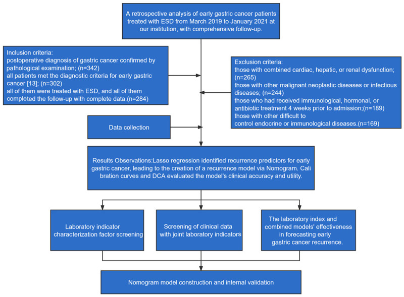
Research flowchart.
Definition of relapse
Relapse is defined as the emergence of new cancerous tissue, confirmed by endoscopy and biopsy. This includes cancerous lesions found at the margins of the original ESD resection or within the area of the original resection [11].
Patient grouping
According to the inclusion-exclusion criteria, we collected 169 cases that met the criteria for this study. According to the definition of recurrence, these patients were categorized into a relapse group (n=45) and a non-recurrence group (n=124).
Data collection
Baseline information and laboratory values were collected from patient follow-up records, outpatient review records, and electronic medical record systems. Baseline data included gender, age, history of smoking, alcohol consumption, diabetes, hypertension, lymph node metastasis, clinical stage, tumor size, and degree of infiltration. Laboratory parameters included carcinoembryonic antigen (CEA), cancer antigen 19-9 (CA19-9), alpha-fetoprotein (AFP), gastrin 17, pepsinogen I, and pepsinogen II, all of which are commonly used for gastric cancer screening in The First People’s Hospital of Xianyang. In addition, the patient’s examination data were obtained on day 1 after admission.
Indicator test methods
Serum gastrin 17 (MB-1448B), pepsinogen I (MB-15603A) and pepsinogen II (MB-15605B) were detected by enzyme-linked immunosorbent assay (ELISA), and the test kits were purchased from Jiangsu Enzymatic Bio-technology Co. CA199, CEA and AFP were detected by electrochemiluminescence, the instrument was Roche CO-BAS6000 biochemical analyzer, and the reagents were provided by Roche.
Follow-up
Gastroscopy was performed at 1, 6, 12, 24, and 36 months postoperatively to monitor for recurrence.
Observation of outcomes
Primary outcomes: Lasso regression was used to analyze the factors predicting recurrence in both the laboratory indicator model and the combined model. A nomogram was utilized to construct the recurrence prediction model, with calibration curves and decision curves (DCA) assessing the model’s clinical accuracy and benefit rate.
Secondary outcomes: Differences in baseline data and laboratory index results were compared between the relapse group and the non-recurrence group.
Statistical analysis
Data were processed using SPSS 26.0 software. Data distribution was analyzed using the K-S test. Measurement data conforming to normal distribution was expressed as mean ± standard deviation (Meas ± SD); and the inter-group comparisons were conducted using the independent samples t-test, while the intra-group comparisons were made using the paired t-test, denoted by t. Non-normally distributed data were displayed by the interquartile range and tested using a non-parametric test, denoted by Z. Count data were compared using the χ2 test. Lasso regression was used to screen factors indicative of recurrence, and receiver operating characteristic (ROC) curves were used to analyze the clinical efficacy of the model and to plot the area under the curve (AUC). AUC values ranged from 0 to 1, with 1 indicating a perfect model and 0.5 a non-discriminatory model. Delong’s test was used to determine differences between ROC curves. P < 0.05 was considered with statistical difference.
Results
Comparison of clinical data
Comparison of baseline data between the two groups showed that the proportions of patients with tumors ≥ 5 cm and with an intramucosal infiltration were higher in the relapse group than in the non-recurrence group (all P < 0.01, Table 1).
Table 1.
Comparison of baseline data
| Factor | Relapse group (n=45) | Non-recurrence group (n=124) | χ2-value | P-value |
|---|---|---|---|---|
| Gender | ||||
| Male | 24 | 63 | 0.084 | 0.771 |
| Female | 21 | 61 | ||
| Age | ||||
| ≥ 60 years | 34 | 100 | 0.521 | 0.470 |
| < 60 years | 11 | 24 | ||
| Smoking history | ||||
| Yes | 7 | 16 | 0.198 | 0.657 |
| No | 38 | 108 | ||
| History of alcohol abuse | ||||
| Yes | 12 | 25 | 0.817 | 0.366 |
| No | 33 | 99 | ||
| History of diabetes | ||||
| Yes | 10 | 35 | 0.609 | 0.435 |
| No | 35 | 89 | ||
| History of hypertension | ||||
| Yes | 11 | 38 | 0.617 | 0.432 |
| No | 34 | 86 | ||
| Clinical staging | ||||
| Yes | 34 | 74 | 3.609 | 0.057 |
| No | 11 | 50 | ||
| Tumor size | ||||
| ≥ 5 cm | 36 | 56 | 16.157 | < 0.001 |
| < 5 cm | 9 | 68 | ||
| Degree of infiltration | ||||
| Submucosal | 11 | 63 | 9.322 | 0.002 |
| Intramucosal | 34 | 61 |
Comparison of laboratory indicators
Comparison of the laboratory indices between the two groups revealed that CEA, CA199, AFP, gastrin 17, pepsinogen I, and pepsinogen II were statistically higher in the relapse group than those in the non-recurrence group (all P < 0.01, Table 2).
Table 2.
Comparison of laboratory indicators
| Norm | Relapse group (n=45) | Non-recurrence group (n=124) | t-value | P-value |
|---|---|---|---|---|
| CEA (μg/L) | 86.24±8.56 | 78.18±8.00 | 5.502 | < 0.001 |
| CA199 (U/mL) | 93.29±10.50 | 86.29±11.29 | 3.752 | < 0.001 |
| AFP (μg/L) | 10.19±2.82 | 8.68±2.32 | 3.212 | 0.002 |
| Gastrin 17 (pmol/L) | 28.55±4.68 | 23.05±4.44 | 6.841 | < 0.001 |
| Pepsinogen I (μg/L) | 34.73±6.10 | 28.66±5.66 | 5.827 | < 0.001 |
| Pepsinogen II (μg/L) | 17.96±5.23 | 13.11±5.53 | 5.241 | < 0.001 |
Note: CEA, Carcinoembryonic Antigen; CA19-9, Cancer Antigen 19-9; AFP, Alpha-Fetoprotein.
Screening of factors characterizing laboratory indicators
Using Lasso regression, we screened six laboratory-related indicators in recurrent patients. The lambda value was set at 0.1 se (0.057335), showing that all 6 indicators (CEA, CA199, AFP, gastrin 17, pepsinogen I, and pepsinogen II) were associated with recurrence in patients with early gastric cancer (Figure 2).
Figure 2.
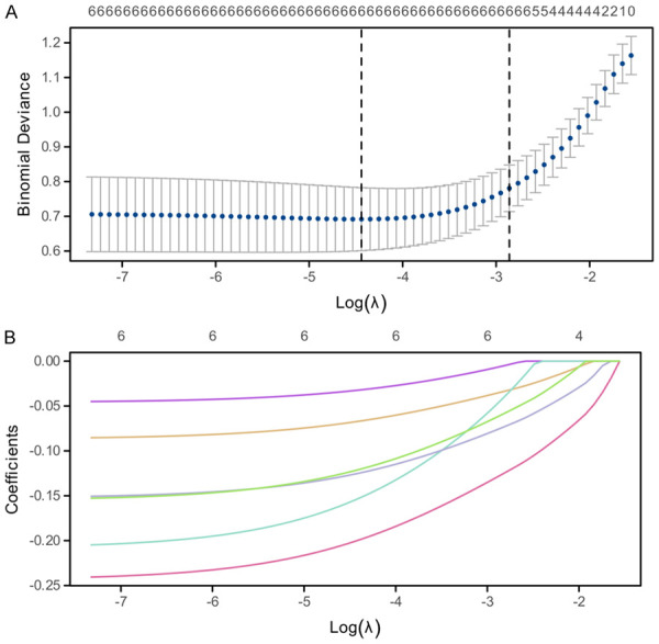
Factor analysis of laboratory indicators for recurrence prediction. A. Lasso paths for feature selection. Each point indicates the model deviation from its baseline performance at different values of λ. The black dashed line marks the value of λ for the optimal model selection. B. Variation of variable coefficients with regularisation parameter λ. Each curve represents the coefficient of a variable as λ increases.
Screening of clinical data with joint laboratory indicators
We performed a Lasso regression analysis on 16 characteristics combining laboratory-related indicators and clinical data of recurrent patients. The lambda value was set at 0.1 se (0.039519), showing that all 8 (CEA, CA199, AFP, gastrin 17, pepsinogen I and pepsinogen II, tumor size, and degree of infiltration) indices were associated with recurrence in patients with early gastric cancer (Figure 3).
Figure 3.
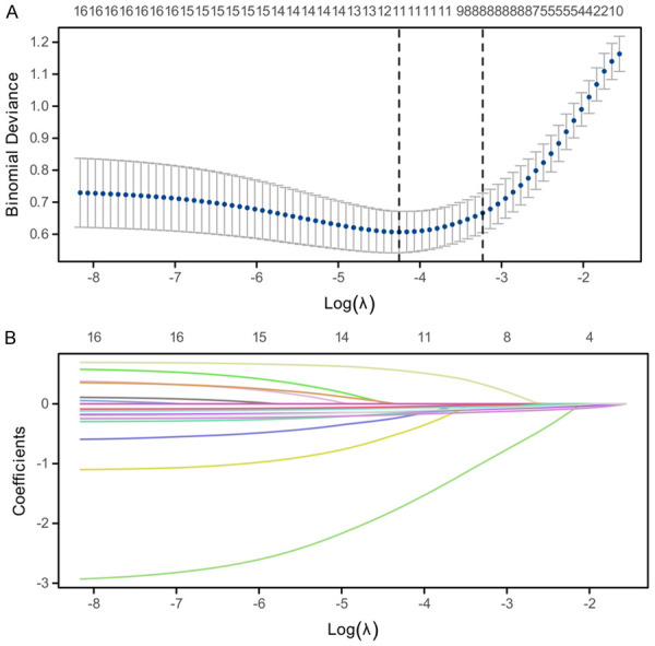
Factor analysis of clinical data combined with laboratory indicators for recurrence prediction. A. Lasso path for feature selection. Each point indicates the model deviation from its baseline performance at different values of λ. The black dashed line marks the value of λ for the optimal model selection. B. Variation of variable coefficients with regularisation parameter λ. Each curve represents the coefficient of a variable as λ increases.
Predictive value of a laboratory indicator model and combined model in predicting patients with early gastric cancer recurrence
We constructed the laboratory indicator and joint models based on lasso regression. Laboratory index model: Riks = 11.2546847 + CEA × -0.034765536 + CA199 × -0.006104295 + AFP × -0.044313554 + Gastrin17 × -0.127225202 + Pepsinogen I × -0.074936115 + Pepsinogen II × -0.060496374. Combined model formula: Riks = 14.74828518 + tumour size × -0.940422867 + degree of infiltration × 0.302826963 + CEA × -0.044950287 + CA199 × -0.018649921 + AFP × -0.073484088 + gastrin 17 × -0.134067176 + Pepsinogen I × -0.088991936 + Pepsinogen II × -0.074111522. It was found that the laboratory index model scores and the combined model scores of patients in the relapse group were statistically lower than those of patients in the non-recurrence group (P < 0.0001, Figure 4A, 4B). To further determine the difference between the two models, we compared them using ROC curves. The results showed that the AUC of the laboratory index model was 0.925, whereas the AUC of the combined model was 0.958 (P=0.011, Figure 4C).
Figure 4.

Clinical efficacy assessment of the laboratory biomarker model and the combined model. A. Comparison of per-patient scores based on the biomarker model. B. Comparison of per-patient scores based on the combined model. C. Comparison of ROC curves between biomarker model and combined model.
Nomogram model construction and internal validation
With the results of Lasso regression analysis and ROC curve analysis, we constructed a nomogram comprising tumor size, degree of infiltration, CEA, CA199, AFP, Gastrin 17, and levels of Pepsinogen I & II (Figure 5A). This nomogram model provided highly accurate prediction, with an AIC of 85.592, indicating a good model fit, and a C-index of 0.966, with a confidence interval of 0.939 to 0.993, indicating that the model had a very high discriminative power (Figure 5B). In addition, using decision curve analysis (DCA), the model showed a net benefit of up to 73.37% at different thresholds compared to no intervention, reflecting the potential application of the model in clinical decision making (Figure 5C).
Figure 5.
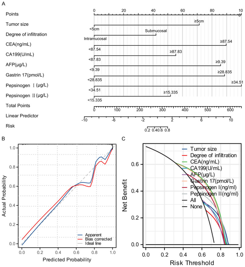
Nomogram model construction and internal validation. A. Nomogram showing the contribution of the different variables to the final risk prediction, where the score for each variable can be read directly from the graph and accumulated to obtain a total score, which is then converted to a predicted probability. B. Calibration curves, where the agreement between the probabilities predicted by the model and the actual observed probabilities is represented by the blue line, which is closer to the ideal case (red line). C. Net benefit of using the model compared to no strategy under different threshold choices.
Discussion
With economic development and increased health awareness, an increasing number of gastric cancer patients are being diagnosed at an early stage, leading to a rise in the adoption of endoscopic resection [14]. This approach has become the standard treatment for early gastric cancer in Korea and Japan. It is also recommended by international guidelines such as the European Society for Gastrointestinal Endoscopy and the National Comprehensive Cancer Network due to its advantages of minimal trauma, rapid recovery, low complications, short hospital stay, and high postoperative quality of life [15]. However, because endoscopic resection preserves the entire stomach, there is a risk of heterochronic gastric cancer developing during the postoperative period. Despite the improvement of endoscopic treatment in China and the increasing popularity of endoscopic surgery for early gastric cancer, the definitive risk factors for heterochronic gastric cancer remain unclear.
Recent advances in data analytics have catalyzed the development of numerous predictive models that provide decision support and predictive insights, particularly for tumor recurrence prediction [16]. Research suggests that models based on individual metrics can achieve high predictive accuracy. For example, Shan et al. found that combining serum tumor markers with dual-source CT significantly improved diagnostic accuracy for lung cancer [17]. Similarly, Hufnagel et al. suggested that using machine learning to select a specific set of proteins could be a predictive tool for assessing the severity of early COVID-19 progression [18]. In addition, Soeda et al. found that certain microRNAs could serve as biomarkers for predicting peritoneal recurrence and prognosis in stage II/III gastric cancer patients [19]. However, these investigations often overlook the comparison of such models with those incorporating a broader range of information, particularly clinical data. Our results underscore that the AUC for models relying solely on indicators was significantly lower than that of combined models, suggesting that while individual metrics may provide some predictive accuracy, integration with clinical data significantly improves model accuracy [20,21].
This improvement is likely due to the fact that clinical data provides a more comprehensive view of the disease, enabling more accurate prediction of disease progression. While individual indicators may reflect only one aspect of the disease, where incorporating clinical data introduces a multi-dimensional perspective, taking into account various factors to refine the model’s ability to generalize and the accuracy of predictions [22]. For example, while a tumor marker may indicate the presence of cancer, integrating this with clinical insights such as tumor size, location, and genetic information can lead to a more accurate prediction of treatment response and disease prognosis [23]. Thus, optimizing the effectiveness of a predictive model necessitates constructing a diversified model that incorporates both laboratory and clinical patient characteristics, thereby improving clinical decision support and facilitating personalized treatment recommendations.
The nomogram model excels at translating complex statistical analyses into an intuitive and straightforward graphical representation, which greatly enhances the interpretability and usability of the model [24]. This model integrates various factors, assigns a score to each, and calculates a patient’s disease risk or prognosis by aggregating these scores. This approach facilitates a rapid assessment of a patient’s condition by clinicians and provides an individualized risk assessment that can guide specific clinical decisions. In our research, the Nomogram model demonstrated exceptional accuracy and utility in predicting the recurrence risk in patients with early gastric cancer by integrating clinical and laboratory data [25]. The model was analyzed by Lasso regression analysis and ROC curve analysis, and finally CEA, CA199, AFP, gastrin 17, pepsinogen I and pepsinogen II, tumor size and degree of infiltration were selected as risk factors for recurrence after ESD in early gastric cancer. According to the results, we can analyze the risk factors into two parts: pathological data and laboratory indicators. First, from the perspective of pathological data, tumor size and depth of infiltration are important factors affecting recurrence. Tumor size reflects its growth rate and invasiveness, and larger tumors may cover a larger area of gastric mucosa, which may retain tumor cells even after ESD treatment, increasing the risk of recurrence [9,26]. Similarly, the depth of infiltration indicates the extent to which tumor cells penetrate the gastric wall, and deeper infiltration may involve more lymphatic and blood vessels, facilitating the spread of tumor cells and the likelihood of recurrence [27]. In addition, laboratory markers, such as gastrin-related markers, CEA, CA19-9, and AFP, provide a direct indication of tumor burden and risk of recurrence [28-30]. Elevated levels of these biomarkers tend to be strongly associated with tumor activity, increased burden, and recurrence potential.
Based on this, we constructed the nomogram model, which performed well based on the AIC and C-indices, and demonstrated its potential for clinical decision support. Previously, a study by Bae et al. [31] constructed a predictive model for extragastric recurrence after radical resection of early gastric cancer, and their model had an AUC of 0.851 in predicting recurrence in patients. In addition, Okuno et al. [32] constructed a model for recurrence prediction of early gastric cancer by liquid biopsy characterization, reported an AUC of 0.860. Our model, with an AUC of 0.958, indicates higher accuracy and reliability in predicting postoperative recurrence in patients with early gastric cancer, underscoring its superiority and potential in clinical application.
The choice of Lasso regression over logistic regression to construct our predictive model was primarily due to its superior performance in handling high-dimensional data containing numerous predictor variables. Lasso introduces a regularization term that effectively performs variable selection, emphasizing variables that contribute most significantly to the prediction target while minimizing the impact of less relevant variables. This method helps prevent overfitting and improves the generalization ability of the model [33,34]. Our comparative analysis showed that the combined model integrating clinical data significantly outperformed models solely based on laboratory indicators alone in predicting the risk of recurrence for early gastric cancer patients, as evidenced by a higher AUC. This suggests that while laboratory indicators may provide some predictive accuracy, the predictive ability of the model is greatly enhanced by the combination of clinical data, providing more accurate and personalized support for clinical decision making.
This study represents a step forward in predicting the recurrence risk in early gastric cancer patients, albeit with several limitations. Conducted as a single-center retrospective analysis, the study design inherently carries the potential for information and selection bias, thereby limiting the broad applicability of its findings. Although it made strides in improving predictive accuracy by incorporating clinical data and laboratory markers, it may not have fully accounted for all relevant predictive factors, including lifestyle and genetic determinants. The modest sample size and the single-center nature of the study underscore the need to confirm the findings in a larger population and in multiple centers. Future research should aim to increase the cohort size, integrate a more comprehensive set of predictors, and use a multicenter approach to strengthen the model’s accuracy and clinical utility.
Conclusion
In our research, we carefully evaluated and compared the effectiveness of predictive models that use either biomarkers or a comprehensive integration of clinical data in predicting disease outcomes. The results of our study clearly indicate that the holistic model, which integrates both biomarkers and comprehensive clinical information, outperforms its counterparts that solely rely on laboratory indicators. This integrated approach not only achieves superior accuracy in predicting disease progression, but also significantly advances the prognostic evaluation and management of disease. As a result, it helps to significantly improve patient outcomes and overall quality of life.
Disclosure of conflict of interest
None.
References
- 1.Joshi SS, Badgwell BD. Current treatment and recent progress in gastric cancer. CA Cancer J Clin. 2021;71:264–279. doi: 10.3322/caac.21657. [DOI] [PMC free article] [PubMed] [Google Scholar]
- 2.Zong L, Abe M, Seto Y, Ji J. The challenge of screening for early gastric cancer in China. Lancet. 2016;388:2606. doi: 10.1016/S0140-6736(16)32226-7. [DOI] [PubMed] [Google Scholar]
- 3.Wender RC, Brawley OW, Fedewa SA, Gansler T, Smith RA. A blueprint for cancer screening and early detection: advancing screening’s contribution to cancer control. CA Cancer J Clin. 2019;69:50–79. doi: 10.3322/caac.21550. [DOI] [PubMed] [Google Scholar]
- 4.Hu C, Yuan L, Cheng X. Current status of early gastric cancer screening research. Cancer Biol Med. 2024;21:218–222. doi: 10.20892/j.issn.2095-3941.2023.0377. [DOI] [PMC free article] [PubMed] [Google Scholar]
- 5.Fu XY, Mao XL, Chen YH, You NN, Song YQ, Zhang LH, Cai Y, Ye XN, Ye LP, Li SW. The feasibility of applying artificial intelligence to gastrointestinal endoscopy to improve the detection rate of early gastric cancer screening. Front Med (Lausanne) 2022;9:886853. doi: 10.3389/fmed.2022.886853. [DOI] [PMC free article] [PubMed] [Google Scholar]
- 6.Kamiya A, Katai H, Ishizu K, Wada T, Hayashi T, Otsuki S, Yamagata Y, Yoshikawa T, Sekine S, Nishi T, Kawasaki Y, Ito T, Domoto H. Recurrence after ESD curative resection for early gastric cancer. Surg Case Rep. 2021;7:5. doi: 10.1186/s40792-020-01089-0. [DOI] [PMC free article] [PubMed] [Google Scholar]
- 7.Goto O, Kaise M, Iwakiri K. What is new with endoscopic treatments for early gastric cancer in the “post-ESD era”? Digestion. 2022;103:92–98. doi: 10.1159/000518892. [DOI] [PubMed] [Google Scholar]
- 8.Duan K, Li D, Shi D, Pei J, Ren J, Li W, Dong A, Chen T, Zhou J. Risk factors and timing of additional surgery after noncurative ESD for early gastric cancer. Can J Gastroenterol Hepatol. 2022;2022:3421078. doi: 10.1155/2022/3421078. [DOI] [PMC free article] [PubMed] [Google Scholar]
- 9.Ryu DG, Kim SJ, Choi CW, Park SB, Nam HS, Lee SH, Hwang SH. Local recurrence after endoscopic submucosal dissection of early gastric cancer. J Clin Med. 2023;12:2018. doi: 10.3390/jcm12052018. [DOI] [PMC free article] [PubMed] [Google Scholar]
- 10.Bae JS, Chang W, Kim SH, Choi Y, Kong SH, Lee HJ, Yang HK, Kwak Y, Kim HH, Han JK. Development of a predictive model for extragastric recurrence after curative resection for early gastric cancer. Gastric Cancer. 2022;25:255–264. doi: 10.1007/s10120-021-01217-1. [DOI] [PubMed] [Google Scholar]
- 11.Imaoka H, Toiyama Y, Okigami M, Yasuda H, Saigusa S, Ohi M, Tanaka K, Inoue Y, Mohri Y, Kusunoki M. Circulating microRNA-203 predicts metastases, early recurrence, and poor prognosis in human gastric cancer. Gastric Cancer. 2016;19:744–753. doi: 10.1007/s10120-015-0521-0. [DOI] [PubMed] [Google Scholar]
- 12.Zhang Y, Huang F, Xu N, Wang J, Li D, Yin L. Overexpression of serum extracellular vesicle microRNA-215-5p is associated with early tumor recurrence and poor prognosis of gastric cancer. Clinics (Sao Paulo) 2021;76:e2081. doi: 10.6061/clinics/2021/e2081. [DOI] [PMC free article] [PubMed] [Google Scholar]
- 13.Oh SE, An JY, Choi MG, Lee JH, Sohn TS, Bae JM. Comparisons of remnant primary, residual, and recurrent gastric cancer and applicability of the 8th AJCC TNM classification for remnant gastric cancer staging. Eur J Surg Oncol. 2020;46:2236–2242. doi: 10.1016/j.ejso.2020.06.032. [DOI] [PubMed] [Google Scholar]
- 14.Noh J, Park KB, Kwon OK. The prognostic value of postoperative tumor marker conversion for gastric cancer. Korean J Clin Oncol. 2020;16:119–126. doi: 10.14216/kjco.20018. [DOI] [PMC free article] [PubMed] [Google Scholar]
- 15.Yashima K, Shabana M, Kurumi H, Kawaguchi K, Isomoto H. Gastric cancer screening in Japan: a narrative review. J Clin Med. 2022;11:4337. doi: 10.3390/jcm11154337. [DOI] [PMC free article] [PubMed] [Google Scholar]
- 16.Li X, Xie G, Zhai J, He Y, Wang T, Wang Y, Shen L. Association of serum Interleukin-8 level with lymph node metastasis and tumor recurrence in gastric cancer. Front Oncol. 2022;12:975269. doi: 10.3389/fonc.2022.975269. [DOI] [PMC free article] [PubMed] [Google Scholar]
- 17.Shan Y, Yin X, Zhao N, Wang J, Yang S. Comparison of serum tumor markers combined with dual-source CT in the diagnosis of lung cancer. Minerva Med. 2023;114:795–801. doi: 10.23736/S0026-4806.21.07124-X. [DOI] [PubMed] [Google Scholar]
- 18.Hufnagel K, Fathi A, Stroh N, Klein M, Skwirblies F, Girgis R, Dahlke C, Hoheisel JD, Lowy C, Schmidt R, Griesbeck A, Merle U, Addo MM, Schröder C. Discovery and systematic assessment of early biomarkers that predict progression to severe COVID-19 disease. Commun Med (Lond) 2023;3:51. doi: 10.1038/s43856-023-00283-z. [DOI] [PMC free article] [PubMed] [Google Scholar]
- 19.Soeda N, Iinuma H, Suzuki Y, Tsukahara D, Midorikawa H, Igarashi Y, Kumata Y, Horikawa M, Kiyokawa T, Fukagawa T, Fukushima R. Plasma exosome-encapsulated microRNA-21 and microRNA-92a are promising biomarkers for the prediction of peritoneal recurrence in patients with gastric cancer. Oncol Lett. 2019;18:4467–4480. doi: 10.3892/ol.2019.10807. [DOI] [PMC free article] [PubMed] [Google Scholar]
- 20.Luzzago S, Colombo A, Mistretta FA, Alessi S, Di Trapani E, Summers PE, Piccinelli ML, Raimondi S, Vignati S, Clemente A, Rosati E, di Meglio L, d’Ascoli E, Scarabelli A, Zugni F, Belmonte M, Maggioni R, Ferro M, Fusco N, de Cobelli O, Musi G, Petralia G. Multiparametric MRI-based 5-year risk prediction model for biochemical recurrence of prostate cancer after radical prostatectomy. Radiology. 2023;309:e223349. doi: 10.1148/radiol.223349. [DOI] [PubMed] [Google Scholar]
- 21.Li J, Luo J, Liu G, Yan S. Influencing factors and risk prediction model for cervical cancer recurrence. Zhong Nan Da Xue Xue Bao Yi Xue Ban. 2022;47:1711–1720. doi: 10.11817/j.issn.1672-7347.2022.210722. [DOI] [PMC free article] [PubMed] [Google Scholar]
- 22.Kumata Y, Iinuma H, Suzuki Y, Tsukahara D, Midorikawa H, Igarashi Y, Soeda N, Kiyokawa T, Horikawa M, Fukushima R. Exosome-encapsulated microRNA-23b as a minimally invasive liquid biomarker for the prediction of recurrence and prognosis of gastric cancer patients in each tumor stage. Oncol Rep. 2018;40:319–330. doi: 10.3892/or.2018.6418. [DOI] [PubMed] [Google Scholar]
- 23.Li Y, Yang Y, Lu M, Shen L. Predictive value of serum CEA, CA19-9 and CA72.4 in early diagnosis of recurrence after radical resection of gastric cancer. Hepatogastroenterology. 2011;58:2166–2170. doi: 10.5754/hge11753. [DOI] [PubMed] [Google Scholar]
- 24.Chen D, Lai J, Cheng J, Fu M, Lin L, Chen F, Huang R, Chen J, Lu J, Chen Y, Huang G, Yan M, Ma X, Li G, Chen G, Yan J. Predicting peritoneal recurrence in gastric cancer with serosal invasion using a pathomics nomogram. iScience. 2023;26:106246. doi: 10.1016/j.isci.2023.106246. [DOI] [PMC free article] [PubMed] [Google Scholar]
- 25.Feng B, Shi J, Huang L, Yang Z, Feng ST, Li J, Chen Q, Xue H, Chen X, Wan C, Hu Q, Cui E, Chen Y, Long W. Robustly federated learning model for identifying high-risk patients with postoperative gastric cancer recurrence. Nat Commun. 2024;15:742. doi: 10.1038/s41467-024-44946-4. [DOI] [PMC free article] [PubMed] [Google Scholar]
- 26.Kim TK, Kim GH, Park DY, Lee BE, Jeon TY, Kim DH, Jo HJ, Song GA. Risk factors for local recurrence in patients with positive lateral resection margins after endoscopic submucosal dissection for early gastric cancer. Surg Endosc. 2015;29:2891–2898. doi: 10.1007/s00464-014-4016-6. [DOI] [PubMed] [Google Scholar]
- 27.Lin JX, Wang ZK, Wang W, Desiderio J, Xie JW, Wang JB, Lu J, Chen QY, Cao LL, Lin M, Tu RH, Zheng CH, Li P, Parisi A, Zhou ZW, Huang CM. Risk factors of lymph node metastasis or lymphovascular invasion for early gastric cancer: a practical and effective predictive model based on international multicenter data. BMC Cancer. 2019;19:1048. doi: 10.1186/s12885-019-6147-6. [DOI] [PMC free article] [PubMed] [Google Scholar]
- 28.Han XL, Yi CL, Ma JD, He Y, Wu LM, Wang YF, Yang HJ, Liang DY, Shi JF. Clinical value of pepsinogen in the screening, prevention, and diagnosis of gastric cancer. Lab Med. 2022;53:71–77. doi: 10.1093/labmed/lmab035. [DOI] [PubMed] [Google Scholar]
- 29.Zheng CY, Wu J, Chen CS, Huang ZN, Tang YH, Qiu WW, He QC, Lin GS, Chen QY, Lu J, Wang JB, Lin JX, Cao LL, Lin M, Tu RH, Xie JW, Li P, Huang CM, Zheng YH, Zheng CH. A scoring model for predicting early recurrence of gastric cancer with normal preoperative tumor markers: a multicenter study. Eur J Surg Oncol. 2023;49:107094. doi: 10.1016/j.ejso.2023.107094. [DOI] [PubMed] [Google Scholar]
- 30.Liu Y, Chen S, Shen W, Qu X, Li S, Shi Y. Construction and validation of a gastric cancer diagnostic model based on blood groups and tumor markers. J Cancer. 2024;15:729–736. doi: 10.7150/jca.88190. [DOI] [PMC free article] [PubMed] [Google Scholar]
- 31.Bae JS, Chang W, Kim SH, Choi Y, Kong SH, Lee HJ, Yang HK, Kwak Y, Kim HH, Han JK. Development of a predictive model for extragastric recurrence after curative resection for early gastric cancer. Gastric Cancer. 2022;25:255–264. doi: 10.1007/s10120-021-01217-1. [DOI] [PubMed] [Google Scholar]
- 32.Okuno K, Watanabe S, Roy S, Kanda M, Tokunaga M, Kodera Y, Kinugasa Y, Goel A. A liquid biopsy signature for predicting early recurrence in patients with gastric cancer. Br J Cancer. 2023;128:1105–1116. doi: 10.1038/s41416-022-02138-1. [DOI] [PMC free article] [PubMed] [Google Scholar]
- 33.Kawaguchi ES, Darst BF, Wang K, Conti DV. Sign-based shrinkage based on an asymmetric LASSO penalty. J Data Sci. 2021;19:429–449. doi: 10.6339/21-JDS1015. [DOI] [PMC free article] [PubMed] [Google Scholar]
- 34.Ye J, Shao X, Yang Y, Zhu F. Predicting the negative conversion time of nonsevere COVID-19 patients using machine learning methods. J Med Virol. 2023;95:e28747. doi: 10.1002/jmv.28747. [DOI] [PubMed] [Google Scholar]