The incidence of tinea capitis in children living in inner cities in the United Kingdom is rising and is reaching epidemic proportions in London. The organism responsible for the infection is also changing. There has been a dramatic increase in infection with Trichophyton tonsurans, particularly among Asian and Afro-Caribbean patients. Fungal infections of the scalp can cause kerions—pus filled inflammatory swellings that may look like bacterial abscesses. Many children with scalp infections who present at accident and emergency departments are being triaged to surgical teams rather than dermatology units. We report two children who underwent incision and drainage of their kerions under local and general anaesthesia. This treatment was unnecessary and inappropriate and carried the risks associated with general anaesthesia and surgery. We recommend that children who present at emergency departments with boggy, pus filled swellings on the scalp should be seen first at dermatology units, where appropriate investigations and antifungal treatment can be instigated.
Case reports
Case 1
An 11 year old white boy presented to the accident and emergency department. He had had a scaly eruption affecting the right parietal area of the scalp for six weeks. He had been prescribed potent topical steroids, Fucidin cream, and oral amoxycillin, but none had been effective. Within the previous two weeks the scaly area had become acutely inflamed. A boggy, tender swelling (7 cm diameter) had developed, accompanied by localised alopecia and cervical lymphadenopathy. The boy was otherwise well and was attending school.
In the emergency department, the swelling was incised and drained under local anaesthesia with ethyl chloride. The next day, the boy underwent a diagnostic incisional biopsy and drainage procedure under general anaesthesia. A wound swab grew Staphylococcus aureus only. Two days later he was referred to the dermatology department, where a clinical diagnosis of a fungal kerion was made. Treatment was begun with oral griseofulvin (15 mg/kg), flucloxacillin (500 mg four times a day), daily ketoconazole shampoo, and terbinafine cream (to be applied twice a day). Subsequent mycological hair culture confirmed an endothrix infection with Trichophyton tonsurans. The kerion resolved within two weeks and hair regrowth was noted three months later.
Case 2
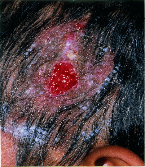
A 10 year old Asian boy presented to the accident and emergency department. He had a four week history of a pus filled swelling on the left temple region of the scalp but was otherwise healthy. Although the swelling had begun as a small pustule, it had gradually increased in size and had become much bigger within the past week. Treatment with oral co-fluampicil and topical sodium fusidate ointment had made no difference. Physical examination showed a boggy, tender swelling (3 cm diameter) on the scalp, accompanied by localised alopecia and cervical lymphadenopathy. The boy was admitted to hospital, and three days later the swelling was incised and drained under general anaesthesia. He was discharged home with a 10 day course of oral flucloxacillin and an open appointment to attend as necessary. Fourteen days later he was referred to the dermatology department by the district nurse, who was concerned that the wound was not healing. A clinical diagnosis of a fungal kerion was made. Although Wood's light examination was negative, mycological culture of plucked hair confirmed T tonsurans infection. The boy was treated with oral griseofulvin (10 mg/kg) and topical ketoconazole shampoo and terbinafine cream (twice daily). At follow up two weeks later, the lesion was healing (fig 1). Full resolution with hair regrowth occurred within two months.
Figure 1.
Fungal kerion on the scalp two weeks after beginning treatment with griseofulvin
Discussion
Tinea capitis is a common and easily transmitted childhood fungal scalp infection. Infection rates have reached epidemic proportions in London1 and are particularly high in black and Asian boys aged 1-10 years.2 Changing patterns of infection with different anthropophilic and zoophilic fungal species are well documented; the current London (and United Kingdom) outbreak results largely from the anthropophilic T tonsurans species.3 The Communicable Disease Surveillance Centre has recorded a 25-fold rise in cases of T tonsurans infection since 1995, and a further doubling of incidence was seen within the first half of 1999 (unpublished data). Further supportive evidence of treatment for the increase in tinea capitis comes from the 100% increase in prescriptions for griseofulvin in the London boroughs of Redbridge and Waltham Forest over the past year (unpublished data). Investigation of children with alopecia should include plucked hairs and scalp brushings for mycological analysis and clinical examination with Wood's light. (Fluorescence will not be seen, however, with the endothrix T tonsurans.) Because the positivity rates from mycological microscopy examination vary, three months' treatment with oral griseofulvin (10-15 mg/kg) is indicated once a confident clinical diagnosis is made. Griseofulvin is the only currently licensed treatment for tinea capitis in children, although many studies are now reporting the results of shorter treatment regimens with the newer antifungal agents terbinafine and itraconazole.4,5
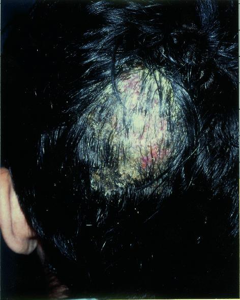
The progression from a scaly eruption to a kerion depends on the nature of the infective organism and host factors.6 Surgical drainage remains an essential part of the treatment of bacterial abscesses. However, scalp abscesses are extremely rare unless there is immune deficiency or penetrating trauma and are usually associated with severe pain and constitutional upset.7,8 Surgical drainage of a fungal kerion in the absence of other medical or surgical problems should therefore not be undertaken. We have gained support from our surgical, paediatric, and casualty departments in circulating a letter and clinical photograph (fig 2) advising all medical and nursing staff to refer children with boggy scalp swellings to our dermatology unit before considering surgical intervention. We recommend that a child with a suspected kerion or scalp abscess should be assessed initially by a dermatologist.
Figure 2.
Fungal kerion—a large pus filled swelling on the scalp
Acknowledgments
We thank Mrs Heather Walker, head of Drug Information Services, for her help in retrieving prescribing data.
Children with suspected kerion or scalp abscess should be assessed first by a dermatologist
Footnotes
Competing interests: None declared.
References
- 1.Hay RJ, Clayton YM, De Silva N, Midgley G, Rossor E. Tinea capitis in south-east London—a new pattern of infection with public health implications. Br J Dermatol. 1996;135:955–958. doi: 10.1046/j.1365-2133.1996.d01-1101.x. [DOI] [PubMed] [Google Scholar]
- 2.Fuller LC, Child FC, Higgins EM. Tinea capitis in south east London: an outbreak of Trichophyton tonsurans infection. Br J Dermatol. 1997;136:139. doi: 10.1111/j.1365-2133.1997.tb08771.x. [DOI] [PubMed] [Google Scholar]
- 3.Leeming JG, Elliott TS. The emergence of Trichophyton tonsurans tinea capitis in Birmingham UK. Br J Dermatol. 1995;133:929–931. doi: 10.1111/j.1365-2133.1995.tb06928.x. [DOI] [PubMed] [Google Scholar]
- 4.Gupta AK, Hofstader SL, Adam P, Summerbell RC. Tinea capitis: an overview with emphasis on management. Pediatr Dermatol. 1999;16:171–189. doi: 10.1046/j.1525-1470.1999.00050.x. [DOI] [PubMed] [Google Scholar]
- 5.Elewski BE. Treatment of tinea capitis: beyond griseofulvin. J Am Acad Dermatol. 1999;40(suppl):S27–S30. doi: 10.1016/s0190-9622(99)70394-4. [DOI] [PubMed] [Google Scholar]
- 6.Sohnle PG. Dermatophytosis. In: Cox RA, editor. Immunology of fungal diseases. Boca Raton, FL: CDC Press; 1989. pp. 1–27. [Google Scholar]
- 7.Nandwani R, Shanson DC, Fisher M, Nelson MR, Gazzard BG, et al. Mycobacterium kansasii scalp abscesses in an AIDS patient. J Infect. 1995;31:79–80. doi: 10.1016/s0163-4453(95)91705-5. [DOI] [PubMed] [Google Scholar]
- 8.Narotam PK, van Dellen JR, du Trevou MD, Gouws E. Operative sepsis in neurosurgery: a method of classifying surgical cases. Neurosurgery. 1994;34:409–415. doi: 10.1227/00006123-199403000-00004. [DOI] [PubMed] [Google Scholar]


