Abstract
An SK6 cell line (SK6c26) which constitutively expressed the glycoprotein Erns of classical swine fever virus (CSFV) was used to rescue CSFV Erns deletion mutants based on the infectious copy of CSFV strain C. The biochemical properties of Erns from this cell line were indistinguishable from those of CSFV Erns. Two Erns deletion mutants were constructed, virus Flc23 and virus Flc22. Virus Flc23 encoded only the utmost N- and C-terminal amino acids of Erns (deletion of 215 amino acids) to retain the original protease cleavage sites. Virus Flc22 is not recognized by a panel of Erns antibodies, due to a deletion of 66 amino acids in Erns. The Erns deletion mutants Flc22 and Flc23 could be rescued in vitro only on the complementing SK6c26 cells. These rescued viruses could infect and replicate in SK6 cells but did not yield infectious virus. Virus neutralization by Erns-specific antibodies was similar for the wild-type virus and the recombinant viruses, indicating that Erns from SK6c26 cells was incorporated in the viral particles. Pigs vaccinated with Flc22 or Flc23 were protected against a challenge with a lethal dose of CSFV strain Brescia. This is the first demonstration of trans-complementation of defective pestivirus RNA with a pestiviral structural protein and opens new ways to develop nontransmissible modified live pestivirus vaccines. In addition, the absence of (the antigenic part of) Erns in the recombinant viral particles can be used to differentiate between infected and vaccinated animals.
Classical swine fever virus (CSFV) is the causative agent of classical swine fever (CSF), an economically important disease of domestic pigs and wild boar. The disease is highly contagious and often fatal. It is characterized by fever and hemorrhages and can run an acute, chronic, or even subclinical course. Although effective live-attenuated vaccines are available, vaccination is not allowed in the European Union, since vaccinated and infected pigs are serologically indistinguishable. To control outbreaks of the disease, infected and suspected herds are slaughtered and quarantine restrictions are imposed. This can cause large economic losses; e.g., during the 1997 to 1998 CSF epizootic in the Netherlands, more than 10 million pigs were slaughtered and destroyed. The use of so-called marker vaccines, which makes discrimination between vaccinated and infected animals possible, might contribute to controlling the disease during a CSF epizootic.
CSFV belongs, together with bovine viral diarrhea virus (BVDV) and border disease virus, to the Pestivirus genus of the Flaviviridae family (33). The pestiviruses are structurally, genetically, and antigenically closely related. CSFV is restricted to swine, whereas BVDV and border disease virus have been isolated from several species such as cattle, sheep, swine, giraffes, and deer (21). Pestiviruses are small, enveloped, plus-strand RNA viruses, and the genome, varying in length from 12.5 to 16.5 kb, contains a single large open reading frame. The open reading frame is translated into a polyprotein that is processed into mature proteins by viral and host cell proteases (14). The envelope of the pestivirus virion contains three glycoproteins, Erns, E1, and E2 (28).
Animals infected with a pestivirus develop antibodies against the structural proteins Erns and E2 and the nonstructural protein NS3. Monoclonal antibodies (MAbs) directed against NS3 recognize pestivirus conserved epitopes, whereas MAbs against Erns and E2 can be used to discriminate between pestivirus species as well as between strains of one species (4, 32, 36). Glycoprotein E2 is the most immunogenic protein of CSFV. Subunit vaccines based on E2 are protective and induce high titers of neutralizing antibodies (1, 8, 31), whereas pigs immunized with Erns, the second immunogenic protein of CSFV, were protected even though neutralizing antibodies were not detected (10). However, since these dead subunit vaccines consist mostly of only one protein, live-attenuated vaccines are often preferred since they are more efficient in generating a protective immune response. Also, a live virus vaccine will be easier and less costly to produce. Recently, CSFV infectious DNA copies have been described (16, 19, 23), enabling the construction of a genetically modified live vaccine against CSF. We have constructed an infectious DNA copy based on the live-attenuated vaccine strain C (19). Viruses derived from this infectious clone have retained the biological and immunogenic properties of the parent strain C in rabbits and pigs (3).
In this study, we used our infectious clone to construct CSFV Erns deletion mutants; in this paper, we present the first demonstration of trans-complementation by an SK6 cell line constitutively expressing Erns. Complemented viruses can infect target cells but are unable to infect new cells due to the lack of Erns. Thus, such viruses are very safe as vaccines since they cannot spread and are thus nontransmissible from inoculated to contact animals. Furthermore, since the genomes of the mutants will encode E2 but not for Erns after infection, these viruses can potentially be used as marker vaccines against CSF.
MATERIALS AND METHODS
Cells and viruses.
Swine kidney (SK6) cells were grown in Eagle's basal medium containing 5% fetal bovine serum, glutamine (0.3 mg/ml), and the antibiotics penicillin (200 U/ml), streptomycin (0.2 mg/ml), and mycostatin (100 U/ml). Fetal bovine serum was tested for the absence of BVDV and BDV antibodies as described previously (18). Virus stocks were prepared by passaging the virus 8 to 10 times on SK6c26 cells. The virus titers obtained ranged from 105.0 to 105.8 50% tissue culture infective doses (TCID50)/ml. Since CSFV tends to be associated with the host cells, lysates were used for reinfection of fresh cells and as vaccine inoculation unless indicated otherwise. These lysates were prepared by freezing and thawing cell culture twice and clarification.
Construction and characterization of an SK6 cell line (SK6c26) expressing Erns.
The Erns gene of CSFV strain C was amplified by PCR with primers p974 (5′ AAG AAA AGA TCT AAA GCC CTA TTG GCA TGG 3′) and p976 (5′ TT GTT ACA GCT GCA TAT GTA CCC TAT TTT GCT TG 3′). After BglII digestion, the PCR fragment was ligated into the vector pPRKc16, which was digested with SalI, filled in, and subsequently digested with BglII. The resulting plasmid, pPRKc26, contains the Erns gene of CSFV strain C under the control of the transcription and translation signals of expression vector pEVhisD12 (22) and the histidinol dehydrogenase gene (hisD), which was used as a selective marker. SK6 cells persistently expressing Erns were established by hisD selection as described (30). A second round of cloning was performed to ensure clonality. The SK6 cell line constitutively expressing Erns was designated SK6c26.
Erns expression of the cell line SK6c26 was studied in an immunoperoxidase staining with Erns-specific MAbs C5 specific for strain C, (34), 140.1 and 137.5 directed against Erns of CSFV strains C and Brescia (A. J. de Smit, personal communication), and a polyclonal rabbit serum, R716 (7). The amount of Erns in the SK6c26 cells was determined by an enzyme-linked immunosorbent assay (ELISA), and its RNase activity was measured as described previously (7).
Construction of recombinant CSFV deletion Erns mutants.
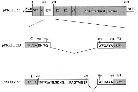
A mutant lacking the Erns gene was constructed, but the 5′ and 3′ bases encoding the five utmost N-terminal amino acids and the six utmost C-terminal amino acids of Erns were retained for intact protease cleavage sites. Two complementary oligomers, the forward oligomer p1135 (5′ CCG AAA ATA TAA CTC AAT GGT TTG GCG CTT ATG 3′) and the reverse oligomer p1136 (5′ CAT AAG CGC CAA ACC ATT GAG TTA TAT TTT CGG 3′), were phosphorylated, hybridized, and inserted via ligation in an alkaline phosphatase-treated, StuI-digested vector pPRKc5. PPRKc5 is a pEVhisD12 derivative which contains the nucleotide sequence of the autoprotease and structural genes of CSFV strain C, without Erns (Npro-C-E1-E2) but with a unique StuI site at the position where Erns was deleted (7). The resulting construct, pPRKc48, harbors the five utmost N-terminal amino acids and the six utmost C-terminal amino acids of Erns. This corresponds to a deletion of amino acids 273 to 488 of CSFV strain C (19).
A deletion of amino acids 422 to 488 in Erns of strain C was accomplished by PCR amplification of the Erns gene, using the forward primer p974 and the reverse primer p1120 (5′ GAC GGA TTC GGC ATA GGC GCC AAA TTG GCT CTC TAT AAC TGT AAC 3′). The HA epitope, amino acid sequence YPYDVPDYA (37), was constructed by annealing p1124 (5′ GAC AGA TCT ATC GAT TAC CCA TAC GAT GTT CCA GAT 3′) and p1125 (5′ GAC GTC GAC GGA TCC AGC GTA ATC TGG AAC ATC 3′) (the HA sequence is underlined) and filling in the 5′ single-stranded nucleotides in a PCR with Vent polymerase (New England Biolabs). The HA epitope PCR product was digested with ClaI and SalI, and the Erns PCR product was digested with BglII and NarI. The two digested PCR products were ligated via a three-point ligation into plasmid pPRKc16 that was digested with BglII and SalI, resulting in plasmid pPRKc43. After PCR amplification of pPRKc43 with the forward primer p935 (5′ CCG AAA ATA TAA CTC AAT GGT 3′) and the reverse primer p925 (5′ CAT AAG CGC CAA ACC AGG TT 3′), the PCR product was phosphorylated and ligated into the alkaline phosphatase-treated, StuI-digested vector pPRKc5, resulting in plasmid pPRKc50. Clones in which the mutated Erns gene was inserted in the correct orientation were transfected to SK6 cells and tested for expression of E2 by immunostaining with antibodies against E2-specific MAbs b3 and b4 (35).
A ClaI-NgoMI fragment of pPRKc48 and pPRKc50 was isolated and ligated into the ClaI-NgoMI-digested vector pPRKflc2 (previously named pPRKflc133) (19), and the resulting full-length cDNA CSFV strain C Erns mutants were named pPRKflc23 and pPRKflc22, respectively. A schematic representation of these constructs is shown in Fig. 1.
FIG. 1.
Schematic representation of the full-length DNA of pPRKflc2, pPRKflc23 and pPRKflc22. pPRKflc2 is the wild-type full-length DNA copy of CSFV strain C. The amino acid sequence numbering is for the open reading frame of the CSFV strain C (19). The starts of the Erns and E1 proteins are based on N-terminal sequencing (24), but the carboxy termini of the capsid and Erns proteins have not yet been determined. Npro, autoprotease; C, capsid protein; Erns, E1 and E2 envelope proteins; p7, nonstructural protein p7; 5′, 5′ noncoding region; 3′, 3′ noncoding region.
Regeneration of recombinant viruses.
Plasmids pPRKflc22 and pPRKflc23 were purified on columns (Qiagen) and linearized with XbaI. RNA transcription was performed as described previously (19). RNA (1 μg) was transfected with Lipofectin (Gibco BRL) to SK6c26 cells grown in 10-cm2 tissue culture plates. RNA transfection was performed in duplicate. Four days after transfection, one sample was immunostained with MAb b3 specific for E2. When the E2 immunostaining was negative, the duplicate sample was passaged and split into two samples. One of these samples was used for immunostaining 4 days after passaging. From monolayers, which showed E2 expression, supernatant was applied onto fresh SK6c26 or SK6 cells to determine the presence of infectious virus. After 4 days, the monolayers were immunostained as described above.
Characterization of recombinant Erns viruses.
The growth kinetics of the viruses was determined in SK6c26 cells. Subconfluent monolayers in M24 wells (2 cm2) were infected at a multiplicity of infection of 0.05. Viruses were adsorbed for 1.5 h. Before the cells were supplied with fresh medium, the first sample was collected; this was defined as time zero. At 0, 1, 2, 3, 4, 5, 6, and 7 days after infection, the monolayers were frozen-thawed twice and clarified by centrifugation for 10 min at 5,000 × g at 4°C. Virus titers (log TCID50 per milliliter) of total lysates (cell lysates plus supernatant) were determined on SK6c26 cells.
The virus neutralization index (log reduction of virus titer [TCID50/milliliter] by a neutralizing serum) was determined at a 1:250 dilution of serum 716 specifically directed against Erns of CSFV strain C and at a 1:1,000 dilution of pig serum 539 specifically directed against E2 of CSFV strain Brescia (7). The virus stocks of Flc2, Flc22, and Flc23 were subjected to titer determination by end-point dilution in the presence or absence of these CSFV neutralizing antibodies.
The Erns genes of Flc22 and Flc23 were sequenced. Therefore, cytoplasmic RNA of SK6c26 cells infected with these viruses was isolated using the RNeasy total-RNA isolation kit (Qiagen). DNA fragments covering the Erns genes were analyzed by reverse transcription-PCR (RT-PCR) using primers p1154 (5′ GTT ACC AGT TGT TCT GAT GAT 3′) and p305 (5′ GGG GTG CAG TTG TTG TAT CCA 3′) amplifying nucleotide sequences 865 to 1920, analyzed on a 1.5% agarose gel in 1× Tris-acetate-EDTA (TAE), and purified on Costar Spin-X columns. An RT-PCR of the E2 gene was performed with primer pair p307 (TGG AAT GTT GGC AAA TAT GT) and p304 (CAC TTA CCT AT[A,G] GGG TAG TGT GG) amplifying nucleotide position 2200 to 3174. Sequences of the purified PCR fragments were determined by PCR cycle sequencing using the Big Dye dRhodamine terminator ready-reaction cycle-sequencing kit (Perkin-Elmer) as specified by the manufacturer, with flanking primers and analyzed on a 310 ABI PRISM genetic analyzer.
Vaccination and challenge of pigs.
Four pigs born from conventional sows free of antibodies against pestiviruses were divided into two groups of two pigs; one group was vaccinated with virus Flc23, while the other group was vaccinated with virus Flc22. The pigs were vaccinated by several routes of inoculation and received, via each route, 2 ml of vaccine corresponding to 2 × 10⁵ TCID50; in total, each pig received 8 ml of vaccine, corresponding to 8 × 10⁵ TCID50. The pigs were sedated, placed on their backs, and inoculated with a virus suspension in phosphate-buffered saline into the nostrils, intravenously, and intradermally. In addition, the vaccines Flc22 and Flc23 were administered intramuscularly with a double-oil-water emulsion, since a recently performed study with an attenuated pseudorabies virus vaccine administered intramuscularly with a water-oil emulsion (20) protected pigs significantly better against challenge with virulent pseudorabies virus than when the adjuvant was omitted.
The pigs were challenged intranasally with 100 50% lethal doses (100 LD50) of CSFV strain Brescia 456610 (27) 4 weeks after vaccination. This inoculum was made by dilution from a virus stock of Brescia. Negative control pigs that were inoculated with the same LD50 derived from this stock solution before and after this pilot study all died within 14 days after infection (1, 27). All pigs were observed daily for signs of disease, and their body temperatures were measured.
Blood samples were collected in EDTA on the day before challenge (day −1) and on days 2, 6, 9, 12, and 15 after challenge to monitor changes of leukocyte and thrombocyte numbers in the blood. A decrease in the number of leukocytes (leukopenia) and thrombocytes (thrombocytopenia) and viremia is one of the signs of CSF. Normal cell counts of leukocytes and thrombocytes in conventional swine range between 11 × 109 and 23 × 109/liter and between 320 × 109 and 720 × 109/liter, respectively. Leukopenia and thrombocytopenia were defined as cell or platelets counts lower than the minimum numbers mentioned above, preferably for more than 1 day (less than 8 × 10⁹/liter and 200 × 109/liter, respectively.
Peripheral blood leukocytes were extracted from EDTA-treated blood samples taken to monitor viremia, and virus was isolated as described previously (3). Serum blood samples of all pigs were collected at 1-week intervals for 5 weeks after challenge. Samples were assayed in a CSFV-specific virus neutralization test (neutralizing peroxidase-linked assay) (26), in a Ceditest ELISA for detecting CSFV specific antibodies against E2 (2), and in a Ceditest ELISA for the detection of antibodies against Erns (A. J. de Smit, personal communication).
At postmortem, tissue samples were collected from the tonsils, spleen, kidneys, and ileum and were tested by direct immunofluorescence for the presence of viral antigen.
RESULTS
Establishment of an SK6 cell line expressing CSFV Erns.
SK6 cells were transfected with plasmid pPRKc26 harboring the CSFV strain C Erns gene and the histidinol (hisD) resistance gene. After 3 weeks, SK6 cells surviving histidinol selection were tested for the expression of Erns by immunostaining with MAb C5 specific for CSFV Erns. Positive cells were cloned twice to ensure clonality. The SK6c26 cell line showed detectable expression of Erns in the cytoplasm in more than 95% of the cells, whereas the parental SK6 cells showed no expression (data not shown). Continuous passaging of the SK6c26 cells in the presence of histidinol retained persistent expression in more than 95% of the cells for at least 10 months (46 passages). Passaging in the absence of histidinol for 10 passages resulted in a slight decrease of Erns-stained cells to approximately 80%.
The cell line SK6c26 was further characterized with respect to the biochemical characteristics of the Erns produced (Table 1). The cell line could be immunostained with the same Erns antibodies (MAb C5, 140.1, and 137.5 and polyclonal serum R716) as could SK6 cells infected with CSFV strain C (Flc2). The amount of Erns quantified in the SK6c26 cell lysate was 5 ng of Erns per 105 cells, as determined by ELISA and extrapolation from a standard curve of immunoaffinity-purified Erns (5). By this quantification method, CSFV-infected SK6 cells contained 15 ng of Erns per 105 cells. In addition, the RNase activity of the SK6c26 cells and CSFV-infected SK6 cells was determined, and the two systems were shown to have comparable RNase activity. The Erns protein of the stable cell line had a similar mobility to the wild-type Erns as determined by sodium dodecyl sulfate-polyacrylamide gel electrophoresis (results not shown) and was efficiently dimerized like the Erns found in virions (28). Thus, in conclusion, the SK6c26 line expresses Erns that is indistinguishable from CSFV-encoded Erns.
TABLE 1.
Characterization of Erns expressed by SK6c26 cells
| Cell line | Immunostaining with Erns-specific antibody:
|
Amt of Erns (ng/105 cells)a | RNase activity (A260 min−1 mg−1)b | Dimerizationc | |||
|---|---|---|---|---|---|---|---|
| MAb C5 | MAb 140.1 | MAb 137.5 | R 716 | ||||
| SK6c26 | + | + | + | + | 5 | 130 | + |
| SK6 Flc2 infectedd | + | + | + | + | 15 | 171 | + |
| SK6 | − | − | − | − | <0.1 | 0 | − |
The amount of Erns in the cell lysates per 105 cells was extrapolated from a standard curve prepared from an immunoaffinity-purified preparation of Erns produced in insect cells (5).
The RNase activity was determined as absorbance at 260 nm (A260) units per milligram of Erns per use as described previously (7).
Detection of dimers of Erns by nonreducing sodium dodecyl sulfate-polyacrylamide gel electrophoresis.
SK6 cells were infected with virus Flc2 with a multiplicity of infection of 0.05 and analyzed 4 days after infection.
Construction and recovery of C strain CSFV Erns recombinant viruses Flc22 and Flc23.
Two full-length CSFV recombinants were constructed. The first Erns recombinant contained a C-terminal deletion of 66 amino acids from position 422 to 488. This Erns deletion mutant was first constructed with a C-terminal HA tag in the expression vector pEVhisD12 (pPRKc43). The latter HA tag was incorporated as a control for the correct open reading frame. Immunostaining of transient-expression products of this plasmid in SK6 cells was positive only with antibodies against the HA tag and not with antibodies against Erns, including neutralizing MAb C5. Transient expression of plasmid pPRKc47, encoding the entire Erns gene with the C-terminal HA tag, was positive with both the Erns and the HA antibodies (data not shown). Thus, the deleted C-terminal amino acids (422 to 488) are essential for recognition of the antigenic site(s) on Erns. The recombinant Erns sequence of pPRKc43 without the HA epitope was transferred to the full-length infectious copy of the CSFV strain C pPRKflc2 (19), yielding the full-length clone pPRKflc22 (Fig. 1).
The second Erns recombinant full-length clone, pPRKflc23, lacked the whole Erns gene but retained only the N- and C-terminal utmost amino acids of Erns in order to retain the original sequences around the cleavage sites (24). This clone contains a deletion of a.a. 273 to 488 (Fig. 1).
Linearized full-length cDNAs of pPRKflc22 and pPRKflc23 were transfected into an SK6 cell line constitutively expressing the bacteriophage T7 RNA polymerase (30). Transient expression of E2 was detected by immunostaining; however, no infectious recombinant virus was recovered, even after repeated passaging of transfected cells (data not shown). Apparently, Erns is required for the formation of such a virus. To investigate whether Erns can complement in trans, in vitro-transcribed RNAs of the plasmids pPRKflc22 and pPRKflc23 were transfected in the cell line SK6c26, which constitutively expressed Erns. At 4 days after transfection, immunostaining of the monolayers with an E2-specific MAb, b3, was negative. The amount of E2 protein of the recombinant viruses might be too small for immunostaining. Therefore, the transfected cells were passaged to obtain higher titers of viruses. E2 could be detected from the wild-type construct (pPRKflc2) two passages after transfection, while virus derived from pPRKflc22 and pPRKflc23 could be detected after four passages. Three to five additional passages were required to obtain a virus titer of approximately 105.5 TCID50/ml, and this stock was used for further characterization of the viruses, which were named Flc22 and Flc23 for clones pPRKflc22 and pPRKflc23, respectively.
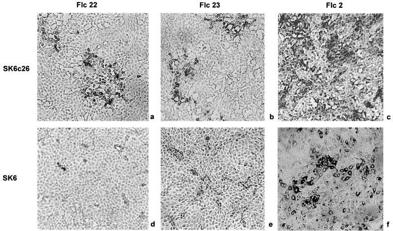
Supernatants from SK6c26 cells infected with Flc22 and Flc23 were used for infection of SK6c26 and SK6 cells. Four days after infection, for both viruses approximately 30% of the SK6c26 cells were positive by E2 immunoperoxidase staining (Fig. 2a and b) whereas infection of SK6 cells resulted in only single cells stained or pairs of single cells being stained (Fig. 2d and e). This indicates that the supernatants from SK6c26 cells contain infectious viruses that can infect and replicate in SK6 cells. However, there is no cell-to-cell spread or secondary infection of the mutated viruses in these cells, taking into account that cells infected with CSFV divide normally (once in 24 h). SK6 cells infected with the viruses Flc22 and Flc23 could be immunostained with MAbs directed against E2 but not with Erns-specific antibodies (Table 2). As a control, infection of SK6 cells with wild-type Flc2 grown on SK6c26 cells resulted in a positive immunostaining for both E2 and Erns (Table 2), and secondary infection was observed (Fig. 2). The numbers of wild-type-virus-infected SK6 and SK6c26 cells were comparable, although infected SK6 cells showed a weaker immunostaining (Fig 2c and f). With supernatant or cell lysates of SK6 cells infected with Flc22 and Flc23, neither SK6 nor SK6c26 cells could be infected. Thus, to obtain infectious virus, the recombinant viral genomes of viruses Flc22 and Flc23 require complementation of full-length Erns, which can be delivered in trans by the Erns-expressing SK6c26 cells.
FIG. 2.
Infection of CSFV in SK6 and SK6c26 cells with a multiplicity of infection of 0.05 and detection by immunostaining with MAb b3 against E2. SK6c26 cells infected with Flc22 (a), Flc23 (b), and Flc2 (c) SK6 cells infected with Flc22 (d), Flc23 (e), and Flc2 (f) are shown. Immunostaining was performed 4 days after infection.
TABLE 2.
Characterization of recombinant CSFV on SK6 and SKc26 monolayers
| Virusa | Reactivity in immunoperoxidase monolayer assay
|
Recovery of virus from:
|
|||
|---|---|---|---|---|---|
| SK6c26 (anti-E2)b | SK6 (anti-E2)b | SK6 (anti-Erns)c | SK6c26 | SK6 | |
| Flc2 (wild type) | + | + | + | + | + |
| Flc22 (Δ422–488) | + | + | − | + | − |
| Flc23 (Δ273–488) | + | + | − | + | − |
Amino acid numbering of CSFV strain C (19).
Immunostaining with MAb b3.
Immunostaining with MAbs C5, 140.1, and 137.5 and polyclonal antibody R716.
Characterization of recombinant CSFV viruses Flc22 and Flc23.
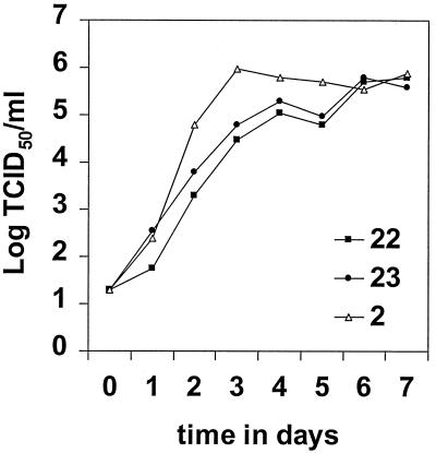
Growth kinetics of Flc22 and Flc23 and wild-type Flc2 in the complementing cell line SK6c26 were determined. As shown in Fig. 3, the multistep growth curves of the recombinant viruses Flc22 and Flc23 were very similar but showed a slower growth compared to the parent virus, Flc2. Titers between 105.0 and 105.8 TCID50/ml were found for the recombinant viruses after 6 days, whereas the parent strain Flc2 reached this titer within 3 days.
FIG. 3.
Growth kinetics of the recombinant CSFV Flc22 and Flc23 and the wild-type virus Flc2. Subconfluent monolayers of SK6c26 cells were infected at a multiplicity of infection of 0.1. Viruses were adsorbed for 1.5 h. Virus titers of the total lysates at various time points were determined by end-point dilution on SK6c26 cells.
To determine whether Erns was incorporated into the viral envelope, virus stocks of Flc2, Flc22, and Flc23 were subjected to titer determination in the presence of CSFV neutralizing antibodies (Table 3). All recombinant viruses were neutralized to the same extent as the parent virus Flc2 with both the Erns-specific and E2-specific neutralizing polyclonal antibodies. For Flc22, Erns on the viral envelope could be derived from the SK6c26 complementing cell line as well as from the recombinant Erns protein encoded by the viral genome, but the latter recombinant Erns is not recognized by the polyclonal serum R716 used for neutralizing the viruses. Thus, it remains unclear whether this recombinant Erns is incorporated into the viral membrane. The similar neutralization index obtained with this polyclonal serum suggests that the amount of Erns derived from the complementing cell line in the viral envelopes of Flc22 was comparable to that of Flc2 and Flc23.
TABLE 3.
Neutralization of CSF viruses by antibodies
| Virusa | Virus neutralizing reduction (log TCID50/ml) with serumb:
|
|
|---|---|---|
| 716 (directed against Erns)c | 539 (directed against E2)d | |
| Flc2 (wild type) | 3.0 | 1.75 |
| Flc22 (Δ422–488) | 3.0 | 1.75 |
| Flc23 (Δ273–488) | 3.25 | 1.0 |
Amino acid numbering of CSFV strain C (19).
Log TCID50 per milliliter reduction of CSFV titers due to the presence of serum.
Rabbit serum prepared against Erns of CSFV strain C.
Pig serum specifically directed against E2 of CSFV strain Brescia.
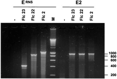
After viruses Flc22 and Flc23 were passaged on the complementing SK6c26 cells 10 times, cellular RNA from infected SK6c26 cells was analyzed by RT-PCR with CSFV-specific primers flanking either the Erns or E2 gene. The PCR fragments with primers flanking the Erns gene were of the expected sizes of 857, 401, and 1,055 nucleotides, respectively, for Flc22, Flc23, and Flc2, whereas the E2 gene products were of the expected size of 974 bp for all viruses (Fig. 4). The amplification products of the Erns gene were sequenced, and the obtained sequences were unchanged, indicating that no mutations, no reversion to the wild type, and no recombination with the Erns gene in the cell line could be detected.
FIG. 4.
RT-PCR of SK6c26 cells infected with viruses Flc22, Flc23, and Flc2 using primers flanking Erns and E2. −, Negative control (mock-infected SK6c26 cells); M, 200-bp marker.
Immunization and challenge of pigs.
To establish the vaccine properties of the Erns deletion mutant viruses, a pilot immunization and challenge experiment was performed. Two pigs in one group were vaccinated with strain Flc22, and two pigs in another group were vaccinated with strain Flc23. Each pig was vaccinated via several routes of inoculation (intramuscularly, intravenously, intradermally, and intratracheally), since we anticipated that the viruses are nontransmissible and the route of application might be important for efficacy. The pigs were challenged intranasally with 100 LD50 of CSFV strain Brescia 456610 (27) 4 weeks after vaccination.
After immunization, none of the animals developed clinical signs or fever. After challenge, all the pigs developed a mild fever (40 to 41°C) for 3 days. None of the vaccinated pigs in either group developed leukopenia or thrombocytopenia, although 6 days after challenge a slight drop in the thrombocyte and leukocyte counts was observed for three of the four pigs. After challenge, no virus could be isolated from the leukocytes throughout the experiment. Moreover, the organs of all pigs were IFT negative at the end of the experiment, indicating the clearance of CSFV.
After vaccination of the pigs with viruses Flc22 and Flc23, no CSFV-specific antibodies were detected by the E2 ELISA and the Erns ELISA until the time of challenge (Table 4). This finding was consistent with the NPLA results: all vaccinated pigs remained negative for neutralizing antibodies against CSFV up to the day of challenge 4 weeks after vaccination. After challenge, maximum inhibition percentages were observed in the E2 ELISA in all inoculated pigs, indicating a strong booster effect. Also, all four pigs seroconverted in the Erns ELISA. In the NPLAs, all inoculated pigs showed high titers against CSFV. These results clearly show that both Flc22 and Flc23 protect pigs against a lethal challenge with the virulent strain Brescia. Moreover, these data indicate that the use of these mutant viruses enables us to discriminate between infected and vaccinated animals based on a CSFV-specific Erns ELISA.
TABLE 4.
Serological response after immunization and challenge of pigsa
| Assay | Virus | Pig | Response on day postchallenge:
|
||||||
|---|---|---|---|---|---|---|---|---|---|
| −28 | −15 | −1 | 6 | 12 | 19 | 33 | |||
| Ceditest ELISA anti-E2b | Flc23 | 469 | − | − | − | + | + | + | + |
| 476 | − | − | − | + | + | + | + | ||
| Flc22 | 477 | − | − | ± | + | + | + | + | |
| 478 | − | − | ± | + | + | + | + | ||
| Ceditest ELISA anti-Ernsc | Flc23 | 469 | − | − | − | − | + | + | + |
| 476 | − | − | − | − | + | + | + | ||
| Flc22 | 477 | − | − | − | − | + | + | + | |
| 478 | − | − | − | − | + | + | + | ||
| NPLA for CSFV Brescia-specific neutralizing antibodiesd | Flc23 | 469 | <10 | <10 | 15 | 80 | >1,280 | >1,280 | >1,280 |
| 476 | <10 | <10 | <10 | 80 | >1,280 | >1,280 | >1,280 | ||
| Flc22 | 477 | <10 | <10 | 10 | 80 | >1,280 | >1,280 | >1,280 | |
| 478 | <10 | <10 | <10 | 480 | >1,280 | >1,280 | >1,280 | ||
Pigs were immunized on day −28 with 8 × 10⁵ TCID50 of virus. On day 0, the pigs were challenged intranasally with 100 LD50 of CSFV strain Brescia 46610.
The Ceditest E2 ELISA specifically detects antibodies against envelope protein E2 of CSFV. Test results are expressed as the percent inhibition of a standard signal; <30% is negative (−), 30 to 50% inhibition is doubtful (±), and >50% inhibition is positive (+).
The Ceditest Erns ELISA specifically detects antibodies against envelope protein Erns of CSFV. Test results are expressed as the percent inhibition of a standard signal; <50% is negative (−), ≥50% is positive (+).
The range of observed NPLA titers of sera are expressed as the reciprocal of the serum dilution neutralizing 100 TCID50 of CSFV strain Brescia.
DISCUSSION
An SK6 cell line (SK6c26) expressing Erns was established, enabling complementation of CSFV Erns deletion mutants. The biochemical properties of Erns from this cell line were indistinguishable those of from CSFV Erns, which is identified as an RNase (5, 25). It is striking that this cell line expresses a protein that hydrolyzes host cell RNAs. This implies either compartmentalization of Erns in the cell or a high substrate specificity of Erns (7, 38). The amount of Erns produced in the SK6c26 cells was of the same order of magnitude as in SK6 cells infected with the wild-type virus (Flc2) and was therefore expected to be sufficient to complement a CSFV Erns deletion mutant in trans.
A CSFV strain C Erns deletion mutant was constructed which retained the utmost terminal amino acids of Erns (pPRKflc23) to circumvent the formation of a capsid-E1 junction, which might not be suitable for proteolytic cleavage. Also, the protease involved in cleavage between Erns-E1 is yet unknown (24). A second recombinant strain C (pPRKflc22) was constructed lacking 66 amino acids in the C-terminal end of Erns (Δ422–488). This Erns deletion mutant lacks antigenic epitopes, since our Erns-specific antibodies did not recognize it. This is in agreement with previous studies which showed that these antibodies do not inhibit RNase activity (7) and that the active domains of the Erns RNase are located in the N-terminal half of the protein (5, 25).
Defective interfering particles of BVDV and CSFV lacking the Npro, the four structural proteins, and NS2 are strictly dependent on complementation by a helper virus for packaging (14, 15). Thus, all proteins encoded by the structural region can be complemented in trans. trans-complementation of a single protein in the nonstructural regions in the NS1 or NS5 gene has been reported for the flaviviruses yellow fever virus and Kunjin virus, which also belong to the Flaviviridae family (9, 13). This is the first study showing that the glycoprotein Erns is indispensable to obtain infectious virus and that it can be delivered in trans by a complementing cell line. Infectious virus was obtained only when RNAs derived from pPRKflc22 and pPRKflc23 were complemented in trans by the SK6c26 cells. The complemented recombinant viruses could infect and replicate in SK6 cells, but no infectious virus was produced in these cells, since no spread of virus was observed.
On the one hand, the finding that the recombinant viruses Flc22 and Flc23 and the wild-type virus Flc2 have comparable neutralization indices with a serum specific for Erns indicates that the amounts of Erns on the viral envelope of these viruses are comparable. Also, similar maximum virus titers on SK6c26 cells (105.0 to 105.8 TCID50/ml) were obtained for these three viruses. However, on the other hand, the wild-type virus Flc2 reached these titers within 3 days whereas the recombinant viruses Flc22 and Flc23 required almost 6 days. Presumably, in-cis interactions between the structural proteins are more effective for virion assembly, although trans-complementation of Erns by the SK6c26 cells still appears to be quite efficient. Another explanation for the retarded growth of the recombinant viruses might be that only one of the cleavage sites at the capsid protein/Erns site or at the Erns/E1 site is cleaved.
Ten-fold-lower titers were obtained for CSFV strain C on SK6c26 cells (105.5 TCID50/ml) compared to the parental SK6 cells (106.5 TCID50/ml). The stable incorporation of the foreign gene in the SK6c26 cells might affect the metabolism of the cells. However, the growth of transmissible gastroenteritis encephalitis virus (TGEV) virus was similar in both cell types (data not shown). Hulst and Moormann (6) showed that purified glycoprotein Erns, synthesized in insect cells, was able to inhibit CSFV infection in SK6 cells. Possibly, the lower titers for strain C on SK6c26 cells were caused by inhibition of infection by the Erns produced by the SK6c26 cell line.
The glycoprotein Erns possesses the unique feature of RNase activity, and inactivation of this activity by a single-amino-acid substitution of the catalytic residues in CSFV strain C resulted in vitro in a cytopathogenic virus (7). In this study, no cytopathogenic effects or apoptosis were observed with cells infected with the Erns deletion mutant Flc22 or Flc23. The titers of the Erns mutants obtained in this study might be too low to induce apoptosis. Otherwise, deletion of the Erns gene in Flc23 might have a different effect on virus-host interaction from that of inactivation of the RNase activity by a single-amino-acid substitution. Furthermore, we do not know whether the C-terminal deletion in Erns of Flc22 inactivates the RNase activity of this protein. This question is now under investigation as part of a study to delinate the functional properties of Erns in relation to the virus-host interaction.
Homologous RNA recombination yielding wild-type virus has been described for other plus-strand RNA viruses like alphaviruses (12) and may carry a potential risk for the use of nonspreading live vaccines. However, no recombination occurred between the Erns genes of the complementing cell line and the deletion mutant genomes after 10 serial passages of defective virus, as analyzed by RT-PCR and sequencing. This is in agreement with complementation studies of nonstructural proteins of flaviviruses YF1 and Kunjin virus, where no recombination was detected after three serial passages of these viruses (9, 13). However, RNA recombination has been described for BVDV strains, including insertions of cellular sequences (e.g., ubiquitin) and genetic rearrangements like deletions, duplications, or even point mutations (14). These recombination events result in cytopathogenic viruses. However, the incidence of these recombination events is very low and is observed in animals that are persistently infected with BVDV and are highly viremic. While cytopathogenic isolates are frequently observed for BVDV, cytopathogenic CSFV isolates are quite rare in vivo and in vitro (11, 15, 17, 29) and no recombination between cellular sequences and the CSFV genome have been described. Nevertheless, although no recombination was observed after 10 serial passages of the mutated CSF viruses, stocks of virus vaccines should always be examined carefully on this point.
The pilot vaccination and challenge experiment showed that the Erns deletion mutants could protect pigs against a lethal challenge of strain Brescia. Since the recombinant viruses could infect host cells only once, the route of vaccination might be important for protection. Therefore, vaccination was performed simultaneously via multiple inoculation routes. Further research will first focus on the appropriate route of vaccination. A remarkable finding is that vaccinated pigs were negative for CSFV neutralizing antibodies and that no E2 and Erns antibodies could be detected by ELISA until challenge, 28 days postvaccination. The absence of detectable amounts of antibodies can be explained by the low titers of induced antibodies that occur because the complemented viruses can infect target cells only once. König et al. also showed that pigs immunized with Erns were protected even though neutralizing antibodies were not detected (10). In contrast, Terpstra and Wensvoort (27) reported that pigs with a neutralization antibody titer of >32 induced by the live C-strain vaccine were protected against clinical signs of CSF and Bouma et al. (1) showed that pigs with neutralization antibody titers of >50 at 21 days postvaccination induced by an E2 subunit vaccine were considered to be protected. Our data suggest that pigs can be protected against a lethal challenge even though no antibodies against CSFV are detected.
No differences were found between virus Flc23, lacking the whole Erns gene, and Flc22, lacking the antigenic part of Erns, in their in vitro properties, such as growth and neutralization by Erns-specific antibodies, and in their in vivo properties, such as induction of protection against a lethal challenge. Thus, the presence of the N-terminal part of Erns in the mutated genome confers no additional advantage over that of the deletion mutant lacking the whole Erns gene. We focused on the constructing of Erns deletion mutants because E2 is the most powerful protein in the induction of neutralizing antibodies (1, 10, 31).
The vaccination and challenge experiments indicated that the Erns deletion mutants could be useful for further studies on the development of a CSF marker vaccine. However, the strength of a marker vaccine is dependent not only on its immunogenic properties but also on the performance of a diagnostic test that can differentiate between infected and vaccinated animals. Therefore, we have developed an ELISA specific for CSFV Erns (A. J. de Smit, personal communication). However, repeated vaccination of an animal with an Erns deletion mutant virus is needed to demonstrate the suitability of such a test.
Recently we have constructed two CSF marker vaccines based on chimeric CSFV viruses, in which the E2 or Erns gene of the C strain was replaced by that of BVDV type II strain 5250 (H. G. P. van Gennip, submitted for publication). These chimeric viruses protected against a lethal challenge of CSFV strain Brescia and could be serologically differentiated from the wild-type C strain. However, a live genetically engineered chimeric virus, although attenuated, could face problems regarding acceptance as vaccine. The described CSFV deletion mutants described here are safer as vaccines than are the chimeric viruses, since these deletion mutants are nontransmissible.
Although live vaccines are generally considered to have the best immunological properties, the use of a conventional live vaccine, besides the lack of marker properties, can have the disadvantage that the vaccine virus can spread through an animal and is eventually transmitted to a second contact animal. Our results represent a significant advance in the area of the development of a live-attenuated nontransmissible CSF marker vaccine. The possibility of trans-complementing the CSFV glycoprotein Erns also opens up new opportunities for the development of safe, nontransmissible marker vaccines for other pestiviruses, such as BVDV, based on deletion of an antigenic structural protein.
REFERENCES
- 1.Bouma A, de Smit A J, de Kluijver E P, Terpstra C, Moormann R J. Efficacy and stability of a subunit vaccine based on glycoprotein E2 of classical swine fever virus. Vet Microbiol. 1999;66:101–114. doi: 10.1016/s0378-1135(99)00003-6. [DOI] [PubMed] [Google Scholar]
- 2.Colijn E O, Bloemraad M, Wensvoort G. An improved ELISA for the detection of serum antibodies directed against classical swine fever virus. Vet Microbiol. 1997;59:15–25. doi: 10.1016/s0378-1135(97)00178-8. [DOI] [PubMed] [Google Scholar]
- 3.De Smit, A. J., H. G. P. Van Gennip, G. K. W. Miedema, P. A. Van Rijn, C. Terpstra, and R. J. M. Moormann. Recombinant pestiviruses derived from the Chinese vaccine strain (C-strain) of classical swine fever retain their avirulent and immunological characteristics. Vaccine, in press. [DOI] [PubMed]
- 4.Edwards S, Moennig V, Wensvoort G. The development of an international reference panel of monoclonal antibodies for the differentiation of hog cholera virus from other pestiviruses. Vet Microbiol. 1991;29:101–108. doi: 10.1016/0378-1135(91)90118-y. [DOI] [PubMed] [Google Scholar]
- 5.Hulst M M, Himes G, Newbigin E, Moormann R J. Glycoprotein E2 of classical swine fever virus: expression in insect cells and identification as a ribonuclease. Virology. 1994;200:558–565. doi: 10.1006/viro.1994.1218. [DOI] [PubMed] [Google Scholar]
- 6.Hulst M M, Moormann R J. Inhibition of pestivirus infection in cell culture by envelope proteins Erns and E2 of classical swine fever virus: Erns and E2 interact with different receptors. J Gen Virol. 1997;78:2779–2787. doi: 10.1099/0022-1317-78-11-2779. [DOI] [PubMed] [Google Scholar]
- 7.Hulst M M, Panoto F E, Hoekman A, van Gennip H G, Moormann R J. Inactivation of the RNase activity of glycoprotein E(rns) of classical swine fever virus results in a cytopathogenic virus. J Virol. 1998;72:151–157. doi: 10.1128/jvi.72.1.151-157.1998. [DOI] [PMC free article] [PubMed] [Google Scholar]
- 8.Hulst M M, Westra D F, Wensvoort G, Moormann R J. Glycoprotein E1 of hog cholera virus expressed in insect cells protects swine from hog cholera. J Virol. 1993;67:5435–5442. doi: 10.1128/jvi.67.9.5435-5442.1993. [DOI] [PMC free article] [PubMed] [Google Scholar]
- 9.Khromykh A A, Kenney M T, Westaway E G. trans-complementation of flavivirus RNA polymerase gene NS5 by using Kunjin virus replicon-expressing BHK cells. J Virol. 1998;72:7270–7279. doi: 10.1128/jvi.72.9.7270-7279.1998. [DOI] [PMC free article] [PubMed] [Google Scholar]
- 10.Konig M, Lengsfeld T, Pauly T, Stark R, Thiel H J. Classical swine fever virus: independent induction of protective immunity by two structural glycoproteins. J Virol. 1995;69:6479–6486. doi: 10.1128/jvi.69.10.6479-6486.1995. [DOI] [PMC free article] [PubMed] [Google Scholar]
- 11.Kosmidou A, Buttner M, Meyers G. Isolation and characterization of cytopathogenic classical swine fever virus (CSFV) Arch Virol. 1998;143:1295–1309. doi: 10.1007/s007050050376. [DOI] [PubMed] [Google Scholar]
- 12.Lai M M. RNA recombination in animal and plant viruses. Microbiol Rev. 1992;56:61–79. doi: 10.1128/mr.56.1.61-79.1992. [DOI] [PMC free article] [PubMed] [Google Scholar]
- 13.Lindenbach B D, Rice C M. trans-complementation of yellow fever virus NS1 reveals a role in early RNA replication. J Virol. 1997;71:9608–9617. doi: 10.1128/jvi.71.12.9608-9617.1997. [DOI] [PMC free article] [PubMed] [Google Scholar]
- 14.Meyers G, Thiel H-J. Molecular characterization of pestiviruses. Adv Virus Res. 1996;47:53–118. doi: 10.1016/s0065-3527(08)60734-4. [DOI] [PubMed] [Google Scholar]
- 15.Meyers G, Thiel H J. Cytopathogenicity of classical swine fever virus caused by defective interfering particles. J Virol. 1995;69:3683–3689. doi: 10.1128/jvi.69.6.3683-3689.1995. [DOI] [PMC free article] [PubMed] [Google Scholar]
- 16.Meyers G, Thiel H J, Rumenapf T. Classical swine fever virus: recovery of infectious viruses from cDNA constructs and generation of recombinant cytopathogenic defective interfering particles. J Virol. 1996;70:1588–1595. doi: 10.1128/jvi.70.3.1588-1595.1996. [DOI] [PMC free article] [PubMed] [Google Scholar]
- 17.Mittelholzer C, Moser C, Tratschin J D, Hofmann M A. Generation of cytopathogenic subgenomic RNA of classical swine fever virus in persistently infected porcine cell lines. Virus Res. 1997;51:125–137. doi: 10.1016/S0168-1702(97)00081-6. [DOI] [PMC free article] [PubMed] [Google Scholar]
- 18.Moormann R J, Warmerdam P A, van der Meer B, Schaaper W M, Wensvoort G, Hulst M M. Molecular cloning and nucleotide sequence of hog cholera virus strain Brescia and mapping of the genomic region encoding envelope protein E1. Virology. 1990;177:184–198. doi: 10.1016/0042-6822(90)90472-4. [DOI] [PubMed] [Google Scholar]
- 19.Moormann R J M, van Gennip H G P, Miedema G K W, Hulst M M, van Rijn P A. Infectious RNA transcribed from an engineered full-length cDNA template of the genome of a pestivirus. J Virol. 1996;70:763–770. doi: 10.1128/jvi.70.2.763-770.1996. [DOI] [PMC free article] [PubMed] [Google Scholar]
- 20.Nauwynck H J, Zonnekeyn V, Pensaert M B. Virological protection of sows upon challenge with Aujeszky's disease virus after multiple vaccinations with attenuated or inactivated vaccines. Zentbl Veterinaermed Reihe B. 1997;44:609–615. doi: 10.1111/j.1439-0450.1997.tb01014.x. [DOI] [PubMed] [Google Scholar]
- 21.Paton D J, Simpson V, Done S H. Infection of pigs and cattle with bovine viral diarrhoea virus on a farm in England. Vet Rec. 1992;131:185–188. doi: 10.1136/vr.131.9.185. [DOI] [PubMed] [Google Scholar]
- 22.Peeters B, de Wind N, Broer R, Gielkens A, Moormann R. Glycoprotein H of pseudorabies virus is essential for entry and cell-to-cell spread of the virus. J Virol. 1992;66:3888–3892. doi: 10.1128/jvi.66.6.3888-3892.1992. [DOI] [PMC free article] [PubMed] [Google Scholar]
- 23.Ruggli N, Tratschin J D, Mittelholzer C, Hofmann M A. Nucleotide sequence of classical swine fever virus strain Alfort/187 and transcription of infectious RNA from stably cloned full-length cDNA. J Virol. 1996;70:3478–87. doi: 10.1128/jvi.70.6.3478-3487.1996. [DOI] [PMC free article] [PubMed] [Google Scholar]
- 24.Rumenapf T, Unger G, Strauss J H, Thiel H J. Processing of the envelope glycoproteins of pestiviruses. J Virol. 1993;67:3288–3294. doi: 10.1128/jvi.67.6.3288-3294.1993. [DOI] [PMC free article] [PubMed] [Google Scholar]
- 25.Schneider R, Unger G, Stark R, Schneider-Scherzer E, Thiel H J. Identification of a structural glycoprotein of an RNA virus as a ribonuclease. Science. 1993;261:1169–1171. doi: 10.1126/science.8356450. [DOI] [PubMed] [Google Scholar]
- 26.Terpstra C, Bloemraad M, Gielkens A L. The neutralizing peroxidase-linked assay for detection of antibody against swine fever virus. Vet Microbiol. 1984;9:113–120. doi: 10.1016/0378-1135(84)90026-9. [DOI] [PubMed] [Google Scholar]
- 27.Terpstra C, Wensvoort G. The protective value of vaccine-induced neutralising antibody titres in swine fever. Vet Microbiol. 1988;16:123–128. doi: 10.1016/0378-1135(88)90036-3. [DOI] [PubMed] [Google Scholar]
- 28.Thiel H J, Stark R, Weiland E, Rumenapf T, Meyers G. Hog cholera virus: molecular composition of virions from a pestivirus. J Virol. 1991;65:4705–4712. doi: 10.1128/jvi.65.9.4705-4712.1991. [DOI] [PMC free article] [PubMed] [Google Scholar]
- 29.Vanderhallen H, Mittelhozer C, Hofmann M A, Koenen F. Classical swine fever virus is genetically stable in vitro and in vivo. Arch Virol. 1999;144:1669–1677. doi: 10.1007/s007050050622. [DOI] [PubMed] [Google Scholar]
- 30.van Gennip H G, van Rijn P A, Widjojoatmodjo M N, Moormann R J. Recovery of infectious classical swine fever virus (CSFV) from full-length genomic cDNA clones by a swine kidney cell line expressing bacteriophage T7 RNA polymerase. J Virol Methods. 1999;78:117–128. doi: 10.1016/s0166-0934(98)00171-2. [DOI] [PubMed] [Google Scholar]
- 31.van Zijl M, Wensvoort G, de Kluyver E, Hulst M, van der Gulden H, Gielkens A, Berns A, Moormann R. Live attenuated pseudorabies virus expressing envelope glycoprotein E1 of hog cholera virus protects swine against both pseudorabies and hog cholera. J Virol. 1991;65:2761–2765. doi: 10.1128/jvi.65.5.2761-2765.1991. [DOI] [PMC free article] [PubMed] [Google Scholar]
- 32.Weiland E, Ahl R, Stark R, Weiland F, Thiel H J. A second envelope glycoprotein mediates neutralization of a pestivirus, hog cholera virus. J Virol. 1992;66:3677–3682. doi: 10.1128/jvi.66.6.3677-3682.1992. [DOI] [PMC free article] [PubMed] [Google Scholar]
- 33.Wengler G, Bradley D W, Colett M S, Heinz F X, Schlesinger R W, Strauss J H. Flaviviridae. In: Murphy F A, Fauquet C M, Bishop D H L, Ghabrial S A, Jarvis A W, Martelli G P, Mayo M A, Summers M D, editors. Virus taxonomy. Sixth report of the International Committee on Taxonomy of Viruses. Vienna, Austria: Springer-Verlag; 1995. pp. 415–427. [Google Scholar]
- 34.Wensvoort G. Ph.D. thesis. Utrecht, The Netherlands: State University Utrecht; 1989. [Google Scholar]
- 35.Wensvoort G. Topographical and functional mapping of epitopes on hog cholera virus with monoclonal antibodies. J Gen Virol. 1989;70:2865–2876. doi: 10.1099/0022-1317-70-11-2865. [DOI] [PubMed] [Google Scholar]
- 36.Wensvoort G, Terpstra C, de Kluijver E P, Kragten C, Warnaar J C. Antigenic differentiation of pestivirus strains with monoclonal antibodies against hog cholera virus. Vet Microbiol. 1989;21:9–20. doi: 10.1016/0378-1135(89)90014-x. [DOI] [PubMed] [Google Scholar]
- 37.Wilson I A, Niman H L, Houghten R A, Cherenson A R, Connolly M L, Lerner R A. The structure of an antigenic determinant in a protein. Cell. 1984;37:767–778. doi: 10.1016/0092-8674(84)90412-4. [DOI] [PubMed] [Google Scholar]
- 38.Windisch J M, Schneider R, Stark R, Weiland E, Meyers G, Thiel H J. RNase of classical swine fever virus: biochemical characterization and inhibition by virus-neutralizing monoclonal antibodies. J Virol. 1996;70:352–358. doi: 10.1128/jvi.70.1.352-358.1996. [DOI] [PMC free article] [PubMed] [Google Scholar]


