Abstract
An earlier report (M. Sakaguchi et al., Vaccine 16:472–479, 1998) showed that recombinant Marek's disease virus type 1 (rMDV1) expressing the fusion (F) protein of Newcastle disease virus (NDV-F) under the control of the simian virus 40 late promoter [rMDV1-US10L(F)] protected specific pathogen-free chickens from NDV challenge, but not commercial chickens with maternal antibodies against NDV and MDV1. In the present study, we constructed an improved polyvalent vaccine based on MDV1 against MDV and NDV in commercial chickens with maternal antibodies. The study can be summarized as follows. (i) We constructed rMDV1 expressing NDV-F under the control of the MDV1 glycoprotein B (gB) promoter [rMDV1-US10P(F)]. (ii) Much less NDV-F protein was expressed in cells infected with rMDV1-US10P(F) than in those infected with rMDV1-US10L(F). (iii) The antibody response against NDV-F and MDV1 antigens of commercial chickens vaccinated with rMDV1-US10P(F) was much stronger and faster than with rMDV1-US10L(F), and a high level of antibody against NDV-F persisted for over 80 weeks postvaccination. (iv) rMDV1-US10P(F) was readily reisolated from the vaccinated chickens, and the recovered viruses were found to express NDV-F. (v) Vaccination of commercial chickens having maternal antibodies to rMDV1-US10P(F) completely protected them from NDV challenge. (vi) rMDV1-US10P(F) offered the same degree of protection against very virulent MDV1 as the parental MDV1 and commercial vaccines. These results indicate that rMDV1-US10P(F) is an effective and stable polyvalent vaccine against both Marek's and Newcastle diseases even in the presence of maternal antibodies.
Marek's disease virus (MDV) is an etiological agent of Marek's disease (MD), a highly contagious malignant T-lymphomatosis of chickens caused by MDV serotype 1 (MDV1) (10, 32, 52). MD represents the first cancer to be prevented and controlled by the use of live attenuated or naturally avirulent vaccines (11, 12). MD vaccine viruses are divided into three categories: attenuated MDV1, naturally apathogenic MDV2, and MDV3, also called herpesvirus of turkeys (HVT), the naturally apathogenic strain (68). The MD vaccine viruses are considered one of the most potent vectors for polyvalent live vaccines expressing foreign antigens related to vaccine-induced immunity against poultry diseases for the following reasons. (i) The viruses induce lifetime protection against MD with just one vaccination (39), (ii) the viruses have a natural host range limited to avian species, and therefore, the vectors would be safe for other domestic animals and people working in the poultry industry, and (iii) techniques for generating recombinant MDVs have been well established (45, 49). Among the vaccine viruses, HVT has been used worldwide both as live vaccine and polyvalent vaccine vector (13, 17, 28, 29, 41, 42, 53). However, attenuated MDV1 strains, such as C/R6 (G. F. de Boer, J. M. A. Pol, and S. H. M. Jeurissen, Proc. 3rd Int. Symp. Marek's Dis., p. 405–413, 1988) and R2/23 (67), are clearly superior to HVT (R. L. Witter, Proc. 19th World's Poult. Congr., p. 298–304, 1992) because the MDV1 vaccine is more efficient than the HVT vaccines, especially against very virulent MDV1 (vvMDV1). Thus, attenuated MDV1 is suitable for construction of a recombinant vaccine against avian diseases.
We have been developing recombinant polyvalent vaccines based on attenuated MDV1 strains. We previously examined 22 sites for insertion of a foreign gene (the Escherichia coli lacZ gene) into the MDV1 genome by homologous recombination and identified several stable sites for expression of the gene in cultured cells (K. Hirai, M. Sakaguchi, H. Maeda, Y. Kino, H. Nakamura, G. S. Zhu, and M. Yamamoto, Proc. 19th World's Poult. Congr., p. 150–155, 1992). Of these sites, those of the US3 and US10 genes and the junction region between the unique short (US) and short inverted repeats were nonessential not only for viral growth in culture but also for vaccine-induced immunity (45, 49, 54). In addition, other groups reported several nonessential sites within US repeat for viral growth in culture (9, 37, 38). Among genes at these insertion sites, the US10 gene appears to be the most stable and not to be connected with vaccinal immunogenicity (45). Based on the information obtained above, we constructed recombinant MDV1 (rMDV1) expressing the fusion (F) protein of the Newcastle disease virus (NDV-F) gene under the control of the simian virus 40 (SV40) late promoter inserted within the US10 gene of MDV1 [rMDV1-US10L(F)] and tested the efficiency of the polyvalent vaccine by using vaccinated chickens challenged with NDV and MDV1 (47). rMDV1 showed almost 100% protective efficacy against NDV and MDV1 challenge in specific-pathogen-free (SPF) chickens lacking maternal antibodies from ND and MD by one-time inoculation, whereas the protective efficacy varied among experiments and decreased on average to 70% in chickens with maternal antibodies even though the challenge experiments were performed at a time when the maternal antibodies would not affect an evaluation of the protective efficacy. In the other systems using rHVT expressing NDV-F under the control of a strong promoter from the Rous sarcoma virus long terminal repeat and several recombinant fowl poxviruses (rFPV) expressing the NDV-F or hemagglutinin-neuraminidase gene, a similar problem with the maternal antibodies was also reported (14, 25, 28, 29, 35, 57, 58). Although it is not known why the recombinant polyvalent vaccines are not completely effective against the avian diseases in the presence of maternal antibodies, it is conceivable that the strong expression of these foreign genes induces a strong host immune reaction against the recombinant vaccines, which results in inhibition of the growth of the recombinant viruses in chickens. The suppression of the growth of vaccine viruses would reduce or redirect the efficiency of the recombinant vaccines in chickens. Therefore, it is hypothesized that the use of an appropriate promoter that regulates the expression of foreign genes would improve the efficiency of the recombinant viruses in chickens with maternal antibodies. In the present study, we attempted to develop a novel recombinant polyvalent vaccine based on the MDV1 background by using the MDV1 glycoprotein B (gB) promoter for expression of NDV-F protein and demonstrated the following. (i) In cell culture, the recombinant rMDV1, in which NDV-F expression is controlled by the MDV1 gB promoter [rMDV1-US10P(F)], expressed less NDV-F than did rMDV1-US10L(F), in which NDV-F cDNA expression is driven by the SV40 late promoter. (ii) In chickens immunized with rMDV1-US10P(F), the immune response against NDV-F and MDV1 was much faster and stronger than that with rMDV1-US10L(F). (iii) As we previously reported (47), the immunization of commercial chickens possessing maternal antibodies against NDV and MDV1 with rMDV1-US10L(F) resulted in only approximately 70% protection against NDV challenge, whereas rMDV1-US10P(F) provided complete protection against NDV challenge in commercial chickens. (iv) The protection efficacy with rMDV1-US10P(F) against vvMDV1 is as good as that with the parent attenuated MDV1. These results indicate that rMDV1-US10P(F) is an effective polyvalent vaccine against ND and MD even in the presence of maternal antibodies to these viruses.
MATERIALS AND METHODS
Viruses and cells.
The avirulent MDV1 CVI988 strain, recombinant viruses, and the virulent MDV1 Alabama strain were propagated in monolayers of primary chicken embryo fibroblasts (CEFs), which were cultured in Eagle's minimum essential medium supplemented with 5% fetal calf serum and antibiotics. The number of passages of parent CVI988 was 26 and those of recombinant viruses used in animal experiments were 13. The RB1B strain of vvMDV1 was propagated in monolayers of primary chicken kidney cells, which were cultured in the same medium as the CEFs. The virulent NDV strain Sato was propagated in growing eggs from SPF chickens.
Construction of plasmids and rMDV1s.
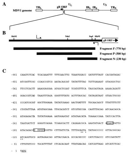
The BamHI-I3 fragment of the MDV1 CVI988 strain containing the gB promoter region was cloned into pUC119 (7, 15, 43). The nucleotide sequence of the gB promoter region was determined by Sawady Technology Sequencing Service (Tokyo, Japan). According to the nucleotide sequence, a PCR primer pair to which the EcoRI site was added at each 5′ end was designed to amplify 550 bp of the MDV1 gB promoter region (Fig. 1B). The PCR product was digested with EcoRI and SspI or with EcoRI and NdeI, and the resulting 500- or 230-bp subfragment was named P or N fragment, respectively (Fig. 1B). A 770-bp subfragment of SfaNI-SfaNI was designated F fragment (Fig. 1B). To construct transfer plasmids for generating rMDV1s, pKA4L(F) (47), which contains NDV-F cDNA (52) under the control of the SV40 late promoter, was used. A HindIII-XhoI fragment of pKA4L(F) was substituted for the F, P, or N fragment, and the resulting plasmids were designated pKA4F(F), pKA4P(F), and pKA4N(F), respectively. rMDV1 was constructed essentially as described previously (45, 49). Briefly, CEFs infected with CVI988 was transfected with a transfer plasmid, pKA4F(F), pKA4P(F), or pKA4N(F), by electroporation. At 7 days after transfection, the plaques were stained with the monoclonal antibody to NDV-F protein 313 (63, 64) and goat anti-mouse immunoglobulin G (IgG) horseradish peroxidase (HRP) conjugate (Bio-Rad Laboratories, Hercules, Calif.) as described previously (47). The positive plaques were collected and plated on fresh CEFs. The cloning procedure was repeated until all of the plaques were stained positively for NDV-F expression. The conditions used for DNA extraction and Southern blot hybridization were essentially as described previously (20).
FIG. 1.
MDV1 gB promoter region. (A) Location of the gB ORF in the UL region of the MDV1 genome. (B) Locations of the fragments F, P, and N upstream of the gB ORF. (C) Sequence upstream of the MDV1 gB ORF. Nucleotide positions are numbered with reference to the translation initiation codon ATG (underlined, designated +1). Predicted CAT and TATA boxes are boxed. The 5′ end of NDV-F mRNA determined by the RACE method is marked by asterisks.
Detection of antigens and antibodies.
To confirm that the transfer plasmids express NDV-F protein, each transfer plasmid was transfected into CEFs by using Lipofectin (GIBCO-BRL, Life Technologies, Inc., Gaithersburg, Md.). Two days after transfection, the cells were fixed with acetone and subjected to an immunofluorescence (IF) test using the monoclonal antibody against NDV-F and anti-mouse IgG labeled with fluorescein isothiocyanate.
To quantitate the expression of NDV-F protein, CEFs transfected with each transfer plasmid were harvested, washed with phosphate-buffered saline (PBS), and added to the wells of 96-well microtiter plates at identical cell concentrations. These plates were dried overnight at 37°C. Then, monoclonal antibody diluted 1:3,000 in PBS containing 5% fetal bovine serum was added to each well, and the plates were incubated overnight at 4°C. After extensive washing of the plates with PBS containing 0.05% Tween 20, anti-mouse IgG labeled with HRP (Bio-Rad Laboratories) at a 1:300 dilution in the same buffer was added, and incubation continued for 1 h at 37°C. After another wash as before, the wells were developed by adding 0.1 ml of the ABTS (2,2′-azino-bis-[3-ethylbenzthiazoline-6-sulfonic acid]; Sigma Chemical Co., St. Louis, Mo.) solution (0.5 mg/ml) and incubating for 30 min at room temperature. The absorbance at 405 and 490 nm was read with a spectrophotometer.
To quantitate the expression of MDV1 antigens, CEFs infected with each virus were harvested, washed with PBS, and added to the wells of 96-well microtiter plates at various cell concentrations. These plates were treated as described above with the serum from an SPF chicken infected with CVI988 (1:300) and anti-chicken IgG labeled with HRP (1:300).
To quantitate the titer of antibody against NDV-F protein in sera from vaccinated chickens, 0.1 ml of the sera from chickens was assayed by the enzyme-linked immunosorbent assay (ELISA) system as described previously (46). Antibodies against MDV1 antigens were detected by ELISA as reported previously (47).
Southern and Northern blot hybridization.
The procedures used for DNA extraction and Southern blot hybridization were essentially as described previously (20, 21). poly(A)+ mRNA was isolated from infected cells by RNA extraction and with mRNA preparation kits (Amersham Pharmacia Biotech, Uppsala, Sweden). RNAs were electrophoretically separated in a denaturing agarose gel containing formaldehyde, transferred to nylon membrane (Boehringer Mannheim, Mannheim, Germany), and hybridized to appropriate DNA probes by using Rapid-hyb buffer (Amersham Pharmacia Biotech). The DNA probes were labeled with 32P by using a DNA labeling system (Amersham Pharmacia Biotech) and purified for removal of unincorporated nucleotides by using NICK Spin Columns (Amersham Pharmacia Biotech).
Determination of the 5′ end of the mRNA.
To determine the 5′ end of NDV-F mRNA from rMDV1-US10P(F), rapid amplification of cDNA ends (RACE) was carried out with 5′-Full RACE Core Set (TaKaRa, Shiga, Japan) according to the manufacturer's instructions. A phosphorylated 15-mer oligonucleotide, 5′-pAAGTAGTCAATGTCC-3′, based on the nucleotide sequence of NDV-F cDNA was used for the synthesis of the first strand of cDNA. The cDNA of the 5′ region of the mRNA was amplified by nested PCR using two pairs of NDV-F-specific primers and cloned into pUC18. For the first PCR, 5′-CAGGGTCAATCATAATCAAGTT-3′ and 5′-CTGCTTTGTCTCCTGTTCC-3′ were used. For the second PCR, 5′-AAGGATAAAGAGGCGTGTGC-3′ and 5′-TTCGGACGGTCAGCATCAG-3′ were used. All primers were synthesized by Amersham Pharmacia custom oligo DNA service (OligoExpress PCR; Amersham Pharmacia Biotech, Tokyo, Japan). Then, the nucleotides of the PCR products were sequenced by the TaKaRa sequencing service.
Animal experiments.
One-day-old conventional chickens, Babcock B-300 with maternal antibodies to MDV1 and NDV, were obtained from Tsuboi Farm (Kumamoto, Japan). Maternal antibodies against NDV were detected by a hemagglutination inhibition (HI) assay according to the method of Hitcher et al. (22). The HI titers of maternal antibodies against NDV chickens used in this study ranged from 4 to 640 (geometric mean = 58.5), and the ELISA value against maternal MDV antibodies ranged from 0.17 to 1.03 (average = 0.60). Vaccinations of 1-day-old conventional chickens with rMDV1s, the parental CVI988 strain, or the NDV B1 strain (The Chemo-Sero-Therapeutic Research Institute, Kumamoto, Japan) and challenge experiments using virulent NDV strain Sato or vvMDV1 strain RB1B were performed as described previously (47). Briefly, 20 1-day-old chickens in each group were vaccinated with 10,000 PFU of the indicated viruses and then challenged with 10,000 minimum lethal doses of the NDV Sato strain at 6 weeks postvaccination. The chickens were examined for the onset of ND daily, 2 weeks after the challenge. For MDV1 challenge experiments, 1-day-old chickens were vaccinated with 10,000 PFU of virus and then challenged with 500 PFU of the vvMDV1 RB1B strain at 7 days postvaccination. Ten weeks after the challenge, the chickens were examined grossly and histopathologically for the presence of MD lesions in the peripheral nerves, brains, and visceral organs. Titers of antibody against NDV-F and MDV1 antigens were chased as described above. The recovery of rMDV1 from vaccinated chickens was examined at 7 weeks after immunization as previously described (45).
RESULTS
MDV1 gB promoter activity for expression of NDV-F cDNA.
Ross et al. have determined the sequence of the putative promoter region (positions −1 to −360 relative to the first nucleotide of the MDV1 gB open reading frame [ORF]) of the MDV1 gB gene of strain RB1B (43). We determined the nucleotide sequence of the region upstream of the gB ORF (−1 to −621) of the MDV1 CVI988 strain and found that the sequence from −1 to −360 was identical to that of RB1B (Fig. 1C). The region includes putative promoter elements and TATA and CAT boxes (CAAT), as suggested by Ross et al. Furthermore, a homologue of herpes simplex virus ICP18.5 was found in the upstream region, as reported for other alpha-herpesviruses (4, 26, 27, 40).
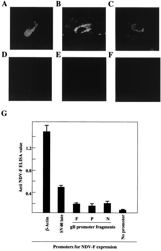
Next, we examined whether the region upstream of the MDV1 gB gene in fact possesses promoter activity by a transient-transfection assay. We selected three putative gB promoter regions, including the putative TATA and CAT boxes: an F fragment of an SfaNI-SfaNI subfragment of 770 bp, a P fragment of an SspI-EcoRI subfragment of 500 bp and an N fragment of an SfaNI-NdeI subfragment of 230 bp (Fig. 1B). The expression cassettes in which NDV-F cDNA is driven by these putative promoter regions were inserted into the US10 region of the transfer vector. CEFs were transfected with the transfer vectors and subjected to an IF test 72 h later. NDV-F expression was detected in the cytoplasm of cells transfected with the transfer vectors driven by the putative gB promoter regions (Fig. 2A to C), whereas no IF was detected in cells transfected with the control transfer vectors in which the putative promoter sequences were inserted in the opposite orientation (Fig. 2D to F). These results indicate that the upstream region of the gB gene has promoter activity and the activity is enough to express NDV-F protein.
FIG. 2.
Expression of the NDV-F gene under the control of putative gB promoters in CEFs. (A to F) IF patterns of CEF cells transfected with insertion vectors. The insertion vector plasmids containing fragments F (A and D), P (B and E), and N (C and F) in the right (A to C) and opposite (D to F) directions were transfected into CEFs. After 72 h, the cells were fixed and subjected to an IF test using anti-NDV-F monoclonal antibody. (G) ELISA analysis of CEF cells transfected with insertion vector plasmids containing various promoters. Four independent experiments were performed.
Next, we investigated the promoter activity for NDV-F expression in these transfer vectors by ELISA. Although a consistent level of NDV-F expression controlled by the sequences within the F, P, and N fragments was detected, the level was much lower than that controlled by the SV40 late and chicken β-actin promoters (Fig. 2G). Therefore, the gB promoter activity to express NDV-F is very weak, compared with those of SV40 late and chicken β-actin promoters.
Constructs of rMDV1 expressing NDV-F under the control of MDV1 gB promoter.
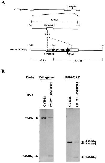
To generate rMDV1 in which NDV-F expression is controlled by MDV1 gB promoter regions, the transfer vectors (Fig. 3A) were transfected into CEFs infected with CVI988. Then, the plaques were immunostained with the monoclonal antibody to NDV-F protein, and the positive plaques were recloned until 100% of the plaques became positive. We successfully isolated three different rMDV1s: rMDV1-US10P(F) with the P fragment, rMDV1-US10N(F) with the N fragment, and rMDV1-US10F(F) with the F fragment. However, we used only rMDV1-US10P(F) since rMDV1-US10F(F) became unstable for expression of NDV-F after the sixth passage (data not shown) and rMDV1-US10N(F) induced very little antibody against NDV-F in sera from inoculated chickens, as described later. rMDV1-US10P(F) expressed NDV-F stably over 10 passages (data not shown). Next, to confirm the insertion of the expression cassette at the predicted site in rMDV1-US10P(F), DNAs extracted from CEFs infected with CVI988 or rMDV1-US10P(F) were digested with PstI and subjected to Southern blot hybridization with the P fragment and US10 ORF sequences as probes (Fig. 3B). Insertion of the sequence of the NDV-F gene with the gB promoter P fragment into the US10 gene was expected to yield one PstI site (Fig. 3A). Therefore, in rMDV1-US10P(F), the P fragment hybridized to a 2.47-kb fragment in addition to an approximately 20-kb fragment that is derived from the gB promoter region located within the unique long (UL) sequence of the MDV1 DNA and also detected in CVI988 (Fig. 3B). The US10 ORF probe hybridized to two PstI fragments of 2.47 and 4.51 kb, but only to the 4.50-kb fragment in CVI988 (Fig. 3B). The possibility of contamination of parental CVI988 with rMDV1-US10P(F) was eliminated by Northern blot analysis, the results of which are shown in Fig. 4. In rMDV1-US10P(F)-infected cells, any transcripts from the US10 gene region that are specific to parental CVI988 were not detectable. Furthermore, that rMDV1-US10P(F) is free from the parental virus was confirmed by PCR analysis of the US10 region in rMDV1-US10P(F) (data not shown). These results indicated that the NDV-F gene controlled by the MDV1-gB promoter was correctly integrated into the MDV1 DNA at the predicted sites by homologous recombination and the recombinant virus was purified.
FIG. 3.
Construction of rMDV1-US10(F). (A) The predicted genomic structures of parental strain CVI988 and rMDV1-US10P(F). (B) Southern hybridization blots. DNA from CEFs infected with parental CVI988 and rMDV1-US10P(F) were digested with PstI and then subjected to Southern blot hybridization. The length (in kilobase pairs) of each hybridized fragment is shown on both sides of the panel for the PstI fragments.
FIG. 4.
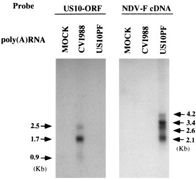
Northern blot hybridization of RNA extracted from CEFs infected with parental CVI988 and rMDV1-US10P(F). The length (in kilobase pairs) of each hybridized fragment is shown on both sides.
Analysis of transcripts from the inserted NDV-F gene in cells infected with rMDV1-US10P(F).
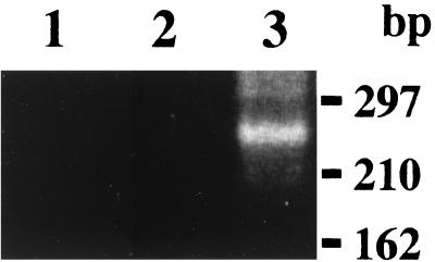
To demonstrate that the expression of NDV-F cDNA in cells infected with rMDV1-US10P(F) is in fact controlled by the MDV1 gB promoter within the P fragment, we carried out a RACE with NDV-F-specific primers. As shown in Fig. 5, an approximately 240-bp fragment was amplified mainly by using RNA from rMDV1-US10P(F)-infected cells and not by using those from mock- or CVI988-infected cells. By cloning and sequencing seven cDNAs obtained from independent PCR amplifications, the transcription initiation sites of the NDV-F expression cassette were mapped as two clusters in the MDV1 gB promoter, approximately 25 and 45 bp downstream of the putative TATA motif (Fig. 1C). The distance between the TATA motif and the transcription initiation sites is similar to that in other eucaryotic genes (6), and the CAAT box is located upstream of the TATA motif. These results indicate that a specific transcript(s) of NDV-F is initiated from the MDV1 gB promoter and the transcript was expressed under the control of the gB promoter. Next, to analyze the transcripts from the inserted NDV-F gene in the rMDV1-US10P(F) DNA, RNAs extracted from cells infected with rMDV1-US10P(F) or the CVI988 strain were subjected to Northern blot hybridization (Fig. 4). The US10 ORF probe hybridized to three transcripts of 2.5, 1.7, and 0.9 kb in RNA extracted from cells infected with CVI988. Consistent with our previous report (48), the probe did not detect any transcripts in cells infected with rMDV1-US10P(F), indicating that no US10 gene product is expressed in cells infected with the virus. The NDV-F cDNA probe hybridized to four transcripts of 4.2, 3.4, 2.6, and 2.1 kb in RNAs extracted from cells infected with rMDV1-US10P(F), but not from cells infected with CVI988. Although we do not know at present which transcripts detected by the NDV-F cDNA probe are driven by the MDV1 gB promoter, these results suggested that a specific transcript(s) encoding the NDV-F ORF is expressed and controlled by the gB promoter because (i) the specific transcript(s) of NDV-F initiated from the gB promoter is detected by RACE as described above and (ii) NDV-F protein is in fact expressed in cells infected with rMDV1-US10P(F) (Table 1).
FIG. 5.
Amplification of the 5′ cDNA of the NDV-F transcript in CEFs infected with rMDV1-US10P(F) by the RACE method. The 5′ cDNA ends of NDV-F mRNAs were amplified by using RNA isolated from mock-infected CEFs (lane 1) or CEFs infected with CVI988 (lane 2) or rMDV1-US10P(F) (lane 3). The sizes of molecular weight markers are shown at the right.
TABLE 1.
Comparison of the amount of NDV-F and MDV antigens expressed by the respective recombinant viruses
| Virus | ELISA value
|
Ratio of NDV/MDV | |
|---|---|---|---|
| NDV | MDV1 | ||
| rMDV1-US10L(F) | 1.309 ± 0.117 | 1.406 ± 0.046 | 0.933 ± 0.103 |
| rMDV1-US10P(F) | 0.263 ± 0.030 | 1.543 ± 0.070 | 0.171 ± 0.026 |
| rMDV1-US10N(F) | 0.275 ± 0.044 | 1.948 ± 0.169 | 0.140 ± 0.013 |
| CVI988 | 0.026 ± 0.004 | 0.935 ± 0.068 | 0.028 ± 0.006 |
Expression of NDV-F protein in CEF cells infected with rMDV1-US10P(F).
To examine the expression of NDV-F and MDV1 antigens in cells infected with rMDV1-US10P(F) and compare it with the expression of other rMDV1s in which NDV-F is driven by other promoters, ELISAs were performed. The results (Table 1) show that rMDV1-US10L(F) with the SV40 late promoter expressed NDV-F well in proportion to MDV1 antigen. In cells infected with rMDV1-US10P(F) and rMDV1-US10N(F), the level of NDV-F protein expression was consistent with that in CVI988-infected cells. However, rMDV1-US10P(F) and rMDV1-US10N(F) expressed much less NDV-F than rMDV1-US10L(F). These results indicate that gB promoter activity to express NDV-F in the context of the MDV1 genome is less than SV40 late promoter activity, but the capability to express NDV-F protein remains.
Immune responses and virus recovery in commercial chickens immunized with rMDV1-US10P(F).
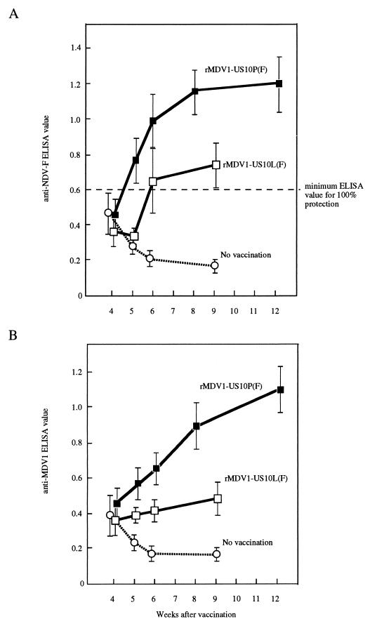
To investigate the immune responses against NDV-F and MDV1 antigens in commercial chickens vaccinated with rMDV1-US10P(F), the sera of chickens were examined weekly for the presence of anti-NDV-F antibody and anti-MDV1 antibodies by ELISA from 4 weeks after inoculation. As shown in Fig. 6A, the titers of antibody against NDV-F in chickens vaccinated with rMDV1-US10P(F) increased from 5 weeks after inoculation, much earlier than with rMDV1-US10L(F). The ELISA values were much higher than the minimum ELISA value of 0.6, which provides 100% protection against NDV challenge (47). Furthermore, the high level of antibody against NDV-F persisted for over 80 weeks (data not shown). By contrast, rMDV1-US10N(F) showed the lowest antibody titer, providing no protection from NDV challenge as described later (data not shown). The antibodies against MDV1 antigens were also examined in sera from commercial chickens vaccinated with rMDV1-US10P(F) from 4 to 12 weeks after immunization. As shown in Fig. 6B, rMDV1-US10P(F) induced higher titers of MDV1 antibodies than rMDV1-US10L(F).
FIG. 6.
Antibody responses of commercial chickens vaccinated with rMDV1-US10P(F) and rMDV1-US10L(F). One-day-old commercial chickens were inoculated with rMDV1-US10P(F) and rMDV1-US10L(F). The sera were tested for the presence of antibodies against NDV-F (A) and MDV1 antigens (B). Data shown are averages and standard errors (n = 20).
Next, to see whether rMDV1-US10P(F) is genetically stable in commercial chickens upon insertion of the NDV-F expression cassette into the viral genome, rMDV1s were recovered from chickens in the seventh week after vaccination and the expression of the NDV-F protein was examined. rMDV1-US10L(F) was not recovered from any chickens tested, while rMDV1-US10P(F) was isolated from all chickens. Furthermore, all the plaques of recovered rMDV1-US10P(F) were found to express NDV-F.
These results indicate that rMDV1-US10P(F) is stable even in vivo and infects persistently with expression of the NDV-F protein in commercial chickens. The difference in frequency of viral isolation between rMDV1-US10P(F) and rMDV1-US10L(F) also suggests that rMDV1-US10P(F) replicates better in commercial chickens with maternal antibodies than does rMDV1-US10L(F).
Protective efficacy of rMDV1-US10P(F) against NDV and MDV1 challenges in commercial chickens.
To test the protective efficacy of rMDV1-US10P(F) against NDV and MDV1, 1-day-old commercial chickens with maternal antibodies against NDV-F and MDV1 antigens were subcutaneously inoculated with rMDV1s once and then challenged with NDV strain Sato at 6 weeks postvaccination or with vvMDV1 strain RB1B at 7 days postvaccination. The results (Tables 2 and 3) were as follows.
TABLE 2.
Protective efficacy of rMDV1-US10P(F) against virulent NDV challenge in commercial chickens
| Virus for vaccination | % Protection (protected/total)
|
|||
|---|---|---|---|---|
| Expt 1 | Expt 2 | Expt 3 | Expt 4 | |
| rMDV1-US10P(F) | 100 (20/20) | 100 (20/20) | 100 (20/20) | 100 (20/20) |
| rMDV1-US10L(F) | NDa | 70 (14/20) | 74 (14/19) | 60 (12/20) |
| rMDV1-US10N(F) | ND | 0 (0/20) | ND | ND |
| CVI988 | 0 (0/20) | ND | ND | ND |
| NDV vaccine B1 | 100 (20/20) | ND | ND | ND |
| None | 0 (0/20) | 0 (0/20) | 5 (1/20) | 0 (0/20) |
ND, not done.
TABLE 3.
Protection of commercial chickens against the vvMDV RB1B strain
| Virus for vaccination | % Protection (protected/total) | Protective indexa |
|---|---|---|
| rMDV1-US10P(F) | 90 (18/20) | 89 |
| Parental strain (CVI988) | 89 (17/19) | 88 |
| Commercial vaccine (Rispens) | 84 (16/19) | 83 |
| Commercial vaccine (HVT) | 44 (8/18) | 41 |
| None | 5 (1/20) | 0 |
The formula is as follows: (% of MD in unvaccinated − % of MD in vaccinated)/% of MD in unvaccinated × 100.
(i) As we reported earlier, the protective efficacy of rMDV1-US10L(F) against NDV varied from 60 to 74% in several experiments (Table 2). In no experiments, however, did we obtain perfect protection against ND by using rMDV1-US10L(F). In contrast, rMDV1-US10P(F) provided complete protection against ND in all series of experiments (Table 2).
(ii) The commercial vaccines Rispens and CVI988, the parental strain of rMDV1-US10P(F), provided not perfect but sufficient protection against vvMDV1 in commercial chickens (Table 3). Similarly, the vaccination of commercial chickens with rMDV1-US10P(F) resulted in 90% protection against vvMDV1. The protection efficacy was as good as that of the parental vaccine strain, CVI988, or the commercial vaccine Rispens (Table 3).
These results indicate that rMDV1-US10P(F) is a reliable and effective polyvalent vaccine against MD and ND for commercial chickens, even those with maternal antibodies.
NDV was barely recovered from the tracheae of commercial chickens vaccinated with rMDV1-US10P(F) after NDV challenge.
Previously reported recombinant vaccines which express NDV-F protein generally provided systemic protection but very poor local protection (28). To see whether vaccination of commercial chickens with rMDV1-US10P(F) induces local immunity against NDV, commercial chickens were mock immunized or immunized with rMDV1-US10P(F). At 7 weeks after vaccination, those chickens were placed with three SPF chickens that had been inoculated with the virulent NDV. The chickens were examined for onset of ND daily until 3 weeks passed and for NDV recovery from the trachea at 9 days after NDV-infected commercial chickens were provided. The results were as follows.
(i) All of the chickens with no vaccination showed the serious symptoms of ND immediately. In contrast, vaccination of chickens with rMDV1-US10(F) provided complete protection against ND.
(ii) Ten vaccinated chickens were examined for NDV recovery from the trachea. NDV was recovered from only one chicken but not from nine chickens, while the virus was easily recovered from all nonvaccinated chickens tested. These results suggest that vaccination with rMDV1-US10(F) induces local immunity against NDV.
DISCUSSION
Virus vectors have been widely studied for efficiency of vaccination and for use as a system of gene transfer into the living body. In the poultry industry, four recombinant viruses (rFPV [1, 5, 8, 14, 18, 25, 31, 34, 35, 59], rHVT [13, 17, 28, 41, 44, 53], adenovirus [51], and rMDV1 [45, 47, 49, 54, 62]) that express foreign antigens of other avian pathogens (including NDV [14, 17, 24, 28–30, 35, 47, 53, 58], MDV [31, 33, 42, 44, 69], infectious bursal disease virus [1, 5, 13, 18, 19, 51, 62], avian influenza virus [2, 3, 5, 56, 59, 61, 65, 66], avian leukosis virus [34], and avian reticuloendotheliosis virus [8]) have been developed, and these viruses showed significant vaccine efficacy against a variety of avian diseases. Further, rHVT, rMDV1, and rFPV expressing NDV antigens have been constructed and used to protect chickens from NDV infection. Although these recombinant viruses showed good vaccine efficacy in SPF chickens without maternal antibody, the vaccine efficacy decreased in commercial chickens with maternal antibody. Therefore, an improved recombinant polyvalent vaccine against ND that overcomes the problem of maternal antibodies has been awaited. MD live vaccine viruses are known to infect persistently within chickens in spite of the presence of neutralizing antibodies in sera and induce a high titer of antibody against MDV. The expression of viral antigens of the vaccines that are the target of the host immune system is regulated by MDV promoters, and therefore, the vaccines are able to escape from the host immune system and establish persistent infection in chickens. In previous MDV-based polyvalent vaccines against NDV infection, heterologous promoters, such as SV40 late promoter and the Rous sarcoma virus long terminal repeat, were used for the expression of NDV antigens. These promoters are known to show very strong activity in various types of cells (16, 55, 60), resulting in high expression levels of NDV antigens in chickens given the vaccines. Conceivably, these vaccines induce a strong immune response against the products of vaccines and are unable to establish themselves, unlike MD vaccines in chickens with maternal antibodies. Therefore, we hypothesized that a promoter from an immunogenic viral protein of MD vaccine virus would regulate the expression of NDV antigens properly and that a vaccine with the promoter would grow in chickens in the same manner as MD vaccine viruses and provide protection against NDV infection. Among MDV promoters, we chose the gB promoter of MDV because (i) MDV gB is one of the viral antigens responsible for virus neutralization (23) and (ii) chickens immunized with the purified protein were protected partially against virulent MDV1 challenge (36). As we expected, the new polyvalent vaccine, rMDV1-US10P(F), showed a more significant and persistent immunogenicity against the NDV-F protein and had more protective efficacy against NDV challenge in commercial chickens with maternal antibodies than rMDV1-US10L(F) with the SV40 late promoter. Although our vaccine is in fact effective against both NDV and MDV infection in chickens with maternal antibodies, the exact mechanism by which rMDV1-US10P(F) shows improved efficacy against NDV infection compared to other MDV-based polyvalent vaccines is unclear at present. Also, we do not know whether expression of NDV-F cDNA is controlled only by the MDV1 gB promoter, because four transcripts were detected by Northern blot hybridization with an NDV-F probe (Fig. 4). Further characterization of this recombinant virus and studies to reveal why the use of the gB promoter for the expression of the NDV-F protein improves vaccine efficacy in the presence of maternal antibodies would be of interest and provide insight into how to develop effective recombinant vaccines.
The key findings of the present study are that (i) our newly developed polyvalent vaccine [rMDV1-US10P(F)] afforded complete protection against NDV challenge even in commercial chickens with maternal antibody following only one vaccination, (ii) the ability of the recombinant vaccine to protect chickens from MDV challenge was as good as that of the parent MDV1 vaccine strain of the recombinant virus, and (iii) rMDV1-US10P(F) is quite stable for the expression of NDV in vitro and in vivo. This vaccine will be useful in the poultry industry, and further, the usage of the gB promoter in the context of the MDV1 genome will be applicable for vaccine antigens for other chicken diseases, including viral, bacterial, and parasitic diseases.
ACKNOWLEDGMENTS
We are grateful to M. Kawakita for the NDV-F cDNA and T. Kohama and S. Umino for the monoclonal antibody against the NDV-F protein. We are also grateful to M. Fujimoto, H. Sakamoto, and K. Naruse for technical assistance in construction of plasmids and rMDV1.
This work was supported in part by a grant-in-aid from the ministry of Education, Science, Sports and Culture of Japan.
REFERENCES
- 1.Bayliss C D, Peters R W, Cook J K, Reece R L, Howes K, Binns M M, Boursnell M E. A recombinant fowlpox virus that expresses the VP2 antigen of infectious bursal disease virus induces protection against mortality caused by the virus. Arch Virol. 1991;120:193–205. doi: 10.1007/BF01310475. [DOI] [PubMed] [Google Scholar]
- 2.Beard C W, Schnitzlein W M, Tripathy D N. Effect of route of administration on the efficacy of a recombinant fowlpox virus against H5N2 avian influenza. Avian Dis. 1992;36:1052–1055. [PubMed] [Google Scholar]
- 3.Beard C W, Schnitzlein W M, Tripathy D N. Protection of chickens against highly pathogenic avian influenza virus (H5N2) by recombinant fowlpox viruses. Avian Dis. 1991;35:356–359. [PubMed] [Google Scholar]
- 4.Beuken E, Slobbe R, Bruggeman C A, Vink C. Cloning and sequence analysis of the genes encoding DNA polymerase, glycoprotein B, ICP18.5 and major DNA-binding protein of rat cytomegalovirus. J Gen Virol. 1996;77:1559–1562. doi: 10.1099/0022-1317-77-7-1559. [DOI] [PubMed] [Google Scholar]
- 5.Boyle D B, Heine H G. Recombinant fowlpox virus vaccines for poultry. Immunol Cell Biol. 1993;71:391–397. doi: 10.1038/icb.1993.45. [DOI] [PMC free article] [PubMed] [Google Scholar]
- 6.Bucher P, Trifonov E N. Compilation and analysis of eukaryotic POL II promoter sequences. Nucleic Acids Res. 1986;14:10009–10026. doi: 10.1093/nar/14.24.10009. [DOI] [PMC free article] [PubMed] [Google Scholar]
- 7.Buckmaster A E, Scott S D, Sanderson M J, Boursnell M E, Ross N L, Binns M M. Gene sequence and mapping data from Marek's disease virus and herpesvirus of turkeys: implications for herpesvirus classification. J Gen Virol. 1988;69:2033–2042. doi: 10.1099/0022-1317-69-8-2033. [DOI] [PubMed] [Google Scholar]
- 8.Calvert J G, Nazerian K, Witter R L, Yanagida N. Fowlpox virus recombinants expressing the envelope glycoprotein of an avian reticuloendotheliosis retrovirus induce neutralizing antibodies and reduce viremia in chickens. J Virol. 1993;67:3069–3076. doi: 10.1128/jvi.67.6.3069-3076.1993. [DOI] [PMC free article] [PubMed] [Google Scholar]
- 9.Cantello J L, Anderson A S, Francesconi A, Morgan R W. Isolation of a Marek's disease virus (MDV) recombinant containing the lacZ gene of Escherichia coli stably inserted within the MDV US2 gene. J Virol. 1991;65:1584–1588. doi: 10.1128/jvi.65.3.1584-1588.1991. [DOI] [PMC free article] [PubMed] [Google Scholar]
- 10.Churchill A E, Biggs P M. Agent of Marek's disease in tissue culture. Nature. 1967;215:528–530. doi: 10.1038/215528a0. [DOI] [PubMed] [Google Scholar]
- 11.Churchill A E, Chubb R C, Baxendale W. The attenuation, with loss of oncogenicity, of the herpes-type virus of Marek's disease (strain HPRS-16) on passage in cell culture. J Gen Virol. 1969;4:557–564. doi: 10.1099/0022-1317-4-4-557. [DOI] [PubMed] [Google Scholar]
- 12.Churchill A E, Payne L N, Chubb R C. Immunization against Marek's disease using a live attenuated virus. Nature. 1969;221:744–747. doi: 10.1038/221744a0. [DOI] [PubMed] [Google Scholar]
- 13.Darteil R, Bublot M, Laplace E, Bouquet J F, Audonnet J C, Riviere M. Herpesvirus of turkey recombinant viruses expressing infectious bursal disease virus (IBDV) VP2 immunogen induce protection against an IBDV virulent challenge in chickens. Virology. 1995;211:481–490. doi: 10.1006/viro.1995.1430. [DOI] [PubMed] [Google Scholar]
- 14.Edbauer C, Weinberg R, Taylor J, Rey-Senelonge A, Bouquet J F, Desmettre P, Paoletti E. Protection of chickens with a recombinant fowlpox virus expressing the Newcastle disease virus hemagglutinin-neuraminidase gene. Virology. 1990;179:901–904. doi: 10.1016/0042-6822(90)90165-n. [DOI] [PubMed] [Google Scholar]
- 15.Fukuchi K, Sudo M, Lee Y S, Tanaka A, Nonoyama M. Structure of Marek's disease virus DNA: detailed restriction enzyme map. J Virol. 1984;51:102–109. doi: 10.1128/jvi.51.1.102-109.1984. [DOI] [PMC free article] [PubMed] [Google Scholar]
- 16.Gorman C M, Merlino G T, Willingham M C, Pastan I, Howard B H. The Rous sarcoma virus long terminal repeat is a strong promoter when introduced into a variety of eukaryotic cells by DNA-mediated transfection. Proc Natl Acad Sci USA. 1982;79:6777–6781. doi: 10.1073/pnas.79.22.6777. [DOI] [PMC free article] [PubMed] [Google Scholar]
- 17.Heckert R A, Riva J, Cook S, McMillen J, Schwartz R D. Onset of protective immunity in chicks after vaccination with a recombinant herpesvirus of turkeys vaccine expressing Newcastle disease virus fusion and hemagglutinin-neuraminidase antigens. Avian Dis. 1996;40:770–777. [PubMed] [Google Scholar]
- 18.Heine H G, Boyle D B. Infectious bursal disease virus structural protein VP2 expressed by a fowlpox virus recombinant confers protection against disease in chickens. Arch Virol. 1993;131:277–292. doi: 10.1007/BF01378632. [DOI] [PubMed] [Google Scholar]
- 19.Heine H G, Hyatt A D, Boyle D B. Modification of infectious bursal disease virus antigen VP2 for cell surface location fails to enhance immunogenicity. Virus Res. 1994;32:313–328. doi: 10.1016/0168-1702(94)90080-9. [DOI] [PubMed] [Google Scholar]
- 20.Hirai K, Ikuta K, Kato S. Comparative studies on Marek's disease virus and herpesvirus of turkey DNAs. J Gen Virol. 1979;45:119–131. doi: 10.1099/0022-1317-45-1-119. [DOI] [PubMed] [Google Scholar]
- 21.Hirai K, Nakajima K, Ikuta K, Kirisawa R, Kawakami Y, Mikami T, Kato S. Similarities and dissimilarities in the structure and expression of viral genomes of various virus strains immunologically related to Marek's disease virus. Arch Virol. 1986;89:113–130. doi: 10.1007/BF01309883. [DOI] [PubMed] [Google Scholar]
- 22.Hitcher S B, Domermuth C H, Purchase H G, Williams J E, editors. Isolation and identification of avian pathogens. The American Association of Avian Pathologists. Endwell, N.Y: Creative Printing Company, Inc.; 1980. [Google Scholar]
- 23.Ikuta K, Ueda S, Kato S, Hirai K. Identification with monoclonal antibodies of glycoproteins of Marek's disease virus and herpesvirus of turkeys related to virus neutralization. J Virol. 1984;49:1014–1017. doi: 10.1128/jvi.49.3.1014-1017.1984. [DOI] [PMC free article] [PubMed] [Google Scholar]
- 24.Iritani Y, Aoyama S, Takigami S, Hayashi Y, Ogawa R, Yanagida N, Saeki S, Kamogawa K. Antibody response to Newcastle disease virus (NDV) of recombinant fowlpox virus (FPV) expressing a hemagglutinin-neuraminidase of NDV into chickens in the presence of antibody to NDV or FPV. Avian Dis. 1991;35:659–661. [PubMed] [Google Scholar]
- 25.McMillen J K, Cochran M D, Junker D E, Reddy D N, Valencia D M. The safe and effective use of fowlpox virus as a vector for poultry vaccines. Dev Biol Stand. 1994;82:137–145. [PubMed] [Google Scholar]
- 26.Messerle M, Keil G M, Schneider K, Koszinowski U H. Characterization of the murine cytomegalovirus genes encoding the major DNA binding protein and the ICP18.5 homolog. Virology. 1992;191:355–367. doi: 10.1016/0042-6822(92)90198-x. [DOI] [PubMed] [Google Scholar]
- 27.Mettenleiter T C, Saalmuller A, Weiland F. Pseudorabies virus protein homologous to herpes simplex virus type 1 ICP18.5 is necessary for capsid maturation. J Virol. 1993;67:1236–1245. doi: 10.1128/jvi.67.3.1236-1245.1993. [DOI] [PMC free article] [PubMed] [Google Scholar]
- 28.Morgan R W, Gelb J, Jr, Pope C R, Sondermeijer P J. Efficacy in chickens of a herpesvirus of turkeys recombinant vaccine containing the fusion gene of Newcastle disease virus: onset of protection and effect of maternal antibodies. Avian Dis. 1993;37:1032–1040. [PubMed] [Google Scholar]
- 29.Morgan R W, Gelb J, Jr, Schreurs C S, Lutticken D, Rosenberger J K, Sondermeijer P J. Protection of chickens from Newcastle and Marek's diseases with a recombinant herpesvirus of turkeys vaccine expressing the Newcastle disease virus fusion protein. Avian Dis. 1992;36:858–870. [PubMed] [Google Scholar]
- 30.Nagy E, Krell P J, Heckert R A, Derbyshire J B. Vaccination of chickens with a recombinant fowlpox virus containing the hemagglutinin-neuraminidase gene of Newcastle disease virus under the control of the fowlpox virus thymidine kinase promoter. Can J Vet Res. 1994;58:306–308. . (Erratum, 59:25, 1995.) [PMC free article] [PubMed] [Google Scholar]
- 31.Nazerian K, Lee L F, Yanagida N, Ogawa R. Protection against Marek's disease by a fowlpox virus recombinant expressing the glycoprotein B of Marek's disease virus. J Virol. 1992;66:1409–1413. doi: 10.1128/jvi.66.3.1409-1413.1992. [DOI] [PMC free article] [PubMed] [Google Scholar]
- 32.Nazerian K, Solomon J J, Witter R L, Burmester B R. Studies on the etiology of Marek's disease. II. Finding of a herpesvirus in cell culture. Proc Soc Exp Biol Med. 1968;127:177–182. doi: 10.3181/00379727-127-32650. [DOI] [PubMed] [Google Scholar]
- 33.Nazerian K, Witter R L, Lee L F, Yanagida N. Protection and synergism by recombinant fowl pox vaccines expressing genes from Marek's disease virus. Avian Dis. 1996;40:368–376. [PubMed] [Google Scholar]
- 34.Nazerian K, Yanagida N. A recombinant fowlpox virus expressing the envelope antigen of subgroup A avian leukosis/sarcoma virus. Avian Dis. 1995;39:514–520. [PubMed] [Google Scholar]
- 35.Ogawa R, Yanagida N, Saeki S, Saito S, Ohkawa S, Gotoh H, Kodama K, Kamogawa K, Sawaguchi K, Iritani Y. Recombinant fowlpox viruses inducing protective immunity against Newcastle disease and fowlpox viruses. Vaccine. 1990;8:486–490. doi: 10.1016/0264-410x(90)90251-g. [DOI] [PubMed] [Google Scholar]
- 36.Ono K, Takashima M, Ishikawa T, Hayashi M, Yoshida I, Konobe T, Ikuta K, Nakajima K, Ueda S, Kato S, et al. Partial protection against Marek's disease in chickens immunized with glycoproteins gB purified from turkey-herpesvirus-infected cells by affinity chromatography coupled with monoclonal antibodies. Avian Dis. 1985;29:533–539. [PubMed] [Google Scholar]
- 37.Parcells M S, Anderson A S, Morgan R W. Characterization of a Marek's disease virus mutant containing a lacZ insertion in the US6 (gD) homologue gene. Virus Genes. 1994;9:5–13. doi: 10.1007/BF01703430. [DOI] [PubMed] [Google Scholar]
- 38.Parcells M S, Anderson A S, Morgan T W. Retention of oncogenicity by a Marek's disease virus mutant lacking six unique short region genes. J Virol. 1995;69:7888–7898. doi: 10.1128/jvi.69.12.7888-7898.1995. [DOI] [PMC free article] [PubMed] [Google Scholar]
- 39.Payne L N, editor. Marek's disease. Boston, Mass: Martinus Nijhoff Publishing; 1985. [Google Scholar]
- 40.Pederson N E, Enquist L W. The nucleotide sequence of a pseudorabies virus gene similar to ICP18.5 of herpes simplex virus type 1. Nucleic Acids Res. 1989;17:3597. doi: 10.1093/nar/17.9.3597. [DOI] [PMC free article] [PubMed] [Google Scholar]
- 41.Reddy S K, Sharma J M, Ahmad J, Reddy D N, McMillen J K, Cook S M, Wild M A, Schwartz R D. Protective efficacy of a recombinant herpesvirus of turkeys as an in ovo vaccine against Newcastle and Marek's diseases in specific-pathogen-free chickens. Vaccine. 1996;14:469–477. doi: 10.1016/0264-410x(95)00242-s. [DOI] [PubMed] [Google Scholar]
- 42.Ross L J, Binns M M, Tyers P, Pastorek J, Zelnik V, Scott S. Construction and properties of a turkey herpesvirus recombinant expressing the Marek's disease virus homologue of glycoprotein B of herpes simplex virus. J Gen Virol. 1993;74:371–377. doi: 10.1099/0022-1317-74-3-371. [DOI] [PubMed] [Google Scholar]
- 43.Ross L J, Sanderson M, Scott S D, Binns M M, Doel T, Milne B. Nucleotide sequence and characterization of the Marek's disease virus homologue of glycoprotein B of herpes simplex virus. J Gen Virol. 1989;70:1789–1804. doi: 10.1099/0022-1317-70-7-1789. [DOI] [PubMed] [Google Scholar]
- 44.Ross N, O'Sullivan G, Coudert F. Influence of chicken genotype on protection against Marek's disease by a herpesvirus of turkeys recombinant expressing the glycoprotein B (gB) of Marek's disease virus. Vaccine. 1996;14:187–189. doi: 10.1016/0264-410x(95)00215-m. [DOI] [PubMed] [Google Scholar]
- 45.Sakaguchi M, Hirayama Y, Maeda H, Matsuo K, Yamamoto M, Hirai K. Construction of recombinant Marek's disease virus type 1 (MDV1) expressing the Escherichia coli lacZ gene as a possible live vaccine vector: the US10 gene of MDV1 as a stable insertion site. Vaccine. 1994;12:953–957. doi: 10.1016/0264-410x(94)90040-x. [DOI] [PubMed] [Google Scholar]
- 46.Sakaguchi M, Nakamura H, Sonoda K, Hamada F, Hirai K. Protection of chickens from Newcastle disease by vaccination with a linear plasmid DNA expressing the F protein of Newcastle disease virus. Vaccine. 1996;14:747–752. doi: 10.1016/0264-410x(95)00254-x. [DOI] [PubMed] [Google Scholar]
- 47.Sakaguchi M, Nakamura H, Sonoda K, Okamura H, Yokogawa K, Matsuo K, Hirai K. Protection of chickens with or without maternal antibodies against both Marek's and Newcastle diseases by one-time vaccination with recombinant vaccine of Marek's disease virus type 1. Vaccine. 1998;16:472–479. doi: 10.1016/s0264-410x(97)80001-1. [DOI] [PubMed] [Google Scholar]
- 48.Sakaguchi M, Urakawa T, Hirayama Y, Miki N, Yamamoto M, Hirai K. Sequence determination and genetic content of an 8.9-kb restriction fragment in the short unique region and the internal inverted repeat of Marek's disease virus type 1 DNA. Virus Genes. 1992;6:365–378. doi: 10.1007/BF01703085. . (Erratum, 7:209, 1993.) [DOI] [PubMed] [Google Scholar]
- 49.Sakaguchi M, Urakawa T, Hirayama Y, Miki N, Yamamoto M, Zhu G S, Hirai K. Marek's disease virus protein kinase gene identified within the short unique region of the viral genome is not essential for viral replication in cell culture and vaccine-induced immunity in chickens. Virology. 1993;195:140–148. doi: 10.1006/viro.1993.1354. [DOI] [PubMed] [Google Scholar]
- 50.Sato H, Oh-hira M, Ishida N, Imamura Y, Hattori S, Kawakita M. Molecular cloning and nucleotide sequence of P, M and F genes of Newcastle disease virus avirulent strain D26. Virus Res. 1987;7:241–255. doi: 10.1016/0168-1702(87)90031-1. [DOI] [PubMed] [Google Scholar]
- 51.Sheppard M, Werner W, Tsatas E, McCoy R, Prowse S, Johnson M. Fowl adenovirus recombinant expressing VP2 of infectious bursal disease virus induces protective immunity against bursal disease. Arch Virol. 1998;143:915–930. doi: 10.1007/s007050050342. [DOI] [PMC free article] [PubMed] [Google Scholar]
- 52.Solomon J J, Witter R L, Nazerian K, Burmester B R. Studies on the etiology of Marek's disease. I. Propagation of the agent in cell culture. Proc Soc Exp Biol Med. 1968;127:173–177. doi: 10.3181/00379727-127-32649. [DOI] [PubMed] [Google Scholar]
- 53.Sondermeijer P J, Claessens J A, Jenniskens P E, Mockett A P, Thijssen R A, Willemse M J, Morgan R W. Avian herpesvirus as a live viral vector for the expression of heterologous antigens. Vaccine. 1993;11:349–358. doi: 10.1016/0264-410x(93)90198-7. [DOI] [PubMed] [Google Scholar]
- 54.Sonoda K, Sakaguchi M, Matsuo K, Zhu G S, Hirai K. Asymmetric deletion of the junction between the short unique region and the inverted repeat does not affect viral growth in culture and vaccine-induced immunity against Marek's disease. Vaccine. 1996;14:277–284. doi: 10.1016/0264-410x(95)00210-r. [DOI] [PubMed] [Google Scholar]
- 55.Sprague J, Condra J H, Arnheiter H, Lazzarini R A. Expression of a recombinant DNA gene coding for the vesicular stomatitis virus nucleocapsid protein. J Virol. 1983;45:773–781. doi: 10.1128/jvi.45.2.773-781.1983. [DOI] [PMC free article] [PubMed] [Google Scholar]
- 56.Swayne D E, Beck J R, Mickle T R. Efficacy of recombinant fowl poxvirus vaccine in protecting chickens against a highly pathogenic Mexican-origin H5N2 avian influenza virus. Avian Dis. 1997;41:910–922. [PubMed] [Google Scholar]
- 57.Taylor J, Christensen L, Gettig R, Goebel J, Bouquet J F, Mickle T R, Paoletti E. Efficacy of a recombinant fowl pox-based Newcastle disease virus vaccine candidate against velogenic and respiratory challenge. Avian Dis. 1996;40:173–180. [PubMed] [Google Scholar]
- 58.Taylor J, Edbauer C, Rey-Senelonge A, Bouquet J F, Norton E, Goebel S, Desmettre P, Paoletti E. Newcastle disease virus fusion protein expressed in a fowlpox virus recombinant confers protection in chickens. J Virol. 1990;64:1441–1450. doi: 10.1128/jvi.64.4.1441-1450.1990. [DOI] [PMC free article] [PubMed] [Google Scholar]
- 59.Taylor J, Weinberg R, Kawaoka Y, Webster R G, Paoletti E. Protective immunity against avian influenza induced by a fowlpox virus recombinant. Vaccine. 1988;6:504–508. doi: 10.1016/0264-410x(88)90101-6. [DOI] [PubMed] [Google Scholar]
- 60.Templeton D, Voronova A, Eckhart W. Construction and expression of a recombinant DNA gene encoding a polyomavirus middle-size tumor antigen with the carboxyl terminus of the vesicular stomatitis virus glycoprotein G. Mol Cell Biol. 1984;4:282–289. doi: 10.1128/mcb.4.2.282. [DOI] [PMC free article] [PubMed] [Google Scholar]
- 61.Tripathy D N, Schnitzlein W M. Expression of avian influenza virus hemagglutinin by recombinant fowlpox virus. Avian Dis. 1991;35:186–191. [PubMed] [Google Scholar]
- 62.Tsukamoto K, Kojima C, Komori Y, Tanimura N, Mase M, Yamaguchi S. Protection of chickens against very virulent infectious bursal disease virus (IBDV) and Marek's disease virus (MDV) with a recombinant MDV expressing IBDV VP2. Virology. 1999;257:352–362. doi: 10.1006/viro.1999.9641. [DOI] [PubMed] [Google Scholar]
- 63.Umino Y, Kohama T, Kohase M, Sugiura A, Klenk H D, Rott R. Protective effect of antibodies to two viral envelope glycoproteins on lethal infection with Newcastle disease virus. Arch Virol. 1987;94:97–107. doi: 10.1007/BF01313728. [DOI] [PubMed] [Google Scholar]
- 64.Umino Y, Kohama T, Sato T A, Sugiura A. Protective effect of monoclonal antibodies to Newcastle disease virus in passive immunization. J Gen Virol. 1990;71:1199–1203. doi: 10.1099/0022-1317-71-5-1199. [DOI] [PubMed] [Google Scholar]
- 65.Webster R G, Kawaoka Y, Taylor J, Weinberg R, Paoletti E. Efficacy of nucleoprotein and haemagglutinin antigens expressed in fowlpox virus as vaccine for influenza in chickens. Vaccine. 1991;9:303–308. doi: 10.1016/0264-410x(91)90055-b. [DOI] [PubMed] [Google Scholar]
- 66.Webster R G, Taylor J, Pearson J, Rivera E, Paoletti E. Immunity to Mexican H5N2 avian influenza viruses induced by a fowl pox-H5 recombinant. Avian Dis. 1996;40:461–465. [PubMed] [Google Scholar]
- 67.Witter R L. Attenuated revertant serotype 1 Marek's disease viruses: safety and protective efficacy. Avian Dis. 1991;35:877–891. [PubMed] [Google Scholar]
- 68.Witter R L, Nazerian K, Purchase H G, Burgoyne G H. Isolation from turkeys of a cell-associated herpesvirus antigenically related to Marek's disease virus. Am J Vet Res. 1970;31:525–538. [PubMed] [Google Scholar]
- 69.Yanagida N, Ogawa R, Li Y, Lee L F, Nazerian K. Recombinant fowlpox viruses expressing the glycoprotein B homolog and the pp38 gene of Marek's disease virus. J Virol. 1992;66:1402–1408. doi: 10.1128/jvi.66.3.1402-1408.1992. [DOI] [PMC free article] [PubMed] [Google Scholar]


