Abstract
A new human endogenous retrovirus (HERV) family, termed HERV-W, was recently described (J.-L. Blond, F. Besème, L. Duret, O. Bouton, F. Bedin, H. Perron, B. Mandrand, and F. Mallet, J. Virol. 73:1175–1185, 1999). HERV-W mRNAs were found to be specifically expressed in placenta cells, and an env cDNA containing a complete open reading frame was recovered. In cell-cell fusion assays, we demonstrate here that the product of the HERV-W env gene is a highly fusogenic membrane glycoprotein. Transfection of an HERV-W Env expression vector in a panel of cell lines derived from different species resulted in formation of syncytia in primate and pig cells upon interaction with the type D mammalian retrovirus receptor. Moreover, envelope glycoproteins encoded by HERV-W were specifically detected in placenta cells, suggesting that they may play a physiological role during pregnancy and placenta formation.
About 1% of the human genome is human endogenous retrovirus (HERV) sequences (58). Some HERVs are transcribed, and HERV proteins as well as replication-defective virus particles have been detected in several tissues, under either pathological (6, 48) or physiological (32, 38) circumstances. However, the biological significance of HERV expression awaits clarification (26, 30, 31). We have recently described a new family of HERVs, termed HERV-W (4). A sequential multiprobe screening of a human DNA library showed that the human genome does not contain a replication-competent HERV-W provirus (F. Besème, J.-L. Blond, O. Bouton, and F. Mallet, unpublished data). The phylogenetic distribution of HERV-W sequences indicated that its ancestor entered the genomes of higher primates 25 to 40 million years ago, after the divergence of Old World and New World monkeys (57).
We have previously shown that HERV-W expression in normal tissues, leading to transcription of mRNAs containing gag, pol, and env sequences, is restricted to the placenta, allowing us to clone a cDNA containing a complete env open reading frame (ORF) (4). Although HERVs are frequently expressed in placental and various other tissues (31, 58), few HERVs express complete envelope glycoproteins (Env). The loss of env gene sequence by several HERVs (5) or, alternatively, the selective pressure potentially exerted by evolution to maintain some HERV Env ORFs and restrict their expression in specific tissues suggests that Env may exhibit a positive role, provided adequate control and regulation of expression are supplied by the host. Indeed, a significant physiological potential resides in retroviral envelope glycoproteins (22) and may permit several functions beneficial for the host (26, 31), such as (i) inducing resistance to exogenous retrovirus invasion by receptor interference, (ii) conferring local immunosuppression, or (iii) allowing the formation of syncytia between neighboring cells. For example, ERV-3 envelope glycoproteins are abundantly expressed in placental tissue (7) and have been proposed to participate in syncytiotrophoblast differentiation by fusing the underlying cytotrophoblast cell layer (56). However, the presence of a stop codon before the membrane anchoring domain of ERV-3 env (10) is likely to preclude a cell-cell fusion function. In contrast, the polypeptide putatively encoded by the HERV-W env gene harbors all of the determinants (4) exhibited by bona fide exogenous retrovirus envelopes required to promote membrane fusion, thus suggesting that HERV-W Env may be functional.
In this study, we therefore analyzed the virus-cell and cell-cell fusion properties of HERV-W Env by forcing its expression in vitro. We demonstrate that HERV-W encodes a highly fusogenic membrane glycoprotein able to induce syncytium formation upon interaction with the type D mammalian retrovirus receptor expressed in primate and pig cells. Moreover, we found that HERV-W was expressed in placenta cells, suggesting that it may be involved in normal placenta function.
MATERIALS AND METHODS
Cell lines.
TELCeB6 cells (14), derived from human TE671 cells, express Moloney murine leukemia virus (MLV) Gag and Pol proteins and a nuclear localization signal-lacZ (nls-lacZ) reporter MLV vector. Production of infectious retroviral particles by TELCeB6 cells depends on newly introduced envelope expression vectors. These cells were therefore used to carry out the virus-cell fusion assays. TELac2 cells (54), originally derived from TE671 cells to express the nuclear localization signal-lacZ gene and encoding a nuclear β-galactosidase, were used as effector cells in cell-cell fusion assays after transfection of Env expression vectors.
The receptor-blocked cells used here were a series of MLV vector-packaging cell lines, named TE-FLY, TE-FLY-A, TE-FLY-RD, and TE-FLY-GA (Sylvie Chapel-Fernandes and François-Loïc Cosset, unpublished results), which were derived from TE671 cells. The TE-FLY cell line, which only expresses MLV cores, was generated by introducing the CeB gag-pol gene expression plasmid (14) into TE671 cells. The TE-FLY-A, TE-FLY-RD, and TE-FLY-GA cell lines were of TE-FLY cell subclones engineered to stably express envelope glycoproteins encoded by three types of mammalian retroviruses which recognize a cell surface receptor on TE671 cells. They were obtained upon transfection of the AF, RDF (14), and FBdelPGASAF (Sylvie Chapel-Fernandes and François-Loïc Cosset, unpublished) stable expression vectors, encoding 4070A amphotropic MLV (MLV-A), RD114, and gibbon ape leukemia virus (GALV) envelope glycoproteins, respectively. Stable transfectants were screened for envelope production by immunoblotting, and the cells producing the highest Env levels were retained for receptor interference assays performed as described previously (13).
XC-RDR cells were derived from XC cells by transfection of the pcD3.1VHR16/4 expression plasmid (53) carrying a human cDNA encoding the RDR type D mammalian retrovirus receptor and the neomycin-selectable marker. Stable transfectants were recovered after G418 selection and pooled.
Other cell lines used in this work were as follows: QT6, quail fibrosarcoma cells (ATCC CRL-1708); XC, rat sarcoma cells (ATCC CCL-165); NIH 3T3, mouse fibroblasts; Cear13, a subclone of Chinese hamster ovary cells (ATCC CCL-61) stably expressing the PiT-2 amphotropic MLV receptor (24); CCC, cat kidney cells (ATCC CCL-94); PAE, pig aortic endothelial cells (37); COS-7, African green monkey kidney cells (ATCC CRL-1650); A204, human rhabdomyosarcoma cells (ATCC HTB 82); B-1, human melanoma cells (34); 293, human embryonal kidney cells (ATCC CRL-1573); HeLa, human epithelioid carcinoma cells (ATCC CCL2); TE671, human rhabdomyosarcoma cells (ATCC CRL8805); and A431, human epidermal cells (ATCC CRL1555).
Envelope expression vectors.
An expression vector encoding the HERV-W envelope glycoprotein was derived from the phCMV-G expression plasmid (62), using the human cytomegalovirus early promoter and the rabbit beta-globin intron and polyadenylation sequences. The HERV-W env cDNA (clone PH74; GenBank accession no. AF072506) (4) was inserted between the EcoRI sites of phCMV-G in either a positive or an antisense orientation. The FBARlessSALF expression vector (27), encoding the A-Rless hyperfusogenic mutant amphotropic MLV envelope glycoprotein, in which a premature stop codon was introduced immediately before the R peptide of the cytoplasmic region (46), was used as a positive control in the virus-cell and cell-cell fusion assays.
Transfections and production of viral particles.
Envelope glycoprotein expression plasmids were transfected by calcium phosphate precipitation into TELCeB6 or TELac2 cells as reported elsewhere (12). Transfected cells were grown for 24 to 48 h, and after an overnight production, virus-containing supernatants were collected from confluent Env-transfected TELCeB6 cells in Dulbecco's modified Eagle medium supplemented with 10% fetal calf serum, centrifuged for 10 min at 1,000 × g, filtered through 0.45-μm-pore-size membranes to remove cell debris, and stored at 4°C.
Infection assays.
Target cells were seeded in 24-well plates at a density of 5 × 10⁴ per well and incubated overnight at 37°C. Dilutions of viral supernatant containing 5 μg of Polybrene/ml were added, and the cells were incubated for 3 h at 37°C. Cell supernatants were then removed, and cells were incubated in regular medium for 48 h. Determination of viral titers was performed as previously described and expressed as lacZ infectious units (i.u.) per milliliter of viral supernatant (14).
Antibodies.
Antibodies against MLV proteins employed in these studies were as follows: anti-gp70 (Quality Biotech Inc.), a goat antiserum raised against Rausher leukemia virus gp70, used at a dilution of 1:2,000 for Western blotting analyses; and anti-CA (Quality Biotech Inc.), a goat antiserum raised against the Rausher leukemia virus p30 capsid protein (CA), used at a dilution of 1:10,000 for Western blotting.
Antibodies against the HERV-W transmembrane (TM) envelope subunit were immunopurified from the supernatant of mouse monoclonal hybridoma cell line 6A2B2. To generate 6A2B2 monoclonal antibody, a DNA fragment (ΔSUΔTM) containing amino acids 68 to 446 of the HERV-W envelope ORF and encompassing most of the surface protein (SU) and TM ectodomain (4) was derived from HERV-W Env clone C15 (23) (nucleotides 653 to 1789; GenBank accession no. AF127228), inserted in the pMH79 procaryotic expression vector (9), and expressed in Escherichia coli. Recombinant ΔSUΔTM was purified from bacterial lysates by metal affinity chromatography. Monoclonal antibodies were generated in mice in accordance with standard procedures (19) and double screened with purified recombinant ΔSUΔTM and ΔTM polypeptides (amino acids 308 to 446 of the HERV-W envelope ORF) harvested from bacteria. 6A2B2 monoclonal antibodies were used at a dilution of 1:5,000 for Western blotting and at a 1:100 dilution for immunohistochemistry studies.
Immunoblot analyses.
Env-producing cells were lysed in a 20 mM Tris-HCl buffer (pH 7.5) containing 1% Triton X-100, 0.05% sodium dodecyl sulfate (SDS), 5 mg of sodium deoxycholate/ml, 150 mM NaCl, and 1 mM phenylmethylsulfonyl fluoride. Lysates were incubated for 10 min at 4°C and then were centrifuged for 10 min at 10,000 × g to pellet the nuclei. Supernatants were then frozen at −70°C until further analysis. Virus samples were obtained by ultracentrifugation of the clarified viral supernatants (5 ml) in a Beckman SW41 rotor (150,000 × g, 1 h, 4°C). Pellets were suspended in 50-μl volumes of phosphate-buffered saline and frozen at −70°C.
Immunoblotting was performed in accordance with standard procedures (19). Samples (30 μg for cell lysates or 20 μl for purified virus preparations) were mixed 5:1 (vol/vol) in a 375 mM Tris-HCl (pH 6.8) buffer containing 6% SDS, 30% β-mercaptoethanol, 10% glycerol, and 0.06% bromophenol blue; they were heated at 95°C for 5 min, then run on SDS–12% acrylamide gels. After transfer of proteins to nitrocellulose filters, immunostaining was performed in Tris-buffered saline (pH 7.4) with 5% milk powder and 0.1% Tween 20. The blots were probed with anti-Env or anti-CA antibodies and developed by using horseradish peroxidase-conjugated antibodies (DAKO) and an enhanced chemiluminescence kit (Amersham Life Science).
Immunohistochemistry.
Human adult normal tissue sections from tissue donors' uterus, esophagus, stomach, small intestine, colon, bladder, adipose tissue, and 13-week-old placenta were commercially available on slides (human tissue set 2; Novagen, Inc., Madison, Wis.). Tissues were fixed in 4% paraformaldehyde, embedded in paraffin, and sectioned at a thickness of 5 μm by the manufacturer. Prior to use, the slides were washed three times (5 min each) in xylene to remove paraffin, three times (5 min each) with 100, 90, and 70% ethanol (in that order), and hydrated in distilled water before tissue treatment. The slides were then stained with 6A2B2 monoclonal antibodies by an immunoperoxidase procedure, using the Novostain Super ABC Kit (Novocastra Laboratories Ltd., Newcastle upon Tyne, United Kingdom), and were counterstained with Mayer's hematoxylin solution by standard procedures (19).
Cell-cell fusion assays.
After transfection, cells were harvested by trypsinization and sparsely seeded in 2.2-cm-diameter wells at a density of 5 × 10⁴/well. After adhesion of the transfected cells, indicator cells (3 × 10⁵/well) were overlaid. The determination of the fusion activity of the transfected envelope glycoproteins was performed after 36 h of coculture. The fusion index (1) was defined as (N − S)/T, where N is the number of nuclei in the syncytia, S is the number of syncytia, and T is the total number of nuclei counted. Results are expressed as percentages of the fusion indices. Cocultures were stained, as previously reported (27), by incubating the cells with 5-bromo-4-chloro-3-indolyl-β-d-galactopyranoside (X-Gal; Sigma) substrate for 4 h at 37°C to reveal β-galactosidase activity and to visualize the nuclei of the producer cells and then adding May-Grünwald and Giemsa solutions (Merck) in accordance with the manufacturer's recommendations.
RESULTS
HERV-W Env cannot be incorporated on MLV particles.
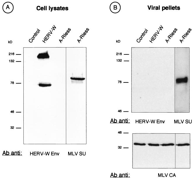
To test the virus-cell fusion capacity of HERV-W Env, we sought to generate retroviral pseudotypes in which MLV core particles were coated with HERV-W envelope glycoproteins. Thus, TELCeB6 cells, derived from human TE671 cells and constitutively expressing MLV Gag-Pol proteins and a lacZ gene-carrying MLV-derived retroviral vector (14), were transiently transfected with plasmids expressing either an HERV-W env cDNA (4) in a positive or antisense orientation or, as a positive control, the A-Rless amphotropic MLV envelope glycoprotein. Expression of HERV-W Env in transfected cells was assessed by immunoblotting with a monoclonal antibody which could recognize the TM and precursor proteins encoded by the HERV-W env gene (Fig. 1A). No staining occurred in lysates of cells transfected with either control plasmid. In contrast, two bands, corresponding to molecular masses of ca. 75 and 200 kDa, were readily detected in TELCeB6 cells transfected with the HERV-W Env expression vector (Fig. 1A), thus demonstrating that the HERV-W env gene had retained a coding capacity and could be expressed ex vivo. While the 200-kDa band might have represented HERV-W Env trimers, the lower-molecular-mass band most likely corresponded to monomeric HERV-W Env precursors and was about 5 kDa smaller than the size of Env precursors that we previously obtained in cell-free transcription-translation assays in the presence of canine microsomes (4), probably owing to slight differences in glycosylation and/or to removal of the Env signal peptide in vivo. No band migrating at the position of TM (ca. 27 kDa) could be detected, suggesting the absence of, or at least inefficient, precursor cleavage.
FIG. 1.
Detection of envelope glycoprotein in HERV-W Env-transfected cells. TELCeB6 cells were transfected with plasmids expressing either the HERV-W env cDNA in the antisense (Control) or positive (HERV-W) orientation or a hyperfusogenic mutant amphotropic MLV envelope glycoprotein (A-Rless). Two days later, the Env-transfected cells were lysed and their supernatants were harvested, filtered, and ultracentrifuged at 150,000 × g to pellet the viral particles. Immunoblots of cell lysates (A) and viral pellets (B) were probed with antibodies (Ab) against HERV-W Env, MLV SU, or MLV CA protein, as indicated. The positions of molecular mass markers are shown to the left of the gels (in kilodaltons).
To test for the formation of infectious MLV/HERV-W hybrid viral particles, the supernatant of Env-transfected TELCeB6 cells was harvested and used to infect an array of target cell types, including human cells. Although MLV vector particles pseudotyped with A-Rless envelope glycoproteins could readily infect the target cells, with infectious titers of up to 5 × 10⁴ lacZ i.u./ml, viral particles released by HERV-W Env-transfected TELCeB6 cells were not infectious in all tested cell types, including cells of primate and nonprimate species (data not shown). We therefore sought to determine whether HERV-W Env could be incorporated on MLV particles. Thus, the supernatant of Env-transfected cells was ultracentrifuged and the Env content of the viral pellet, reflecting incorporation on virions (27), was analyzed by immunoblotting (Fig. 1B). No HERV-W envelope glycoprotein could be detected on MLV particles, although in a side-to-side comparison, A-Rless envelope glycoprotein was readily incorporated on virions upon transfection in TELCeB6 cells (Fig. 1B). These data therefore indicated that the absence of infectivity of HERV-W Env pseudotypes was most likely due to the inability of HERV-W envelope glycoproteins to be incorporated on MLV particles.
HERV-W Env is a highly fusogenic membrane glycoprotein.
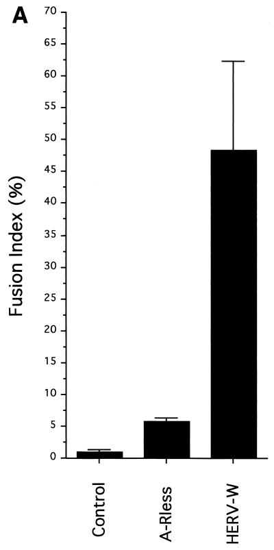
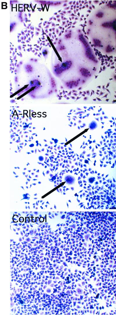
When HERV-W Env was expressed in TELCeB6 cells, a strong cytopathic effect, most likely due to cell-cell fusion, was observed in the transfected-cell culture. As expected from this preliminary observation, cell-cell fusion assays performed by transiently expressing HERV-W Env in parental TE671 human cells also resulted in numerous multinucleated giant cells, or syncytia, 1 to 2 days after transfection (Table 1). These results indicated that HERV-W envelope glycoprotein could promote homotypic cell-cell fusion. Heterotypic cell-cell fusion assays were then performed by cocultivating TE671 Env-transfected cells with HeLa indicator cells (Fig. 2). At day 2 posttransfection, up to 48% of the nuclei of the coculture were distributed in large syncytia (Fig. 2A). Each syncytium resulted from the fusion of one Env-expressing TE671 cell with 40 target HeLa cells, on average (Fig. 2B). By comparison, the expression of A-Rless, a hyperfusogenic mutant of amphotropic MLV Env (46), resulted in fusion of only 6% of the cell culture (Fig. 2A) and induced the formation of syncytia containing up to 15 nuclei (Fig. 2B). Similar results were obtained when quail QT6 cells, hamster BHK21 cells, mouse NIH 3T3 cells, or rat XC cells were used as Env-producing cells in the cell-cell fusion assays with HeLa target cells (data not shown), thus indicating that the genetic background of the Env-expressing cells did not influence the fusion activity of the HERV-W envelope glycoprotein. Taken together, these results demonstrated that the product of the HERV-W env gene is a highly fusogenic membrane glycoprotein.
TABLE 1.
Fusion host range of the HERV-W envelope glycoprotein
| Species | Target cell line | Envelope glycoprotein expresseda
|
|
|---|---|---|---|
| HERV-W | A-Rless | ||
| Quail | QT6 | − | − |
| Rat | XC | − | ++ |
| Mouse | NIH 3T3 | − | ++ |
| Hamster | Cear13 | − | ++ |
| Cat | CCC | − | ++ |
| Pig | PAE | + | + |
| Monkey | COS-7 | ++ | + |
| Human | A204 | + | + |
| B-1 | + | + | |
| 293 | ++ | + | |
| HeLa | ++ | + | |
| A431 | + | + | |
| TE671 | ++ | + | |
Fusogenic envelope glycoprotein was transiently expressed in cell-cell fusion assays. After 36 h of coculture with the indicated target cells, the fusion indices were determined: −, less than 1% fusion (background levels); +, 1 to 10% fusion; and ++, >10% fusion.
FIG. 2.
Formation of syncytia by HERV-W envelope glycoprotein. TELac2 cells, derived from TE671 human rhabdomyosarcoma cells and constitutively expressing a nuclear β-galactosidase, were transfected with plasmids expressing either the HERV-W env cDNA in the positive (HERV-W) or antisense (Control) orientation or a hyperfusogenic mutant amphotropic MLV envelope glycoprotein (A-Rless). Transfected cells were overlaid with HeLa indicator cells. The determination of the fusion activity of the transfected envelope glycoproteins was performed after 36 h of coculture. (A) Results are expressed as percentages of the fusion indices (means ± standard deviations; n = 5). (B) Cocultures were stained with X-Gal substrate to reveal β-galactosidase activity and to visualize the nuclei of the producer cells (arrows) and then with May-Grünwald and Giemsa solutions. Magnification, ×250.
To further characterize the fusogenic properties of the HERV-W envelope glycoprotein, heterotypic cell-cell fusion assays were performed by cocultivating HERV-W Env-transfected TE671 cells with a panel of target cell types derived from different animal species (Table 1). No syncytium formation was detected with QT6 quail cells, 3T3 mouse cells, XC rat cells, Cear13 (CHO-PiT-2) hamster cells, or CCC cat cells, although the last four cell types exhibited high cell-cell fusion activities upon expression of A-Rless amphotropic envelope glycoprotein. In contrast, HERV-W envelope glycoprotein mediated a significant fusogenic activity with PAE pig cells and was highly fusogenic with all simian and human cells tested (Table 1). Since the fusogenicity of retroviral envelopes is activated upon their interaction with specific cell surface receptors (22), these data indicated that HERV-W envelope glycoproteins could functionally interact with a receptor expressed on primate and pig cells but not one expressed on avian, rodent, or feline cells.
HERV-W Env interacts with the type D mammalian retrovirus receptor.
Retroviruses that infect human cells fall into different groups, determined by the nature of the receptors with which they interact (50). To determine whether HERV-W Env would promote cell-cell fusion upon interaction with one of the previously identified retrovirus receptors (49), we designed a fusion assay by using an array of receptor-blocked indicator cells. In such cells, the accessibility to either of these receptors was competitively decreased by endogenous expression of a panel of retrovirus envelope glycoproteins, thus resulting in receptor blockage. Hence, any reduction of the HERV-W Env fusion activity on some of these receptor-blocked cells would indicate a putative receptor on the parental cells. TE671 human cells were chosen because they express the receptors for several retrovirus groups (43), including that of HERV-W Env, which is highly fusogenic for these cells (Table 1). Three candidate mammalian retrovirus receptors were investigated: PiT-1 and PiT-2, two independent inorganic-phosphate symporters which are receptors for GALV (39) and amphotropic MLV (36, 55), respectively, and RDR, a neutral-amino acid transporter which is a receptor for RD114 cat endogenous retrovirus and type D simian retroviruses (45, 53). Stable expression vectors encoding the envelope glycoproteins of GALV, amphotropic MLV, and RD114 were individually introduced into TE671 cells (Table 2). As expected, infection of cells of each of the three TE671 subclones could be attained only with retroviral vectors generated with envelope glycoproteins different from that expressed in target cells (Table 2), thus demonstrating the strong and specific receptor interference achieved in the three different cell lines. When the receptor-blocked sublines as well as the parental TE671 cells were used in cell-cell fusion assays with HERV-W fusogenic glycoproteins, a strong reduction of syncytium formation was found only in cells expressing RD114 envelope glycoproteins, in which accessibility to RDR was blocked (Table 2). The absence of syncytia in the latter cells was not due to a loss of their intrinsic capacity to form syncytia, since they were as easily fused by amphotropic A-Rless envelopes as were the parental TE671 cells. Therefore, since HERV-W envelope glycoprotein could similarly fuse parental TE671 cells as well as PiT-1- and PiT-2-blocked cells, but not RDR-blocked cells (Table 2), these data suggested that HERV-W might recognize and interact with the type D mammalian retrovirus receptor expressed in human cells.
TABLE 2.
Fusion activity of HERV-W Env in receptor-blocked TE671 cells
| Type of assay | Retrovirusa or glycoproteinb | Resultscd for receptor-blocked target cellse
|
|||
|---|---|---|---|---|---|
| None | PiT-2 | PiT-1 | RDR | ||
| Virus-cell fusion | MLV-Aac | 107 | 5 × 10² | 107 | 107 |
| GALVac | 106 | 106 | 5 × 101 | 106 | |
| RD114ac | 107 | 107 | 107 | 2 × 10² | |
| Controlacf | <1 | <1 | <1 | <1 | |
| Cell-cell fusion | A-Rlessbd | 43.4 | 0.8 | 34.1 | 47.6 |
| HERV-Wbd | 79.0 | 74.5 | 77.15 | 0.48 | |
| Controlbd | 0.1 | 0.4 | 0.2 | 0.3 | |
Retrovirus from which Env was derived.
Fusogenic envelope glycoprotein.
Titers (as lacZ i.u. per milliliter) of lacZ retrovirus vectors pseudotyped with the indicated envelope glycoproteins were determined on the receptor-blocked target cells. These pseudotyped lacZ vectors were harvested from the supernatants of TELCeB6 cells stably transfected with expression plasmids encoding the envelope glycoproteins derived from the indicated retroviruses.
Fusion indices (as percentages) determined after transfection of the indicated fusogenic envelope glycoproteins by using the receptor-blocked TE671 cells as target cells. The background value of syncytium formation was provided by transfecting a plasmid expressing the HERV-W env gene in the antisense orientation (control).
The target cells were TE671 subclones stably expressing the envelope glycoproteins derived from the MLV-A, GALV, and RD114 retroviruses that respectively recognize the PiT-2, PiT-1, and RDR receptors, thus blocking the accessibility of exogenously presented retroviral envelopes to these receptors.
Control, no Env expression plasmid was transfected into TELCeB6 cells.
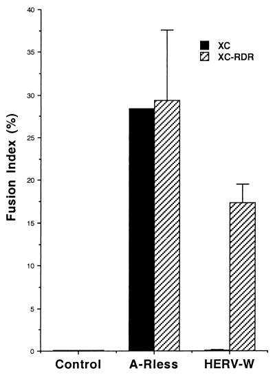
To confirm this possibility, XC cells, which cannot be fused by HERV-W Env (Table 1), were transfected by a stable expression vector encoding the human allele of RDR (53). These cells were then employed as target cells in a cell-cell fusion assay using HERV-W envelope glycoprotein. Compared to that in parental XC cells, formation of syncytia was readily detected in RDR-transfected cells (Fig. 3). Such a high level of fusogenic activity was not due to a nonspecifically increased fusogenicity of the population of RDR-transfected XC cells, since A-Rless envelope glycoprotein could similarly fuse both the parental and the RDR-transfected XC cells (Fig. 3). Altogether, these data indicated that HERV-W envelope glycoprotein mediates cell-cell fusion upon interaction with the type D mammalian retrovirus receptor, consistent with the presence of sequence homologies between HERV-W envelope proteins and those of simian type D retroviruses (4).
FIG. 3.
Cell-cell fusion assays in RDR-transfected XC cells. A plasmid encoding RDR, the human type D mammalian retrovirus receptor, was expressed in XC rat cells. The A-Rless or HERV-W fusogenic envelope glycoprotein was transiently expressed in RDR-transfected XC cells (hatched bars) as well as in parental XC cells (black bars). The results are expressed as percentages of the fusion indices (means ± standard deviations n = 3).
HERV-W is expressed in placenta cells.
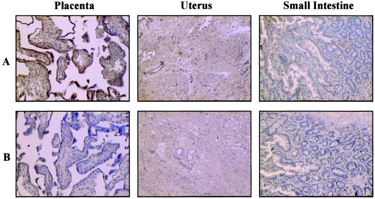
Previous results from our laboratory indicated that among 48 different human tissues analyzed by Northern blotting and/or RNA dot blotting, HERV-W Env mRNA is specifically expressed in the placenta (4). To verify expression of HERV-W envelope glycoprotein in this tissue, the HERV-W Env monoclonal antibody was used to probe histological preparations by in situ staining of 13-week placenta tissue and of various human normal adult tissues: uterus, esophagus, stomach, small intestine, colon, bladder, and adipose. As expected, no staining occurred in the different adult tissues. However, in agreement with the placenta-specific expression of HERV-W mRNAs, staining of the placenta tissue occurred and was characterized by areas of positive staining in the cytotrophoblast and of a more marked staining in the syncytiotrophoblast cell layer (Fig. 4).
FIG. 4.
In situ detection of HERV-W envelope glycoprotein in tissue sections. The tissue sections originated from human tissue donors' 13-week placenta, uterus, and small intestine. Slides were labeled (A) or not (B) with the 6A2B2 monoclonal antibody and revealed by immunoperoxidase staining and hematoxylin counterstaining. Magnification, ×100.
DISCUSSION
Here we report the placental expression of an HERV-encoded envelope glycoprotein that exhibits all the features of retroviral envelopes necessary to promote cell-cell fusion. The persistence for more than 25 million years of an env gene encoding a complete retroviral envelope glycoprotein in the genomes of Old World primates as well as its tissue-specific expression in human placenta suggests that evolution has retained a function of this protein that is beneficial for the host. Indeed, uncontrolled expression of fusogenic retroviral envelope glycoproteins in vivo or in vitro may cause cell death and tissue damage (40) by a nonapoptotic process (2). Based on in vitro studies, we demonstrate here that the HERV-W envelope glycoprotein can induce the formation of numerous syncytia upon interaction with the recently identified type D mammalian retrovirus receptor, a cell surface molecule whose gene is also transcribed in placenta cells (53). It is therefore conceivable that HERV-W Env plays a physiological role in vivo in placenta development. During pregnancy, the syncytiotrophoblast cell layer, which comes into intimate contact with the maternal blood space, is formed by differentiation and homotypic fusion of the underlying trophoblastic cells. This process is associated with the expression of several types of endogenous retroviral particles in the placenta (58). As such, the ERV-3 envelope glycoprotein has been suggested to play different roles, such as inducing differentiation of the cytotrophoblastic cells (28) or, alternatively, preventing maternal immune rejection of the fetus (56). However, the involvement of ERV-3 Env in placenta development remains questionable since a stop codon occurs before the membrane anchoring domain of ERV-3 Env (10) and since recent results have indicated that the ERV-3 env gene is mutated in about 1% of homozygous individuals (17). In the case of HERV-W, it will be essential to investigate the polymorphism of its env gene(s) and promoter(s), as well as to analyze the host factors that regulate HERV-W expression in vivo, in order to better appreciate the positive selection exerted by evolution to preserve Env functional domains and expression. Nevertheless, the absence of HERV-W sequences in New World monkeys and in other nonprimate placental mammals (57) indicates that syncytiotrophoblast differentiation may also be induced by distinct and/or complementary mechanisms.
The high-level cell-cell fusogenicity of HERV-W Env is striking in comparison to that of the envelope glycoproteins of type C and type D mammalian retroviruses, to which HERV-W is related (4). Indeed, when expressed individually in cell culture, in the absence of other viral components, the envelope glycoproteins of the type C and D retroviruses do not induce the formation of numerous syncytia (8, 27, 46). In contrast to the envelope glycoproteins of exogenous retroviruses that also use the type D mammalian retrovirus receptor, the HERV-W envelope glycoprotein is highly fusogenic in vitro. Fusogenicity of retroviral envelope glycoproteins is regulated at distinct stages of the Env maturation process. First, the Env polyprotein precursor must be cleaved by a trans-Golgi cellular protease in order to release the SU and TM Env subunits and to allow the fusion peptide, located at the amino terminus of TM, to interact with the target cell membrane during retroviral-receptor-mediated activation of Env fusogenicity (22). Thus, fusion-competent retroviral envelope glycoproteins must be found as Env precursors as well as processed SU and TM proteins in producer cells. Analysis of HERV-W Env expression did not allow us to detect the presence of the SU and TM Env subproducts (Fig. 1). This might be due to inefficient cleavage of the HERV-W Env precursor by cellular proteases, which would prevent detection of the processed Env products by Western blot analysis. Undetected or inefficient cleavage of the MLV Env precursor has already been reported in the literature and does not necessarily imply an incapacity to mediate membrane fusion (27, 63); yet, the high membrane fusion activity of HERV-W Env indicates that precursor processing must occur to some degree.
Second, at least for MLVs (44, 46) and for Mason-Pfizer monkey virus, a prototype type D simian retrovirus (8), during or shortly after budding of the viral particles, a 16-amino-acid carboxy-terminal peptide of TM, named R peptide, is cleaved by the viral protease, allowing the envelope glycoprotein to be fusion competent. Thus, the TM carboxy-terminal ends of these Env proteins exert a fusion-inhibitory effect (60, 61), and their removal by the viral protease is necessary for the full fusion activity of the envelope glycoprotein (8, 46). The cytoplasmic tail of HERV-W Env is 35 amino acids longer than that of type D and type C mammalian retroviruses (4). No retroviral-protease cleavage site could be found in HERV-W Env. Thus, in contrast to those of type D and type C mammalian retroviruses, the HERV-W Env cytoplasmic tail may contain a determinant that activates, or at least does not inhibit, fusogenicity. Of note, the cytoplasmic tails of most retrovirus envelope glycoproteins contain a YXXφ tyrosine-based sorting signal (where Y is Tyr, X is any amino acid, and φ is an amino acid with a bulky hydrophobic side chain [Leu, Ile, Phe, Val, or Met]) which plays a key role in subcellular distribution and adaptin-mediated endocytosis of plasma membrane-bound glycoproteins (11). Disruption of this motif in human T-lymphotropic virus type 1 (HTLV-1) Env and in simian immunodeficiency virus Env results in increased cell-cell fusion and/or cell surface expression (3, 15). Interestingly, the YXXφ motif is located in the R peptide for MLVs and Mason-Pfizer monkey virus Env but is missing in HERV-W Env (4). Thus, its removal upon cleavage of the R peptide or, alternatively, its absence in the case of HERV-W Env is likely to result in augmented Env cell surface expression and fusogenicity.
Our data suggest that the lack of infectivity of MLV viral particles generated with HERV-W Env is probably caused by an inability of these envelope glycoproteins to be incorporated on virions. MLV virions have been shown to efficiently incorporate type I glycoproteins from other viruses that harbor short cytoplasmic tails, such as vesicular stomatitis virus (18), Rous sarcoma virus (25), Semliki Forest virus (52), HTLV-1 (16), human foamy virus (29), fowl plague virus (21), paramyxoviruses (20, 51), lymphocytic choriomeningitis virus (35), and Ebola virus (59). Interestingly, incorporation of human immunodeficiency virus envelope glycoproteins that harbor long cytoplasmic tails could be achieved only after truncation of their cytoplasmic domains (33, 47). Similarly, the unusually long cytoplasmic tail of HERV-W Env may explain its lack of incorporation on MLV viral particles. Ongoing studies are now aiming to determine if recombinant HERV-W envelope glycoproteins with shorter cytoplasmic tails can be incorporated on MLV viral particles as well as on virions of exogenous retroviruses that may infect humans. Indeed, since retroviruses are genetically unstable organisms and since they are being used as gene delivery vectors, the outcome of such studies is critical for several reasons: (i) interaction of exogenous retroviruses such as human immunodeficiency viruses, pig endogenous retroviruses (42), or HTLVs with HERVs may by complementation, cross-packaging, and/or recombination give rise to new viruses with altered cell tropisms and/or pathogenicities; and (ii) HERVs may provide core and envelope proteins which perhaps contribute to mobilization and dissemination of retroviral or lentiviral vectors by trans complementation (41). Thus, unravelling the molecular details of the fusogenic property of HERV-W Env glycoproteins and their capacity to (be) transcomplement(ed by) exogenous retroviruses could have implications in ensuring the safety of gene therapy approaches and also in the elucidation of the hitherto poorly understood biological significance of HERV-W protein expression in placenta tissue.
ACKNOWLEDGMENTS
We thank Nadia Piga and Nicole Battail for obtaining the 6A2B2 monoclonal antibody. We thank S. Isaac for help with reading the histological tissue sections. We thank Chet Tailor and David Kabat for generously providing the Cear13 cells and the pcD3.1VHR16/4 plasmid encoding the RDR receptor.
This work was supported by bioMérieux and INSERM.
REFERENCES
- 1.Andersen K B. A domain of murine retrovirus surface protein gp70 mediates cell fusion, as shown in a novel SC-1 cell fusion system. J Virol. 1994;68:3175–3182. doi: 10.1128/jvi.68.5.3175-3182.1994. [DOI] [PMC free article] [PubMed] [Google Scholar]
- 2.Bateman, A., F. Bullough, S. Murphy, L. Emiliusen, D. Lavillette, F.-L. Cosset, R. Cattaneo, S. J. Russell, and R. Vile. Fusogenic membrane glycoproteins as a novel class of genes for the local and immune-mediated control of tumour growth. Cancer Res., in press. [PubMed]
- 3.Berlioz-Torrent C, Shacklett B L, Erdtmann L, Delamarre L, Bouchaert I, Sonigo P, Dokhelar M C, Benarous R. Interactions of the cytoplasmic domains of human and simian retroviral transmembrane proteins with components of the clathrin adaptor complexes modulate intracellular and cell surface expression of envelope glycoproteins. J Virol. 1999;73:1350–1361. doi: 10.1128/jvi.73.2.1350-1361.1999. [DOI] [PMC free article] [PubMed] [Google Scholar]
- 4.Blond J-L, Besème F, Duret L, Bouton O, Bedin F, Perron H, Mandrand B, Mallet F. Molecular characterization and placental expression of HERV-W, a new human endogenous retrovirus family. J Virol. 1999;73:1175–1185. doi: 10.1128/jvi.73.2.1175-1185.1999. [DOI] [PMC free article] [PubMed] [Google Scholar]
- 5.Boeke J D, Stoye J P. Retrotransposons, endogenous retroviruses, and the evolution of retroelements. In: Coffin J M, Hughes S H, Varmus H E, editors. Retroviruses. Cold Spring Harbor, N.Y: Cold Spring Harbor Laboratory Press; 1997. pp. 343–435. [PubMed] [Google Scholar]
- 6.Boller K, Konig H, Sauter M, Mueller-Lantzsch N, Lower R, Lower J, Kurth R. Evidence that HERV-K is the endogenous retrovirus sequence that codes for the human teratocarcinoma-derived retrovirus HTDV. Virology. 1993;196:349–353. doi: 10.1006/viro.1993.1487. [DOI] [PubMed] [Google Scholar]
- 7.Boyd M T, Bax C M R, Bax B E, Bloxam D L, Weiss R A. The human endogenous retrovirus ERV-3 is upregulated in differentiating placental trophoblast cells. Virology. 1993;196:905–909. doi: 10.1006/viro.1993.1556. [DOI] [PubMed] [Google Scholar]
- 8.Brody B A, Rhee S S, Hunter E. Postassembly cleavage of a retroviral glycoprotein cytoplasmic domain removes a necessary incorporation signal and activates fusion activity. J Virol. 1994;68:4620–4627. doi: 10.1128/jvi.68.7.4620-4627.1994. [DOI] [PMC free article] [PubMed] [Google Scholar]
- 9.Cheynet V, Verrier B, Mallet F. Overexpression of HIV-1 proteins in Escherichia coli by a modified expression vector and their one-step purification. Protein Expr Purif. 1993;4:367–372. doi: 10.1006/prep.1993.1048. [DOI] [PubMed] [Google Scholar]
- 10.Cohen M, Powers M, O'Connell C, Kato N. The nucleotide sequence of the env gene from the human provirus ERV3 and isolation and characterisation of an ERV3-specific cDNA. Virology. 1985;147:449–458. doi: 10.1016/0042-6822(85)90147-3. [DOI] [PubMed] [Google Scholar]
- 11.Collawn J F, Lai A, Domingo D, Fitch M, Hatton S, Trowbridge I S. YTRF is the conserved internalization signal of the transferrin receptor, and a second YTRF signal at position 31–34 enhances endocytosis. J Biol Chem. 1993;268:21686–21692. [PubMed] [Google Scholar]
- 12.Cosset F-L, Morling F J, Takeuchi Y, Weiss R A, Collins M K L, Russell S J. Retroviral retargeting by envelopes expressing an N-terminal binding domain. J Virol. 1995;69:6314–6322. doi: 10.1128/jvi.69.10.6314-6322.1995. [DOI] [PMC free article] [PubMed] [Google Scholar]
- 13.Cosset F-L, Ronfort C, Molina R-M, Flamant F, Drynda A, Benchaibi M, Valsesia S, Nigon V-M, Verdier G. Packaging cells for avian leukosis virus-based vectors with various host ranges. J Virol. 1992;66:5671–5676. doi: 10.1128/jvi.66.9.5671-5676.1992. [DOI] [PMC free article] [PubMed] [Google Scholar]
- 14.Cosset F-L, Takeuchi Y, Battini J-L, Weiss R A, Collins M K L. High-titer packaging cells producing recombinant retroviruses resistant to human serum. J Virol. 1995;69:7430–7436. doi: 10.1128/jvi.69.12.7430-7436.1995. [DOI] [PMC free article] [PubMed] [Google Scholar]
- 15.Delamarre L, Pique C, Rosenberg A R, Blot V, Grange M-P, Le Blanc I, Dokhelar M-C. The Y-S-L-I tyrosine-based motif in the cytoplasmic domain of the human T-cell leukemia virus type 1 envelope is essential for cell-to-cell transmission. J Virol. 1999;73:9659–9663. doi: 10.1128/jvi.73.11.9659-9663.1999. [DOI] [PMC free article] [PubMed] [Google Scholar]
- 16.Denesvre C, Carrington C, Corbin A, Takeuchi Y, Cosset F-L, Schulz T, Sitbon M, Sonigo P. TM domain swapping of murine leukemia virus and human T-cell leukemia virus envelopes confers different infectious abilities despite similar incorporation into virions. J Virol. 1996;70:4380–4386. doi: 10.1128/jvi.70.7.4380-4386.1996. [DOI] [PMC free article] [PubMed] [Google Scholar]
- 17.de Parseval N, Heidmann T. Physiological knockout of the envelope gene of the single-copy ERV-3 human endogenous retrovirus in a fraction of the Caucasian population. J Virol. 1998;72:3442–3445. doi: 10.1128/jvi.72.4.3442-3445.1998. [DOI] [PMC free article] [PubMed] [Google Scholar]
- 18.Emi N, Friedmann T, Yee J-K. Pseudotype formation of murine leukemia virus with the G protein of vesicular stomatitis virus. J Virol. 1991;65:1202–1207. doi: 10.1128/jvi.65.3.1202-1207.1991. [DOI] [PMC free article] [PubMed] [Google Scholar]
- 19.Harlow E, Lane D. Antibodies: a laboratory manual. 1st ed. Cold Spring Harbor, N.Y: Cold Spring Harbor Laboratory Press; 1988. [Google Scholar]
- 20.Hatziioannou T, Russell S J, Cosset F-L. Incorporation of simian virus 5 fusion protein into murine leukemia virus particles and its effect on the co-incorporation of retroviral envelope glycoproteins. Virology. 2000;267:49–57. doi: 10.1006/viro.1999.0091. [DOI] [PubMed] [Google Scholar]
- 21.Hatziioannou T, Valsesia-Wittmann S, Russell S J, Cosset F-L. Incorporation of fowl plague virus hemagglutinin into murine leukemia virus particles and analysis of the infectivity of the pseudotyped retroviruses. J Virol. 1998;72:5313–5317. doi: 10.1128/jvi.72.6.5313-5317.1998. [DOI] [PMC free article] [PubMed] [Google Scholar]
- 22.Hunter E, Swanstrom R. Retrovirus envelope glycoproteins. Curr Top Microbiol Immunol. 1990;157:187–253. doi: 10.1007/978-3-642-75218-6_7. [DOI] [PubMed] [Google Scholar]
- 23.Komurian-Pradel F, Paranhos-Baccala G, Bedin F, Ounanian-Paraz A, Sodoyer M, Ott C, Rajoharison A, Garcia E, Mallet F, Mandrand B, Perron H. Molecular cloning and characterization of MSRV-related sequences associated with retrovirus-like particles. Virology. 1999;260:1–9. doi: 10.1006/viro.1999.9792. [DOI] [PubMed] [Google Scholar]
- 24.Kozak S L, Siess D C, Kavanaugh M P, Miller A D, Kabat D. The envelope glycoprotein of an amphotropic murine retrovirus binds specifically to the cellular receptor/phosphate transporter of susceptible species. J Virol. 1995;69:3433–3440. doi: 10.1128/jvi.69.6.3433-3440.1995. [DOI] [PMC free article] [PubMed] [Google Scholar]
- 25.Landau N R, Littman D R. Packaging system for rapid production of murine leukemia virus vectors with variable tropism. J Virol. 1992;66:5110–5113. doi: 10.1128/jvi.66.8.5110-5113.1992. [DOI] [PMC free article] [PubMed] [Google Scholar]
- 26.Larsson E, Andersson G. Beneficial role of human endogenous retroviruses: facts and hypotheses. Scand J Immunol. 1998;48:329–338. doi: 10.1046/j.1365-3083.1998.00428.x. [DOI] [PubMed] [Google Scholar]
- 27.Lavillette D, Maurice M, Roche C, Russell S J, Sitbon M, Cosset F-L. A proline-rich motif downstream of the receptor binding domain modulates conformation and fusogenicity of murine retroviral envelopes. J Virol. 1998;72:9955–9965. doi: 10.1128/jvi.72.12.9955-9965.1998. [DOI] [PMC free article] [PubMed] [Google Scholar]
- 28.Lin L, Rote N S. Expression of endogenous retrovirus ERV-3 induces differentiation in BeWo, a choriocarcinoma model of human placental trophoblast. Placenta. 1999;20:109–118. doi: 10.1053/plac.1998.0337. [DOI] [PubMed] [Google Scholar]
- 29.Lindemann D, Bock M, Schweizer M, Rethwilm A. Efficient pseudotyping of murine leukemia virus particles with chimeric human foamy virus envelope proteins. J Virol. 1997;71:4815–4820. doi: 10.1128/jvi.71.6.4815-4820.1997. [DOI] [PMC free article] [PubMed] [Google Scholar]
- 30.Löwer R. The pathogenic potential of endogenous retroviruses: facts and fantasies. Trends Microbiol. 1999;7:350–356. doi: 10.1016/s0966-842x(99)01565-6. [DOI] [PubMed] [Google Scholar]
- 31.Löwer R, Löwer J, Kurth R. The viruses in all of us: characteristics and biological significance of human endogenous retrovirus sequences. Proc Natl Acad Sci USA. 1996;93:5177–5184. doi: 10.1073/pnas.93.11.5177. [DOI] [PMC free article] [PubMed] [Google Scholar]
- 32.Lyden T W, Johnson P M, Mwenda J M, Rote N S. Ultrastructural characterization of endogenous retroviral particles isolated from normal human placentas. Biol Reprod. 1994;51:152–157. doi: 10.1095/biolreprod51.1.152. [DOI] [PubMed] [Google Scholar]
- 33.Mammano F, Salvatori F, Indraccolo S, De Rossi A, Chieco-Bianchi L, Göttlinger H G. Truncation of the human immunodeficiency virus type 1 envelope glycoprotein allows efficient pseudotyping of Moloney murine leukemia virus particles and gene transfer into CD4+ cells. J Virol. 1997;71:3341–3345. doi: 10.1128/jvi.71.4.3341-3345.1997. [DOI] [PMC free article] [PubMed] [Google Scholar]
- 34.Martin F, Neil S, Kupsch J, Maurice M, Cosset F-L, Collins M. Retrovirus targeting by tropism restriction to melanoma cells. J Virol. 1999;73:6923–6929. doi: 10.1128/jvi.73.8.6923-6929.1999. [DOI] [PMC free article] [PubMed] [Google Scholar]
- 35.Miletic H, Bruns M, Tsiakas K, Vogt B, Rezal R, Baum C, Kühlke K, Cosset F-L, Ostertag W, Lother H, von Laer D. Retroviral vectors pseudotyped with lymphocytic choriomeningitis virus. J Virol. 1999;73:6114–6116. doi: 10.1128/jvi.73.7.6114-6116.1999. [DOI] [PMC free article] [PubMed] [Google Scholar]
- 36.Miller D G, Edwards R H, Miller A D. Cloning of the cellular receptor for amphotropic murine retroviruses reveals homology to that for gibbon ape leukemia virus. Proc Natl Acad Sci USA. 1994;91:78–82. doi: 10.1073/pnas.91.1.78. [DOI] [PMC free article] [PubMed] [Google Scholar]
- 37.Miyazono K, Okabe T, Urabe A, Takaku F, Heldin C H. Purification and properties of an endothelial cell growth factor from human platelets. J Biol Chem. 1987;262:4098–4103. [PubMed] [Google Scholar]
- 38.Nelson J, Leong J A, Levy J A. Normal human placentas contain RNA-directed DNA polymerase activity like that in viruses. Proc Natl Acad Sci USA. 1978;75:6263–6267. doi: 10.1073/pnas.75.12.6263. [DOI] [PMC free article] [PubMed] [Google Scholar]
- 39.O'Hara B, Johann S V, Klinger H P, Blair D G, Rubinson H, Dunn K J, Sass P, Vitek S M, Robbins T. Characterisation of the human gene conferring sensitivity to infection by gibbon ape leukemia virus. Cell Growth Differ. 1990;1:119–127. [PubMed] [Google Scholar]
- 40.Park B H, Matuschke B, Lavi E, Gaulton G N. A point mutation in the env gene of a murine leukemia virus induces syncytium formation and neurologic disease. J Virol. 1994;68:7516–7524. doi: 10.1128/jvi.68.11.7516-7524.1994. [DOI] [PMC free article] [PubMed] [Google Scholar]
- 41.Patience C, Takeuchi Y, Cosset F-L, Weiss R A. Packaging of endogenous retroviral sequences in retroviral vectors produced by murine and human packaging cells. J Virol. 1998;72:2671–2676. doi: 10.1128/jvi.72.4.2671-2676.1998. [DOI] [PMC free article] [PubMed] [Google Scholar]
- 42.Patience C, Takeuchi Y, Weiss R A. Infection of human cells by an endogenous retrovirus of pigs. Nat Med. 1997;3:282–286. doi: 10.1038/nm0397-282. [DOI] [PubMed] [Google Scholar]
- 43.Porter C D, Collins M K L, Tailor C S, Parker M H, Cosset F-L, Weiss R A, Takeuchi Y. Comparison of efficiency of infection of human gene therapy target cells via four different retroviral receptors. Hum Gene Ther. 1996;7:913–919. doi: 10.1089/hum.1996.7.8-913. [DOI] [PubMed] [Google Scholar]
- 44.Ragheb J A, Anderson W F. pH-independent murine leukemia virus ecotropic envelope-mediated cell fusion: implications for the role of the R peptide and p12E TM in viral entry. J Virol. 1994;68:3220–3231. doi: 10.1128/jvi.68.5.3220-3231.1994. [DOI] [PMC free article] [PubMed] [Google Scholar]
- 45.Rasko J E, Battini J L, Gottschalk R J, Mazo I, Miller A D. The RD114/simian type D retrovirus receptor is a neutral amino acid transporter. Proc Natl Acad Sci USA. 1999;96:2129–2134. doi: 10.1073/pnas.96.5.2129. [DOI] [PMC free article] [PubMed] [Google Scholar]
- 46.Rein A, Mirro J, Haynes J G, Ernst S M, Nagashima K. Function of the cytoplasmic domain of a retroviral transmembrane protein: p15E-p2E cleavage activates the membrane fusion capability of the murine leukemia virus Env protein. J Virol. 1994;68:1773–1781. doi: 10.1128/jvi.68.3.1773-1781.1994. [DOI] [PMC free article] [PubMed] [Google Scholar]
- 47.Schnierle B S, Stitz J, Bosch V, Nocken F, Merget-Millitzer H, Engelstadter M, Kurth R, Groner B, Cichutek K. Pseudotyping of murine leukemia virus with the envelope glycoproteins of HIV generates a retroviral vector with specificity of infection for CD4-expressing cells. Proc Natl Acad Sci USA. 1997;94:8640–8645. doi: 10.1073/pnas.94.16.8640. [DOI] [PMC free article] [PubMed] [Google Scholar]
- 48.Seifarth W, Skladny H, Krieg-Schneider F, Reichert A, Hehlmann R, Leib-Mösch C. Retrovirus-like particles released from the human breast cancer cell line T47-D display type B- and C-related endogenous retroviral sequences. J Virol. 1995;69:6408–6416. doi: 10.1128/jvi.69.10.6408-6416.1995. [DOI] [PMC free article] [PubMed] [Google Scholar]
- 49.Sommerfelt M A. Retrovirus receptors. J Gen Virol. 1999;80:3049–3064. doi: 10.1099/0022-1317-80-12-3049. [DOI] [PubMed] [Google Scholar]
- 50.Sommerfelt M A, Weiss R A. Receptor interference groups of 20 retroviruses plating on human cells. Virology. 1990;176:58–69. doi: 10.1016/0042-6822(90)90230-o. [DOI] [PubMed] [Google Scholar]
- 51.Spiegel M, Bitzer M, Schenk A, Rossmann H, Neubert W J, Seidler U, Gregor M, Lauer U. Pseudotype formation of Moloney murine leukemia virus with Sendai virus glycoprotein F. J Virol. 1998;72:5296–5302. doi: 10.1128/jvi.72.6.5296-5302.1998. [DOI] [PMC free article] [PubMed] [Google Scholar]
- 52.Suomalainen M, Garoff H. Incorporation of homologous and heterologous proteins into the envelope of Moloney murine leukemia virus. J Virol. 1994;68:4879–4889. doi: 10.1128/jvi.68.8.4879-4889.1994. [DOI] [PMC free article] [PubMed] [Google Scholar]
- 53.Tailor C S, Nouri A, Zhao Y, Takeuchi Y, Kabat D. A sodium-dependent neutral-amino-acid transporter mediates infections of feline and baboon endogenous retroviruses and simian type D retroviruses. J Virol. 1999;73:4470–4474. doi: 10.1128/jvi.73.5.4470-4474.1999. [DOI] [PMC free article] [PubMed] [Google Scholar]
- 54.Takeuchi Y, Cosset F-L, Lachmann P J, Okada H, Weiss R A, Collins M K L. Type C retrovirus inactivation by human complement is determined by both the viral genome and the producer cell. J Virol. 1994;68:8001–8007. doi: 10.1128/jvi.68.12.8001-8007.1994. [DOI] [PMC free article] [PubMed] [Google Scholar]
- 55.van Zeijl M, Johann S V, Closs E, Cunningham J, Eddy R, Shows T B, O'Hara B. An amphotropic retrovirus receptor is a second member of the gibbon ape leukemia virus receptor family. Proc Natl Acad Sci USA. 1994;91:1168–1172. doi: 10.1073/pnas.91.3.1168. [DOI] [PMC free article] [PubMed] [Google Scholar]
- 56.Venables P J, Brookes S M, Griffiths D, Weiss R A, Boyd M T. Abundance of an endogenous retroviral envelope protein in placental trophoblasts suggests a biological function. Virology. 1995;211:589–592. doi: 10.1006/viro.1995.1442. [DOI] [PubMed] [Google Scholar]
- 57.Voisset C, Blancher A, Perron H, Mandrand B, Mallet F, Paranhos-Baccalà G. Phylogeny of a novel family of human endogenous retrovirus sequences, HERV-W, in humans and different primates. AIDS Res Hum Retrovir. 1999;15:1529–1533. doi: 10.1089/088922299309810. [DOI] [PubMed] [Google Scholar]
- 58.Wilkinson D A, Mager D L, Leong J-A C. Endogenous human retroviruses. In: Levy J A, editor. The Retroviridae. Vol. 3. New York, N.Y: Plenum Press; 1994. pp. 465–534. [Google Scholar]
- 59.Wool-Lewis R J, Bates P. Characterization of Ebola virus entry by using pseudotyped viruses: identification of receptor-deficient cell lines. J Virol. 1998;72:3155–3160. doi: 10.1128/jvi.72.4.3155-3160.1998. [DOI] [PMC free article] [PubMed] [Google Scholar]
- 60.Yang C, Compans R W. Analysis of the cell fusion activities of chimeric simian immunodeficiency virus-murine leukemia virus envelope proteins: inhibitory effects of the R peptide. J Virol. 1996;70:248–254. doi: 10.1128/jvi.70.1.248-254.1996. [DOI] [PMC free article] [PubMed] [Google Scholar]
- 61.Yang C, Compans R W. Analysis of the murine leukemia virus R peptide: delineation of the molecular determinants which are important for its fusion inhibition activity. J Virol. 1997;71:8490–8496. doi: 10.1128/jvi.71.11.8490-8496.1997. [DOI] [PMC free article] [PubMed] [Google Scholar]
- 62.Yee J K, Friedmann T, Burns J C. Generation of high-titer pseudotyped retroviral vectors with very broad host range. Methods Cell Biol. 1994;43:99–112. doi: 10.1016/s0091-679x(08)60600-7. [DOI] [PubMed] [Google Scholar]
- 63.Zavorotinskaya T, Albritton L M. Failure to cleave murine leukemia virus envelope protein does not preclude its incorporation in virions and productive virus-receptor interaction. J Virol. 1999;73:5621–5629. doi: 10.1128/jvi.73.7.5621-5629.1999. [DOI] [PMC free article] [PubMed] [Google Scholar]


