

Incision in the fifth right intercostal space used to perform uniportal VATS lobectomy.
Central Message.
Lobectomy via uniportal video-assisted thoracoscopic surgery (U-VATS) allows a fast recovery with fewer complications. Standardized approaches to U-VATS facilitate learning and promote better outcomes.
Video-assisted thoracoscopic surgery (VATS) has evolved significantly over the past 25 years due to improvements in surgical instruments, camera visualization, and surgeon experience.1 When compared with open lobectomy, VATS lobectomy has better early outcomes without compromising oncologic outcomes.2, 3, 4 Thus, current practice guidelines recommend minimally invasive surgery, either using VATS or robotic surgery, as the standard surgical approach when treating patients with resectable, early-stage lung cancer.2,5
VATS Evolution: From Multiportal to Uniportal
Multiportal VATS procedures typically require the creation of 3 or 4 incisions,1 but in 2006, D'Amico and colleagues6 first described a VATS lobectomy technique using only 2 incisions. Rocco and colleagues7 introduced uniportal video-assisted thoracoscopic surgery (U-VATS) in 2004, reporting on their initial experiences using U-VATS for simple procedures, such as lung biopsy or bleb resection for primary spontaneous pneumothorax. The first uniportal lobectomy was reported in 2011, and U-VATS lobectomy has since gained popularity worldwide, primarily in Europe and Asia.8, 9, 10 A review by Tu and Hsu11 found that more than 9545 U-VATS procedures have been performed worldwide and reported as of late 2015, with most of the lobectomies being performed in China, Spain, or Taiwan. This highlighted the international acceptance of U-VATS and the large experience with U-VATS acquired by surgeons around the globe.11 When the outcomes of uniportal and multiportal VATS lobectomy are compared, the uniportal approach is associated with decreased intraoperative bleeding (P < .001), a faster surgery (P < .001), shorter duration of chest tube drainage (P = .001), less postoperative pneumonia (P = .012), and shorter hospital stay (P < .001).12 Despite these findings, the adoption of U-VATS lobectomy in North America has been slow.
U-VATS: Learning Curve
U-VATS requires precise movements, a profound knowledge of surgical anatomy, and specialized equipment to avoid technical complications, especially vascular accidents.13 These technical hurdles impart a substantial learning curve when adopting U-VATS.
Our implementation of the uniportal technique started in 2014 at the Institut Universitaire de Cardiologie et de Pneumologie de Quebec and included several key elements. First, a senior surgeon performed all VATS surgical procedures using U-VATS. Second, a U-VATS prospective database was created to monitor complications and operative time, and all surgeries were video-recorded for review. Surgical videos were routinely reviewed after every 20 patients to identify problematic steps, such as difficulty dissecting the superior pulmonary vein or difficulty placing surgical instruments in the limited working space.14 This video review was crucial to improve the efficiency of dissection and perioperative outcomes. Using the planned strategy, we developed a standardized U-VATS lobectomy approach for each lung lobe.
During U-VATS adoption, 274 consecutive U-VATS lobectomies performed by a single surgeon were analyzed to evaluate her learning curve. The learning curve was separated into 3 distinct phases, using cubic splines, based on the length of the surgery: an initial phase, a transition phase, and a proficient phase. The initial phase had the longest procedure time (158.8 ± 52.2 minutes) and included the first 60 cases. In the transition phase (procedures 61-140), the procedure time decreased to 145.9 ± 43.8 minutes and in the proficient phase (procedures 14-274), the surgeon had mastered the U-VATS lobectomy technique (Figure 1).15 The proficiency phase had the shortest and least variable procedure times, less intraoperative blood loss, a lower conversion rate to open surgery, and a decreased need for a second incision.14
Figure 1.

Learning curve for uniportal video-assisted thoracoscopic surgery (U-VATS) lobectomy performed by a single surgeon. Cubic spline analysis was used for curve fitting, and 95% confidence intervals are depicted with red dotted lines. From Drevet and Ugalde Figueroa.15
When instructing residents and fellows, the introduction of U-VATS should be preceded by a solid foundation in a multiportal VATS anatomic lung resection and be accompanied by the planned strategy described previously. Whenever feasible, the surgeon should select straightforward cases for the initial phase of the learning curve to gain comfort with the technique.
Indications and Contraindications for U-Vats Lobectomy
U-VATS lobectomy can be safely performed for both benign and malignant disease. There are no absolute contraindications to the U-VATS approach, but similar to multiportal VATS, locally advanced lung cancer is a common relative contraindication to U-VATS lobectomy.14 Complex airway reconstructions, such as sleeve resection or bronchoplasty, can be challenging to perform with U-VATS, but dedicated U-VATS instruments and an experienced surgeon make it possible.15 Previous chest surgery, previous radiation therapy, and incomplete or absent fissures should not be considered contraindications to the U-VATS approach.16
Technical Aspects
Based on our experience, we recommend the following considerations to achieve an optimal setup for U-VATS lobectomy.15
-
1.
Positioning: Patients are positioned in full lateral decubitus with maximum flexion of the table at the mid-chest level to enlarge the intercostal spaces, facilitating working space though a single incision.
-
2.
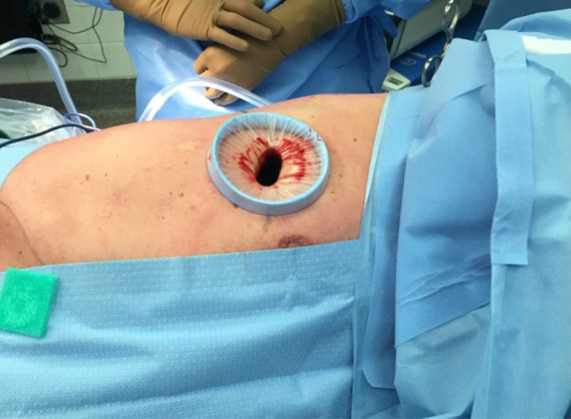
Incision: The incision is made in the fifth intercostal space for upper and middle lobe resections and the sixth intercostal space for lower-lobe resections (Figure 2). The incision is typically 3 to 4 cm and placed in the anterior axillary line. We recommend a wound protector to retract the soft tissue and facilitate placement of multiple instruments through the single incision (Video 1). In female patients, we take care to avoid the breast when placing the incision to reduce postoperative pain.
-
3.
Surgical equipment: A 5-mm 30° camera is recommended for all U-VATS procedures. Our preferred instruments are predominantly sourced from Scanlan instruments and characterized by a slim profile to facilitate the passage of multiple tools through the incision. They are generally 30-cm long. Ideally, the instruments should be bi-articulated with a curved tip.
Figure 2.

Uniportal incision in the fifth right intercostal space with a wound protector.
Surgery
Intercostal nerve blocking is routinely performed before incision in the adjacent interspaces. Epidural analgesia is rarely used and is administered only to patients with a high likelihood of conversion to thoracotomy or with planned chest wall resection. After skin incision, the intercostal muscle is divided with electrocautery to widely open the interspace. The intercostal muscle is divided slightly beyond the edges of the skin incision, and this maneuver promotes better exposure, provides more working space for instrumentation, and facilitates specimen removal. Rib spreading should not be performed at any stage of the surgery.
As a standard for our uniportal surgeries, the surgeon stands in front of the patient's chest, and the assistant is on the opposite side, facing the patient's back. The operating surgeon holds the camera, which allows them to direct the view, and has the assistant hold instrumentation to provide exposure of the target anatomy. When training another surgeon, however, both the teaching surgeon and the learner stand in front of the patient, allowing the same surgical view and improved coordination of movements. Standing in front of the patient enables the learning surgeon to perform procedures safely and learn how to perform movements appropriate for uniportal surgery.
Due to limited working space, the camera and instruments should be deliberately positioned within the incision to minimize collisions. We recommend articulating staplers, if available, and inserting the stapler through the lowest part of the incision, below all other instruments. During stapling, the camera should be positioned above the instruments and stapler to allow the best visualization.
The specifics of dissection and retraction vary based on the tumor location. With the uniportal approach, the order of dissection and division of the pulmonary structures is dependent on the lobe being resected due to the proximity of the hilar structures to the incision. In right upper-lobe, right lower-lobe, and left lower-lobe resections, the pulmonary artery is dissected first (Video 2), followed by the bronchus (Video 3), the vein (Video 4), and lastly the fissure (Video 5; right upper lobectomy: Video 6). For the middle lobectomy, we start with the vein, followed by the lobar bronchus, and artery last. Left upper lobectomy begins with artery dissection followed by vein dissection and dissection of the bronchus (Table 1). Modifications to these steps can be made to account for surgical difficulty, which can increase due to neoadjuvant therapy, calcified adenopathy, vascular injury during dissection, and anatomic variations. Surgical principles, such as dissecting in the optimal plane (vascular sheath or plane of Leriche) to make the vessel dissection simpler and safer, minimizing blunt dissection, and avoiding tension on vascular structures, must always be respected. To divide the fissure, we recommend a stapler or energy-sealing device (eg, LigaSure [Medtronic] or HARMONIC [Ethicon]).
Table 1.
Standardized procedures for U-VATS lobectomy
| Lobe | Steps for U-VATS lobectomy |
|---|---|
| Right upper lobe Right lower lobe Left lower lobe |
|
| Right middle lobe |
|
| Left upper lobe |
|
U-VATS, Uniportal video-assisted thoracoscopic surgery.
After the procedure is complete, a 24-F chest tube is inserted through the same incision used for U-VATS, and operative lung is re-expanded. The muscular layer must be closed carefully around the chest tube to prevent subcutaneous emphysema at the incision site. At the conclusion of bronchoplasty or sleeve resections, however, the chest tube is placed through a separate incision.
Standardization of U-VATS Lobectomy and Lymph Node Dissection
When we began doing U-VATS lobectomies, lymph node dissection was not standardized. Progressively, we moved toward a lobe-specific nodal dissection that consists of at least 6 stations, including at least 3 intrapulmonary or hilar stations (N1) and at least 3 mediastinal stations (N2), depending on the tumor location. Our study showed improved lymph node dissection during the learning curve from the initial phase to proficiency following this standardization.14
In our experience, standardizing U-VATS lobectomy facilitates teaching other surgeons to adopt the technique and maintains the oncologic quality of the lung resections and lymph node dissection. Others have also found value in standardizing their procedure for U-VATS lobectomy. Liu and colleagues17 summarized a series of techniques that they described as the “3-step” method to perform a U-VATS right upper lobectomy. They investigated the feasibility of the 3-step method on patients with lung cancer comparing their outcomes with the outcomes of patients who underwent the same lobectomy but without the 3-step method. In step 1, the mediastinal pleura is cut from the horizontal fissure to the right hilum to identify the middle lobe vein and the upper lobe vein. Next, lymph node dissection (stations 2, 4, and 10) is performed, and the apical-anterior artery is isolated and stapled. In Step 2, the lung is retracted to expose and divide the mediastinal pleura over the upper lobe bronchus. Nodal dissection of station 7 and 11 is performed, and the upper lobe bronchus is divided with a stapler. In step 3, the rest of the structures, including the posterior ascending artery, the fissures, and the vein, could be stapled from the anterior side to the posterior side along the fissures. The operative time for patients undergoing U-VATS lobectomy using the 3-step method was shorter than for patients undergoing U-VATS lobectomy using traditional methods (P < .001). The number of staplers reloads used was also less with the 3-step method than with the traditional method (P = .014).17
A consensus report of U-VATS lobectomy was published in 2019 based on 3 rounds of questionaries completed by 31 international experts with the goal of defining and standardizing U-VATS lobectomy.18 Although a technical quorum was not achieved, it was possible to see trends in the methods used by these experienced surgeons, especially in some perioperative decisions, such as the site of the incision (anterior axillar line, middle-anterior axillar line, or posterior axillar line) and the size of the chest tube placed (24-F or 28-F). This consensus should serve as a valuable guide for surgeons who are learning U-VATS techniques. However, it is important for each surgeon to adapt these guidelines to their individual realities, considering their practice, surgical skills, and experience.18
The consensus report also queried the main reasons for converting to multiportal VATS or open thoracotomy. The need for a bronchovascular sleeve resection was the primary clinical situation in which the experts queried recommended conversion to multiportal VATS with 32% agreement, followed by major bleeding and need for pneumonectomy with 13% each. The main reasons given to convert to open thoracotomy were major bleeding (36% agreement) and the need for a bronchovascular sleeve resection (23% agreement). None of the experts indicated that they consider conversion to multiportal VATS or open thoracotomy in patients with the absence of a fissure, pleural adhesions, chest wall involvement, or poor lung deflation, demonstrating that these technical issues typically do not pose notable difficulties for experienced U-VATS surgeons.18
Bleeding Control
Vascular injury is a rare, but life-threatening complication, during lung resection, and all surgeons learning or performing U-VATS must know how to manage bleeding injuries. When addressing vascular injuries during U-VATS lobectomy, the first step is compressing the bleeding vessel with a sponge stick and removing residual blood from the chest with suctioning to ensure that appropriate control of the bleeding has been obtained. Usually, light-to-moderate compression is sufficient to stop the bleeding. Overcompression should be avoided, as this can enlarge the vascular laceration. With the bleeding under control, the surgeon must keep calm, understand the extent of the laceration, inform the surgical team about the injury, and establish a plan for definitive repair.19
For minor injuries (<6 mm), hemostatic materials, such as a collagen patch or sponge, can be employed. When the bleeding can be resolved with direct compression, hemostatic materials can be used to prevent reopening.20 In situations with injury caused by clip displacement, particularly minor injury, the surgeon may attempt to place a clip if he or she is sure there is enough space to clip the vessel. Blind clipping should be avoided, however, as it could worsen the laceration.21 When bleeding is not controlled despite direct compression or direct repair is not feasible due to inexperience or technical difficulties, the surgeon should not hesitate to convert to a multiportal VATS approach or open thoracotomy depending on the severity of the bleeding complication and surgeon's experience.
Conclusions
U-VATS lobectomy requires thorough knowledge of thoracic anatomy and proven skill with minimally invasive surgical techniques. This article describes aspects of our approach, surgical techniques, and procedure standardization to achieve better outcomes and facilitate the learning process when adopting uniportal surgery.
Conflict of Interest Statement
The authors reported no conflicts of interest.
The Journal policy requires editors and reviewers to disclose conflicts of interest and to decline handling or reviewing manuscripts for which they may have a conflict of interest. The editors and reviewers of this article have no conflicts of interest.
Footnotes
Read at The American Association for Thoracic Surgery International Thoracic Surgical Oncology Summit 2023, New York, New York, September 22-23, 2023.
Supplementary Data
Positioning and port placement. Video available at: https://www.jtcvs.org/article/S2666-2507(24)00066-X/fulltext.
Dissection and division of the anterior truncus artery of the right upper lobe. Video available at: https://www.jtcvs.org/article/S2666-2507(24)00066-X/fulltext.
Dissection and division of the right upper lobe bronchus. Video available at: https://www.jtcvs.org/article/S2666-2507(24)00066-X/fulltext.
Dissection and division of the right upper lobe pulmonary vein. Video available at: https://www.jtcvs.org/article/S2666-2507(24)00066-X/fulltext.
Lung parenchyma division. Video available at: https://www.jtcvs.org/article/S2666-2507(24)00066-X/fulltext.
Uniportal video-assisted thoracoscopic surgery right upper lobectomy. Video available at: https://www.jtcvs.org/article/S2666-2507(24)00066-X/fulltext.
References
- 1.Lewis R.J., Caccavale R.J., Bocage J.P., Widmann M.D. Video-assisted thoracic surgical non-rib spreading simultaneously stapled lobectomy: a more patient-friendly oncologic resection. Chest. 1999;116(4):1119–1124. doi: 10.1378/chest.116.4.1119. [DOI] [PubMed] [Google Scholar]
- 2.National Comprehensive Cancer Network (NCCN) NCCN Clinical Practice Guidelines in Oncology (NCCN Guidelines). Non-small cell lung cancer. Version 5.20. 2023. http://www.ncc.org/professionals/physician_gls/pdf/nscl.pdf
- 3.Scott W.J., Allen M.S., Darling G., et al. Video-assisted thoracic surgery versus open lobectomy for lung cancer: a secondary analysis of data from the American College of Surgeons Oncology Group Z0030 randomized clinical trial. J Thorac Cardiovasc Surg. 2010;139(4):976–983. doi: 10.1016/j.jtcvs.2009.11.059. [DOI] [PubMed] [Google Scholar]
- 4.Lim E., Batchelor T.J.P., Dunning J., et al. Video-assisted thoracoscopic or open lobectomy in early-stage lung cancer. NEJM Evid. 2022;1 doi: 10.1056/EVIDoa2100016. [DOI] [PubMed] [Google Scholar]
- 5.Sihoe A.D. The evolution of minimally invasive thoracic surgery: implications for the practice of uniportal thoracoscopic surgery. J Thorac Dis. 2014;6(S6):S604–S617. doi: 10.3978/j.issn.2072-1439.2014.08.52. [DOI] [PMC free article] [PubMed] [Google Scholar]
- 6.Onaitis M.W., Petersen R.P., Balderson S.S., et al. Thoracoscopic lobectomy is a safe and versatile procedure: experience with 500 consecutive patients. Ann Surg. 2006;244(3):420–425. doi: 10.1097/01.sla.0000234892.79056.63. [DOI] [PMC free article] [PubMed] [Google Scholar]
- 7.Rocco G., Martin Ucar A., Passera E. Uniportal VATS wedge pulmonary resections. Ann Thorac Surg. 2004;77:726–728. doi: 10.1016/S0003-4975(03)01219-0. [DOI] [PubMed] [Google Scholar]
- 8.Gonzalez D., Paradela M., Garcia J., de la Torre M. Single-port video-assisted thoracoscopic lobectomy. Interact Cardiovasc Thorac Surg. 2011;12:514–515. doi: 10.1510/icvts.2010.256222. [DOI] [PubMed] [Google Scholar]
- 9.Ismail M., Helmig M., Swierzy M., et al. Uniportal VATS: the first German experience. J Thorac Dis. 2014;6:S650–S655. doi: 10.3978/j.issn.2072-1439.2014.10.15. [DOI] [PMC free article] [PubMed] [Google Scholar]
- 10.Ng C.S. Uniportal VATS in Asia. J Thorac Dis. 2013;5(suppl 3):S221–S225. doi: 10.3978/j.issn.2072-1439.2013.07.06. [DOI] [PMC free article] [PubMed] [Google Scholar]
- 11.Tu C.C., Hsu P.K. Global development and current evidence of uniportal thoracoscopic surgery. J Thorac Dis. 2016;8(suppl 3):S308–S318. doi: 10.3978/j.issn.2072-1439.2016.02.53. [DOI] [PMC free article] [PubMed] [Google Scholar]
- 12.Bourdages-Pageau E., Vieira A., Lacasse Y., Figueroa P.U. Outcomes of uniportal vs multiportal video-assisted thoracoscopic lobectomy. Semin Thorac Cardiovasc Surg. 2020;32(1):145–151. doi: 10.1053/j.semtcvs.2019.05.021. [DOI] [PubMed] [Google Scholar]
- 13.Andrade H., Vieira A., Figueroa P.U. In: Atlas of Uniportal Video Assisted Thoracic Surgery. Gonzalez-Rivas D., Ng C., Rocco G., D’Amico T., editors. Springer; 2019. Uniportal right upper lobectomy. [Google Scholar]
- 14.Vieira A., Bourdages-Pageau E., Kennedy K., Ugalde P.A. The learning curve on uniportal video-assisted thoracic surgery: an analysis of proficiency. J Thorac Cardiovasc Surg. 2020;159(6):2487–2495.e2. doi: 10.1016/j.jtcvs.2019.11.006. [DOI] [PubMed] [Google Scholar]
- 15.Drevet G., Ugalde Figueroa P. Uniportal video-assisted thoracoscopic surgery: safety, efficacy and learning curve during the first 250 cases in Quebec, Canada. Ann Cardiothorac Surg. 2016;5(2):100–106. doi: 10.21037/acs.2016.03.05. [DOI] [PMC free article] [PubMed] [Google Scholar]
- 16.D'Amico T.A. Operative techniques in early-stage lung cancer. J Natl Compr Canc Netw. 2010;8(7):807–813. doi: 10.6004/jnccn.2010.0057. [DOI] [PubMed] [Google Scholar]
- 17.Liu C., Ran R., Luo L., et al. The three steps method for uniportal video-assisted thoracoscopic right upper lobectomy. J Cardiothorac Surg. 2023;18(1):12. doi: 10.1186/s13019-023-02129-0. [DOI] [PMC free article] [PubMed] [Google Scholar]
- 18.Bertolaccini L., Batirel H., Brunelli A., et al. Uniportal video-assisted thoracic surgery lobectomy: a consensus report from the Uniportal VATS Interest Group (UVIG) of the European Society of Thoracic Surgeons (ESTS) Eur J Cardiothorac Surg. 2019;56:224–229. doi: 10.1093/ejcts/ezz133. [DOI] [PubMed] [Google Scholar]
- 19.Dal Agnol G., Bourdages-Pageau E., Royo-Crespoz I., Ugalde P.A. Management of perioperative complications during uniportal video-assisted thoracoscopic surgery. Video-Assisted Thorac Surg. 2017;2(9):2–3. [Google Scholar]
- 20.Liu L., Mei J., He J., et al. International expert consensus on the management of bleeding during VATS lung surgery. Ann Transl Med. 2019;7(23):712. doi: 10.21037/atm.2019.11.142. [DOI] [PMC free article] [PubMed] [Google Scholar]
- 21.Gonzalez-Rivas D., Stupnik T., Fernandez R., et al. Intraoperative bleeding control by uniportal video-assisted thoracoscopic surgery†. Eur J Cardiothorac Surg. 2016;49:i18. doi: 10.1093/ejcts/ezv333. [DOI] [PubMed] [Google Scholar]
Associated Data
This section collects any data citations, data availability statements, or supplementary materials included in this article.
Supplementary Materials
Positioning and port placement. Video available at: https://www.jtcvs.org/article/S2666-2507(24)00066-X/fulltext.
Dissection and division of the anterior truncus artery of the right upper lobe. Video available at: https://www.jtcvs.org/article/S2666-2507(24)00066-X/fulltext.
Dissection and division of the right upper lobe bronchus. Video available at: https://www.jtcvs.org/article/S2666-2507(24)00066-X/fulltext.
Dissection and division of the right upper lobe pulmonary vein. Video available at: https://www.jtcvs.org/article/S2666-2507(24)00066-X/fulltext.
Lung parenchyma division. Video available at: https://www.jtcvs.org/article/S2666-2507(24)00066-X/fulltext.
Uniportal video-assisted thoracoscopic surgery right upper lobectomy. Video available at: https://www.jtcvs.org/article/S2666-2507(24)00066-X/fulltext.