Abstract
Interleukin-6 (IL-6) is a pleiotropic cytokine and exerts its complex biological functions mainly through three different signal modes, called cis-, trans-, and cluster signaling. When IL-6 binds to its membrane or soluble receptors, the co-receptor gp130 is activated to initiate downstream signaling and induce the expression of target genes. In the liver, IL-6 can perform its anti-inflammatory activities to promote hepatocyte reprogramming and liver regeneration. On the contrary, IL-6 also exerts the pro-inflammatory functions to induce liver aging, fibrosis, steatosis, and carcinogenesis. However, understanding the roles and underlying mechanisms of IL-6 in liver physiological and pathological processes is still an ongoing process. So far, therapeutic agents against IL‑6, IL‑6 receptor (IL‑6R), IL-6-sIL-6R complex, or IL-6 downstream signal transducers have been developed, and determined to be effective in the intervention of inflammatory diseases and cancers. In this review, we summarized and highlighted the understanding of the double-edged effects of IL-6 in liver homeostasis, aging, inflammation, and chronic diseases, for better shifting the “negative” functions of IL-6 to the “beneficial” actions, and further discussed the potential therapeutic effects of targeting IL-6 signaling in the clinics.
Keywords: IL-6; Liver injury; Fibrosis, steatosis, carcinogenesis
Introduction
Interleukin-6 (IL-6) is a small glycoprotein composed of 184 amino acids. Its molecular weight is 21–28 kDa with a four-helix bundle structure, the characteristic of the IL-6 cytokine family [1]. IL-6 is considered as a pleiotropic cytokine for its multiple physio-pathologic functions. Under normal conditions, the levels of IL-6 in the blood and interstitial fluid are extremely low. During aging, inflammation, or other pathological conditions, especially in the liver, IL-6 levels are significantly increased and are crucial for the progression of inflammation, fibrosis and carcinogenesis [1, 2]. However, the deletion of il6 gene also impairs the hepatocyte homeostasis and liver regeneration [3–5]. Understanding the roles and underlying mechanisms of IL-6 in liver physiological and pathological processes is still an ongoing process, and is critical to the development of therapeutic strategies for liver diseases.
IL-6 and its effector signaling
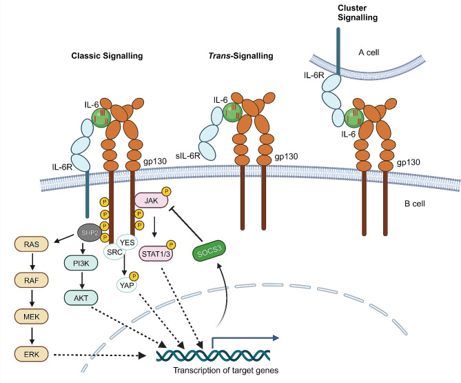
The effector signaling of IL-6 contains three modes (Fig. 1) [6]. IL-6 receptor (IL-6R) is a specialized receptor for IL-6, which is located on the membrane of a set of cell types, such as hepatocytes, immune cells, and some endothelial cells. In addition to IL-6R, IL-6 effector signaling needs another receptor component, 130-kD glycoprotein (gp130) protein. The first signal mode is called classic signaling, also named cis-signaling. The conserved site I of IL-6 cytokine first binds to the membrane bound IL-6R, and then the conserved sites II and III of IL-6 acquire the capability to interact with gp130 and form the complex (Fig. 1) [2], which leads to the dimerization and activation of gp130. The association between the conserved site I of IL-6 and IL-6R is indispensable, as the conserved sites II and III of IL-6 alone are not able to bind gp130 receptor and initiate its dimerization. The second signal mode is IL-6 trans-signaling, for that IL-6 conserved site I first binds a soluble IL-6R (sIL-6R), and then conserved sites II and III bind gp130 on a nearby cell to induce its dimerization and activation (Fig. 1) [7]. For the soluble IL-6 receptor, it is from the membrane bound IL-6R, which is cleaved at the cell surface by A Disintegrin and Metalloprotease17 (ADAM17) and released to tissue interstitial fluid and blood [8]. The third signal mode is cluster signaling. In this mode, IL-6 conserved site I binds a membrane bound IL-6R in A cell, and then the conserved sites II and III within IL-6/IL-6R complex bind gp130 located on the membrane of B cell. The A cell operates as a transmitter cell to activate gp130 downstream signaling of B cell, which is recently described in the dendritic cells (DCs)-mediated activation of IL-6 effector signaling in T cells (Fig. 1) [9, 10]. Collectively, IL-6 trans-signaling and cluster signaling modes can occur in the cells which only express gp130 but not IL-6R, thus amplifying the spectrum of target cells in response to IL-6.
Fig. 1.
Modes of IL-6 signaling and the intracellular signal transduction. The cis-, trans-, and cluster signaling of IL-6 complex. With the activation of IL-6 signaling complex and the phosphorylation of gp130 intracellular domain, JAK-STAT, MAPK, PI3K, and YAP pathways are activated to initiate the transcription of target genes. SOCS3 is a direct target of STAT3 and provides negative feedback to suppress JAK-STAT3 signaling
Upon the formation of IL-6, IL-6R, and dimerized gp130 complex, the intracellular domain of gp130 is phosphorylated. The phosphorylated gp130 then recruits the downstream signal transducers, resulting in the activation of several signaling pathways (Fig. 1) [2, 11]. Janus kinase (JAK)-Signal Transducer and Activator of Transcription (STAT) signaling is the well-established cascade downstream of activated gp130. STAT1 and STAT3 are phosphorylated by JAKs and then form active dimers (homo- and hetero-dimers), which translocate into the nucleus to induce the transcription of IL-6 target genes [12]. The activated gp130 also recruits Src homology 2 domain-containing protein tyrosine phosphatase 2 (SHP2), leading to the activation of downstream extracellular-signal regulated kinase (ERK)/mitogen-activated protein kinase (MAPK) and phosphatidylinositol 3-kinase (PI3K)/AKT pathways. Additionally, YES-associated protein 1 (YAP) and Src family kinase (SRC) are activated by phosphorylated gp130, resulting in YAP phosphorylation and nuclear translocation to initiate downstream transcription [13]. Furthermore, the activation of IL-6 effector signaling can also lead to a negative feedback regulatory system. For example, the expression of suppressor of cytokine signaling 3 (SOCS3) is induced by the activated STAT3, and then binds the phosphorylated domains of gp130 and JAKs to induce their degradation [14]. Based on the three signal modes to activate gp130 and a set of downstream signaling cascades, IL-6 performs its multifaceted functions in tissue homeostasis, regeneration, aging, and inflammation, thus playing critical roles in the physiological and pathological processes.
IL-6 in liver regeneration
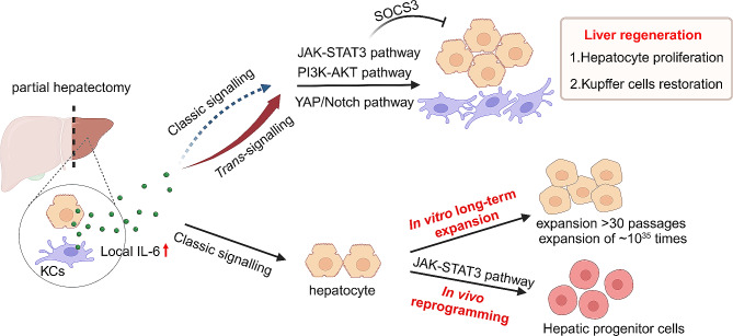
The liver possesses remarkable regeneration capacity under injury conditions. The critical role of the IL-6/IL-6R/gp130-JAK-STAT3 axis has been well-established in the initiation phase of liver regeneration after partial hepatectomy (Fig. 2) [15, 16]. IL-6 is rapidly produced by Kupffer cells, endothelial cells, and hepatocytes after partial hepatectomy, contributing to the following hepatocyte proliferation and parenchyma restoration. The IL-6-promoted liver regeneration was determined in IL-6 knockout mice, as the deletion of il6 impaired the compensatory proliferation of hepatocytes by reducing the downstream STAT3 activation [3, 4], thus promoting liver failure, which could be corrected by the treatment of IL-6. This phenomenon and conclusion have been confirmed in hepatocyte-specific gp130-knockout, hepatocyte-specific STAT3-knockout, or IL-6R deficient mice [17–19]. IL-6 can also activate PI3K/AKT and YAP/Notch pathways independent of the STAT3 pathway to promote hepatocyte compensatory proliferation and liver regeneration [13, 20]. On the other side, the deletion of the negative regulator SOCS3 in hepatocytes resulted in the increased compensatory proliferation and more rapid restoration of liver mass following partial hepatectomy [21]. In addition, except produced by hepatocytes, IL-6 is also produced by myeloid cells, both of them are necessary to restore the number of liver-resident Kupffer cells to maintain tissue homeostasis during liver regeneration [22]. Other sources, including systemic IL-6 or skeletal muscle-derived IL-6, move into the liver and activate its effector signaling to trigger the autophagic flux in hepatocytes, which then contributes to liver regeneration [23, 24].
Fig. 2.
The role of IL-6 signaling in liver homeostasis and regeneration. IL-6 trans-signaling, not classic signaling, is more important for its contribution to liver regeneration. IL-6 classic signaling is significant for the in vitro long-term expansion of hepatocytes and the in vivo reprogramming of hepatocytes into hepatic progenitor cells
Although the classic or cis-signaling of IL-6 is determined to promote the compensatory proliferation of hepatocytes, its trans-signaling in liver regeneration is more important [19, 25–27]. During liver regeneration after 70% hepatectomy, hyper-IL-6, which is a fusion protein of IL-6 and sIL-6R, induced a stronger and a much earlier hepatocyte regeneration effect [25]. Similarly, following D-gal-induced liver damage, the administration of hyper-IL-6 for the activation of IL-6 trans-signaling could rescue animals through an enhanced hepatocyte regeneration [26]. IL-6/sIL-6R double transgenic mice showed massive hepatocyte proliferation even without hepatectomy, whereas IL-6 single transgenic mice did not show such a phenotype [28]. Moreover, it is reported that IL-6 trans-signaling not just accelerate liver regeneration, it is indispensable for the regeneration of liver after partial hepatectomy [19]. This work demonstrated that, during liver regeneration after 70% hepatectomy, sIL-6R can be expressed by not only hepatocytes, but also neutrophils and macrophages, and sIL-6R-mediated IL-6 trans-signaling forms the long-term contribution of IL-6 effects for liver regeneration [19]. Moreover, IL-6 classic signaling is activated in the acute phase-response post liver infection, however, the expression of acute phase protein serum amyloid A2 is similar in wild-type and sIL-6R+/+ (loss of membrane IL-6R) mice, explaining that IL-6 trans-signaling can completely compensate for the loss of IL-6 classic signaling [19]. Indeed, hepatocytes express far more amounts of gp130 on the surface than IL-6R, and hyper-IL-6 thus can lead to a profound downstream effector signaling [5, 27, 29]. Since IL-6 is rapidly internalized whereas the IL-6/sIL-6R complex is not, the IL-6 trans-signal therefore acts much longer [28]. Thus, the clinic use of hyper-IL-6 would be more effective to promote liver regeneration in patients post partial liver resection.
It is incontrovertible that hepatocytes have amazing proliferative capacity in liver regeneration and IL-6 plays a critical role during this process [30]. However, hepatocytes are very difficult to culture or expand in vitro, greatly limiting their clinical application in the therapy of liver diseases. It is reported that IL-6/sIL-6R combined with hepatocyte growth factors (HGF) significantly promoted hepatocyte proliferation after partial hepatectomy [20]. Similarly, a recent study reported that IL-6, co-operated with epidermal growth factor (EGF) and HGF, could promote the long-term expansion (> 30 passages in ~ 150 days with theoretical expansion of ~ 1035 times) of primary mouse hepatocytes in vitro (Fig. 2) [31], which may effectively resolve the limited application of hepatocyte transplantation in liver diseases due to a shortage of enough hepatocytes. In addition, liver progenitor cells are the alternative cell resource to repair the injured liver via their differentiation into hepatocytes [32]. Yet, the activation of liver progenitor cells during liver injury is somewhat restrained, leading to the insufficient contribution to the repairment of liver function. Recent studies suggest that IL-6 is highly expressed and secreted by liver-resident macrophages upon liver damage, and the produced IL-6, as a niche signal, binds the membrane IL-6R and gp130 of hepatocytes and then activates the downstream transcription factor STAT3. Phosphorylated STAT3 associates with the regulatory genomic regions of reprogramming- and progenitor-related genes, which in-turn reprograms the remaining hepatocytes into a progenitor-like state [32–35]. It is unexpected that a single IL-6 cytokine would possess the prominent ability to induce the highly efficient in vivo reprogramming of hepatocytes (Fig. 2). These findings also provide further evidences for that hyper-IL-6 is able to initiate the proliferation of liver progenitor cells in vivo to regenerate the impaired liver [36].
IL-6 in liver aging
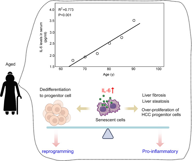
Senescent cells are characterized by the expression of senescence-associated secretory phenotype (SASP) [37, 38], which promotes the development of various chronic diseases. IL-6, one of the major SASP factors, also belongs to the pro-inflammatory cytokines [38]. In the serum of healthy adults, IL-6 is normally lower than 2 pg/mL or undetectable, while a gradual increment of serum IL-6 amounts with advancing ages (Fig. 3) [39, 40]. Moreover, numbers of studies support the potential correlation of IL-6 levels with aging and chronic morbidity [38, 41]. As the data shown in the cohort of end-stage liver diseases, elevated IL-6 levels were highly predictive for mortality. None of the patients with lower IL-6 level (< 5.3 pg/mL) died within one year, but more than half of aged patients died within one year under higher IL-6 level (> 11.6 pg/mL) [41]. When exposed to hepatic ischemia/reperfusion insult, aged liver with high IL-6 in the microenvironment aggravates liver injury such as the intrahepatic tissue damage and inflammation [42]. Moreover, activation of IL-6 effector signaling is positively correlated with age-related dysregulation of lipid metabolism, hepatitis, fibrosis, and xenobiotic detoxification [1, 43]. During the senescence of liver cells, including hepatocytes, cholangiocytes, and stellate cells, IL-6 is produced and participate in the development of liver diseases, especially cancer carcinogenesis and progression [44, 45]. During carcinogenesis, senescence-related IL-6 activates gp130-STAT3 pathway using trans-signaling mode in hepatic progenitor cells or HCC progenitor cells to promote their proliferation, malignant transformation, and hepatocellular-cholangiocarcinoma carcinogenesis. On the other side, IL-6 expressed by senescent cells suppresses the proliferation of senescent hepatocytes, and the hepatic progenitor cells enter compensatory proliferation, thus further promoting hepatocarcinogenesis [44, 45]. Blocking IL-6 trans-signaling using soluble gp130 or clearing senescent cells using a senolytic agent, as well as the depletion of hepatic progenitor cells, result in a significant reduction of hepatocellular-cholangiocarcinoma tumors [44, 45]. As mentioned in the regeneration section, local IL-6 reprograms hepatocytes into hepatic progenitor cells via gp130-STAT3 pathway, and then promotes liver regeneration [33]. However, in chronic senescent conditions, IL-6 is produced by senescent cells to promote the over-proliferation of hepatic progenitor cells and contribute to hepatocellular-cholangiocarcinoma carcinogenesis. A recent study also found that hepatic stellate cells (HSCs) underwent senescence after partial hepatectomy, and elimination of these senescent HSCs impaired liver regeneration. The underlying mechanism was that senescent HSCs expressed and secreted IL-6, binding to membrane IL-6R of hepatocytes to stimulate liver regeneration by the STAT3 pathway and SRC/YAP signal activation [46]. In the aged liver, IL-6 can be expressed and secreted by all senescent liver cells, implying its multifaced actions in the development of age-related diseases (Fig. 3).
Fig. 3.
The role of IL-6 in liver aging. IL-6 concentration in serum is positively- corrected with the increased aging. Increased IL-6 in the liver is mainly expressed and secreted by senescent cells including hepatocytes, macrophages, and endothelial cells, leading to the development of aging-associated chronic liver diseases. On the other side, senescence-secreted high IL-6 can stimulate the reprogramming of cells by the paracrine signaling
Interestingly, IL-6 also plays different roles in the activation of stem cells during aging. When in young mice upon damage, IL-6 is up-regulated and promotes the activation of gland’s stem cells in vivo and in vitro, which is determined in stem cell-derived organoids. However, local IL-6 level is elevated in the aging gland, which does not generate a pituitary stem cell activation but promotes inflammation [47]. Four transcriptional factors abbreviated as OSKM (Oct4, Sox2, Klf4, and c-Myc) are well-established to induce the dedifferentiation and cellular reprogramming in multiple tissues [48]. Actually, OSKM induces two opposite cellular fates, both reprogramming and senescence [49]. However, the OSKM-induced senescence also produces IL-6 to mediate the dedifferentiation and reprogramming, further suggesting the double-edged effects of IL-6 in rejuvenation and aging (Fig. 3) [50].
IL-6 in liver diseases
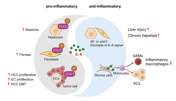
IL-6 signal, also considered as a double-edged sword, performs both pro-inflammatory and anti-proinflammatory roles in the pathogenesis of liver diseases (Fig. 4). During inflammation, the hepatic IL-6 levels can be increased to more than 100 ng/mL [51]. For its pro-inflammatory effect, IL-6 classic signaling especially hepatocyte-specific gp130 activation is crucial for the induction of the acute phase proteins in the liver during host response to the infectious insults [52, 53], and this pro-inflammatory circumstance is quickly resolved when the infection is controlled. If repeated or chronic hepatic IL-6 inflammatory insults are raised, a set of liver diseases will be initiated. Many studies have highlighted that chronic exposure to IL-6 impaired hepatic lipid metabolism [54–56]. In the humanized liver mouse model, the inflammatory effect of IL-6/GP130 pathway promoted hepatic lipid accumulation, suggesting the therapeutic potential of antagonizing GP130 signaling in the treatment of liver steatosis [57]. A recent study used hPSC-derived liver culture to mimic genetic variant-derived NAFLD, and found that IL-6 expression and IL-6/STAT3 activity was elevated during NAFLD development. The dampening of IL-6/STAT3 activity could alleviate the genetic variant-mediated susceptibility to NAFLD [58]. In a mouse model of non-alcoholic steatohepatitis (NASH), knockout of IL-6 or IL-6R also reduced the signs of inflammation during NASH progression [59]. In severe COVID-19 patients, high level of IL-6 was produced in lung and acted on liver sinusoidal endothelial cells by IL-6 trans-signaling, inducing liver inflammation and even liver injury [60]. In the event of hepatocarcinogenesis and progression, increased serum IL-6 level and activated IL-6/STAT3 signaling is highly associated with early tumor recurrence and poor prognosis of HCC patients [61]. For example, inhibition of IL-6 signaling dramatically impedes tumorigenesis and extends the tumor-free survival of patients following surgical partial hepatectomy [62]. Indeed, IL-6 or gp130 knockout mice developed significantly less tumors in the diethylnitrosamine (DEN)-induced HCC model and prolonged survival [63, 64], and we found that the HCC progenitors HcPCs not only autocrined IL-6, but also with promoted responses to IL-6, for their malignant progression to established HCC [65, 66]. In intrahepatic cholangiocarcinoma (ICC), IL-6 increases the expression of circRNA GGNBP2 to encode the protein cGGNBP2-184aa, which in turn forms a positive feedback loop of IL-6/cGGNBP2-184aa/STAT3 to facilitate ICC progression [67]. In addition, HCC-derived fibroblasts can secrete IL-6 and bind adjacent HCC cells to activate IL-6/IL-6R/STAT3 axis, which facilitates epithelial-mesenchymal transition (EMT) of HCC cells and accelerated HCC development [68]. Moreover, increased IL-6 signal with impaired degradation of IL-6 cytokine family signal transducers promotes the proliferation and migration of HCC cells [69]. IL-6 also activates hepatocytes to produce serum amyloid A1 and A2, and forms the pro-metastatic niche to help the metastasis of pancreatic and colorectal cancer cells into the liver [70]. In addition, evidences suggest that IL-6 trans-signaling, but not IL-6 classic signaling, is essential to promote HCC carcinogenesis and progression, and only the activation of membrane-bound IL-6R and gp130 in hepatocytes seems not sufficient for tumor formation [71, 72]. Only the specific inhibition of IL-6 trans-signaling, rather than total inhibition of IL-6 signaling, is sufficient to suppress tumor progression [71, 72]. Together, the pro-inflammatory roles of IL-6 promotes the progression of chronic liver diseases, such as NAFLD, NASH, and HCC.
Fig. 4.
The pro-inflammatory and anti-inflammatory effects of IL-6 in the liver pathological process. The overexpression of IL-6 induced by liver injury presents a double-edged sword effect on the hepatic pathological process. Its pro-inflammatory effect promotes liver steatosis, fibrosis, endothelial cells (ECs) proliferation, and HCC development. On the other side, liver injury or chronic hepatitis are promoted in IL-6 or STAT3 knockout mice, suggesting the anti-inflammatory effect of IL-6. Moreover, IL-6 secreted by liver stroma cells can induce the differentiation of recruited monocytes towards tissue-resident Kupffer cells, but away from SAMs, so as to limit hepatic inflammation
For the anti-inflammatory effect of IL-6, a set of studies in animal models involving chronic hepatitis demonstrate that IL-6 signaling is crucial for ameliorating liver injury and fibrosis. For example, Il6−/− mice developed mature-onset obesity and decreased glucose tolerance, which was partly reversed by IL-6 replacement at low doses [73]. In the mdr2−/− mouse model, the deletion of il6 gene or blockade of IL-6 trans-signaling by the soluble gp130 form exacerbated hepatic steatosis, inflammation, chronic hepatitis, and hepatocarcinogenesis, suggesting the protective role of IL-6 signaling [74]. IL-6 trans-signaling has also been found to have protective roles on acetaminophen-induced acute liver injury in the mice model [75]. It is also reported that high-fat diet (HFD) caused more body-weight gain in il6−/− mice than in wild-type mice [59]. HFD also exaggerated insulin resistance and deterioration of glucose homeostasis in Il6raΔmyel mice, with the underlying mechanism that specific inhibition of IL-6R expression resulted in M1 macrophage polarization and inflammation [76]. For macrophages in the liver, they are divided into the liver-resident Kupffer cells and monocyte-infiltrated macrophages. Tissue resident Kupffer cells’ main function is to maintain homeostasis. Monocyte-infiltrated macrophages are from the differentiation of recruited monocytes when exposed to liver injury [77]. In recent studies, it was found that human liver stromal cells produced and secreted IL-6 cytokine, contributing to the skewing differentiation of monocytes towards tissue-resident Kupffer cells but away from scar-associated macrophages (SAMs) (Fig. 4). Additionally, local IL-6 level is decreased in early-stage human liver diseases as compared to healthy liver tissues, suggesting a protective role for local IL-6 in the healthy liver [78].
Therapeutic strategy of blocking IL-6 signaling
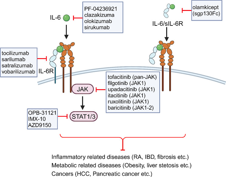
As IL-6 classic and trans-signaling are involved in the development of various diseases, several strategies have been used to inhibit IL-6 effector signaling at different levels in the pre-clinical and clinical settings (Fig. 5; Table 1) [1, 2, 79]. Neutralization of IL-6 inhibits both classic and trans-signaling, and targeting IL-6R blocks all the three modes of IL-6 effects. Moreover, the use of sgp130Fc, targeting IL-6 and sIL-6R complex, selectively suppresses the trans-signaling of IL-6. As IL-6 classic signaling plays crucial roles in the initiation of inflammation and the host defense against pathogen infection, global blockade of IL-6 signaling may reduce the induction of hepatic acute-phase proteins upon infection, such as Listeria monocytogenes [79]. However, the specific blockade of IL-6 trans-signaling using the recombinant sgp130Fc did not interfere with its functions in the defense against pathogens [80].
Fig. 5.
Strategies to specifically block IL-6 signaling. IL-6 signaling can be selectively blocked by antibodies or small molecules targeting the IL-6, IL-6R, sIL-6R, as well as intercellular downstream molecules JAKs and STATs. These neutralizing antibodies or small molecules against IL-6 signaling have been approved for the clinical treatment of inflammatory diseases, metabolic diseases, and cancers
Table 1.
Clinical trials for the effects of IL-6 inhibition
| Target | Drugs | Diseases | Clinical trials |
|---|---|---|---|
| IL-6 | PF-04236921 | Crohn’s disease | Phase II (NCT01345318) |
| olokizumab |
Rheumatoid arthritis Crohn’s disease |
Phase III (NCT03120949) Phase II (NCT01635621) |
|
| siltuximab |
Pancreatic cancer Rheumatoid arthritis |
Phase I (NCT04191421) Phase I (NCT02404558) |
|
| clazakizumab |
COVID-19 Crohn’s disease |
Phase II (NCT04348500) Phase II (NCT01545050) |
|
| IL-6R | tocilizumab |
rheumatoid arthritis COVID-19 Systemic Sclerosis Castleman’s disease |
Phase IV (NCT01331837) Phase III (NCT04356937) Phase III (NCT02453256) Phase II (NCT01441063) |
| sarilumab |
COVID-19 Rheumatoid arthritis |
Phase III (NCT04327388) Phase III (NCT02121210) |
|
| satralizumab | Neuromyelitis Optica | Phase III (NCT02028884) | |
| sIL-6R | olamkicept | Active ulcerative colitis | Phase II (NCT03235752) |
| Pan-JAK | tofacitinib |
Ulcerative colitis Rheumatoid arthritis Crohn’s disease |
Phase III (NCT01458951) Phase IV (NCT02092467) Phase II (NCT01470599) |
| baricitinib | Rheumatoid arthritis | Phase IV (NCT05660655) | |
| JAK1 | upadacitinib |
Rheumatoid arthritis Crohn’s disease |
Phase III (NCT03086343) Phase III (NCT03345836 |
| ruxolitinib | Myelofibrosis | Phase II (NCT01340651) | |
| filgotinib | Crohn’s disease | Phase III (NCT02914600) | |
| itacitinib | HCC | Phase I (NCT04358185) | |
| STAT | OPB-31,121 | HCC | Phase I (NCT01406574) |
| IMX-110 | Pancreatic cancer | Phase I (NCT03382340) |
IL-6 effects can be blocked by a set of antibodies and small molecules, including clazakizumab, sirukumab, olokizumab, and PF-04236921. Clazakizumab and siltuximab directly target the site I of IL-6 for interfering the formation of IL-6 and IL-6R complex, and have been approved for the effectiveness in the treatment of rheumatoid arthritis (RA) and Castleman’s disease in clinical trials [81, 82]. Olokizumab binds to the site III of IL-6, disrupting the recruitment of high-affinity gp130 receptor with IL-6 and IL-6R complex, is also reported to be effective in treating RA patients [83]. Tocilizumab, a humanized monoclonal antibody, is determined to bind to the IL-6 binding site of both membrane and soluble IL-6R, thus blocking classic, trans-, as well as cluster signaling [84, 85]. A series of clinical trials have demonstrated the therapeutic benefit of tocilizumab in chronic inflammatory diseases [86–88]. Indeed, treatment using tocilizumab resulted in the marked reductions of lymphadenopathy and inflammatory parameters in Castleman’s disease [89]. Moreover, fibrosis was also strikingly alleviated after tocilizumab injection in lung complications of systemic sclerosis [90]. Recently, a smart nanosystem of palladium nanoplates loaded with tocilizumab can selectively block IL-6R in the liver, ameliorating anemia with hepcidin production and suppressing cancer progression [91]. Moreover, other IL-6R-blocking antibodies, such as sarilumab and satralizumab, have therapeutic effects on COVID-19, RA, and neuromyelitis optica in phase III studies (Table 1). Soluble gp130Fc, named olamkicept or TJ301, is consists of two soluble human gp130 proteins fused with the Fc of human IgG, and selectively targets the IL-6-sIL-6R complex [79]. As neither IL-6 nor sIL-6R alone can bind to gp130, this sgp130Fc is a selective inhibitor of IL-6 trans-signaling without interfering the classic signaling. Olamkicept has exhibited promising results in the clinical trials [73, 92, 93]. For example, a phase IIa open label trial evaluating olamkicept in patients with active inflammatory bowel disease (IBD) showed that over 20% patients treated with olamkicept were completely alleviated in clinical symptoms and inflammatory markers [93]. Sgp130Fc treatment in HFD-fed mice presented reduced macrophage accumulation in the adipose tissue, resulting in the improved insulin resistance [94]. Furthermore, sgp130Fc administration, but not anti-IL-6, is more likely to improve the survival in the mice models of cecal ligation and puncture, abdominal aortic aneurysms, and acute lung injury [95–97]. In addition, in a mouse model that mimics human NASH-driven HCC, treatment of olamkicept can significantly regress NASH and markedly reduce NASH-driven HCC [98].
IL-6 binding to IL-6R and gp130 results in the activation of intercellular signal transducers including the phosphorylation of JAK, Src, and STAT3. Therefore, these intercellular signal transducers inhibitors can lead to a global blockade of IL-6 signaling. The clinical trials with JAK/STAT inhibitors are underway for the therapy of cancers and inflammatory diseases (Fig. 5) [12, 99]. For example, ruxolitinib, targeting both JAK1 and JAK2 kinases for inhibiting their phosphorylation, was first approved by the FDA in 2011 for the treatment of myelofibrosis [100, 101]. Tofacitinib, competing with the adenosine triphosphate binding site of both JAK1 and JAK3 for blocking their phosphorylation, was first approved by the FDA in 2012 for the treatment of RA [102]. As reported in COVID-19 patients, both sgp130Fc and JAK inhibitors ruxolitinib can block IL-6 trans-signaling with decreased phosphorylation of JAK1 and STAT1/3, resulting in the inhibition of proinflammatory factors production and the alleviation of liver injury [60]. Ruxolitinib also effectively inhibits the JAK/STAT signaling pathway in HCC cells and significantly reduces their proliferation and colony formation [103]. Importantly, evidences from a set of clinical trials showed that the selective JAK1 inhibitors, such as filgotinib and upadacitinib, are more effective than the pan-JAK inhibitor of tofacitinib in the treatment of Crohn’s disease and ulcerative colitis [104–106]. Data from a phase II trial showed that 47% of the Crohn’s disease patients treated with filgotinib achieved the clinical remission compared with 23% in the tofacitinib group [107]. Itacitinib, another JAK1 selective inhibitor, has been investigated in a phase I study of advanced HCC [108]. OPB-31,121, as a STAT3 antagonist targeting its SH2 region, is an orally bioavailable low-molecular-weight compound, and the clinical trial of this drug was conducted in HCC patients [109].
Although the therapeutic effect of intervening IL-6 signaling can be achieved by various inhibitors, ranging from blocking the IL-6 cytokine or its receptor outside of the cell to targeting the kinases and transcription factors inside of the cell, evidences from animal models and pre-clinical studies have demonstrated that inhibition of IL-6 effects also induced adverse effects, thus limiting its use in the clinics in any case [79]. Blocking IL-6 in IBD resulted in severe adverse effects, such as abdominal pain, rather than just ameliorating intestinal inflammation [110]. This problem may be due to the diverse biological effects of the global inhibition of IL-6, and the selective targeting of IL-6 or its downstream effectors may be more important. To date, as evidences from animal models have demonstrated the primary role of IL-6 trans-signaling in inflammatory diseases, olamkicept, selectively targeting sIL-6R but not IL-6 or IL-6R, can effectively discriminate between IL-6 classic and trans-signaling, thus may be more beneficial than the global blockade of IL-6 [111, 112]. Hence, olamkicept, or the developing next-generation selective inhibitors of IL-6 trans-signaling, are considered to be a safe and effective therapeutic strategy for further clinical studies.
Conclusions
IL-6 is a well investigated pleiotropic cytokine with three different signal modes. Membrane IL-6R are mainly expressed in hepatocytes, immune cells, and some endothelial cells, leading to the limit of IL-6 classic signaling. However, the gp130 protein is widely expressed in tissues [78], representing the extensive responses to IL-6 via the trans-signaling activated by IL-6/sIL-6R/gp130 complex. Recently, accumulated evidences have suggested the more important effect of IL-6 trans-signaling on liver regeneration and pathological processes. Selective inhibition of IL-6 trans-signaling rather than the global blockade of IL-6 might therefore be more effective in the treatment of liver pathologies. Interestingly, HHV-8 encodes a viral homolog of human IL-6, called viral IL-6 (vIL-6) [113]. vIL-6, in contrast to hIL-6, can directly bind to and activate gp130 without the need of hIL-6R. With the activation of downstream signaling cascades, vIL-6 can further increase the production of endogenous IL-6 and enhance the acute-phase responses [114, 115].
It is also generally accepted that IL-6 is a double-edged sword factor for its differential functions in hepatic regeneration, aging, and chronic liver diseases. Under physiological conditions, IL-6 signal is critical for liver regeneration and the proliferation of hepatocytes as to its pro-regenerative effect. During aging progression, the IL-6 level was gradually increased, which enhances its pro-inflammatory effects and even promotes the development of inflammation-associated liver diseases. However, on the other side, high IL-6 level produced by senescent cells can also lead to the reprogramming of hepatocytes, and performs its protective effect on the development of liver diseases with anti-inflammatory activities. Therefore, in order to more accurately intervene the IL-6-mediated liver diseases or aging, it is of prior importance to understand when, where, and how IL-6 works in the physiological and pathological processes in the liver.
Acknowledgements
Not applicable.
Abbreviations
- ADAM
A Disintegrin and Metalloprotease
- DC
Dendritic cell
- DEN
Diethylnitrosamine
- EC
Endothelial cell
- EGF
Epidermal growth factor
- EMT
Epithelial-mesenchymal transition
- ERK
Extracellular-signal regulated kinase
- Gp130
130-kD glycoprotein
- HCC
Hepatocellular carcinoma
- HFD
High-fat diet
- HGF
Hepatocyte growth factor
- ICC
Intrahepatic cholangiocarcinoma
- IL-6
Interleukin-6
- IL-6R
IL-6 receptor
- JAK
Janus kinase
- MAPK
Mitogen-activated protein kinase
- NASH
Non-alcoholic steatohepatitis
- PI3K
Phosphatidylinositol 3-kinase
- SAMs
Scar-associated macrophages
- SASP
Senescence-associated secretory phenotype
- SHP-2
Src homology 2 domain-containing protein tyrosine phosphatase 2
- SOCS3
Suppressor of cytokine signaling 3
- SRC
Src family kinase
- vIL-6
Viral IL-6
- YAP
YES-associated protein 1
Author contributions
M.J.W., H.L.Z. and F.C. wrote the paper and contributed equally. X.J.G. performed the statistical analysis of Fig. 3. Q.G.L. designed the Figures. J.H. supervised and revised the paper. All authors have read and approved the final manuscript.
Funding
This work was sponsored by grants from National Key Research and Development Program of China (2023YFC2505900), National Natural Science Foundation of China (32070732, 92269204, 82171755), Shanghai Rising-Star Program (21QA1411400), and Military Outstanding Youth Program (2020QN06119, 01-SWKJYCJJ07).
Data availability
No datasets were generated or analysed during the current study.
Declarations
Ethics approval and consent to participate
Not applicable.
Consent for publication
Not applicable.
Competing interests
The authors declare no competing interests.
Footnotes
Publisher’s Note
Springer Nature remains neutral with regard to jurisdictional claims in published maps and institutional affiliations.
Min-Jun Wang, Hai-Ling Zhang and Fei Chen share co-first authorship.
Contributor Information
Min-Jun Wang, Email: mjwang@smmu.edu.cn.
Jin Hou, Email: houjin@immunol.org.
References
- 1.Forcina L, Franceschi C, Musarò A. The hormetic and hermetic role of IL-6. Ageing Res Rev. 2022;80:101697. doi: 10.1016/j.arr.2022.101697. [DOI] [PubMed] [Google Scholar]
- 2.Giraldez MD, Carneros D, Garbers C, Rose-John S, Bustos M. New insights into IL-6 family cytokines in metabolism, hepatology and gastroenterology. Nat Rev Gastroenterol Hepatol. 2021;18:787–803. doi: 10.1038/s41575-021-00473-x. [DOI] [PubMed] [Google Scholar]
- 3.Blindenbacher A, Wang X, Langer I, Savino R, Terracciano L, Heim MH. Interleukin 6 is important for survival after partial hepatectomy in mice. Hepatology. 2003;38:674–82. doi: 10.1053/jhep.2003.50378. [DOI] [PubMed] [Google Scholar]
- 4.Cressman DE, Greenbaum LE, DeAngelis RA, Ciliberto G, Furth EE, Poli V, et al. Liver failure and defective hepatocyte regeneration in interleukin-6-deficient mice. Science. 1996;274:1379–83. doi: 10.1126/science.274.5291.1379. [DOI] [PubMed] [Google Scholar]
- 5.Schmidt-Arras D, Rose-John S. IL-6 pathway in the liver: from physiopathology to therapy. J Hepatol. 2016;64:1403–15. doi: 10.1016/j.jhep.2016.02.004. [DOI] [PubMed] [Google Scholar]
- 6.Garbers C, Heink S, Korn T, Rose-John S. Interleukin-6: designing specific therapeutics for a complex cytokine. Nat Rev Drug Discov. 2018;17:395–412. doi: 10.1038/nrd.2018.45. [DOI] [PubMed] [Google Scholar]
- 7.Riethmueller S, Somasundaram P, Ehlers JC, Hung CW, Flynn CM, Lokau J, et al. Proteolytic origin of the soluble human IL-6R in vivo and a decisive role of N-glycosylation. PLoS Biol. 2017;15:e2000080. doi: 10.1371/journal.pbio.2000080. [DOI] [PMC free article] [PubMed] [Google Scholar]
- 8.Yousif AS, Ronsard L, Shah P, Omatsu T, Sangesland M, Bracamonte Moreno T, et al. The persistence of interleukin-6 is regulated by a blood buffer system derived from dendritic cells. Immunity. 2021;54:235–46. doi: 10.1016/j.immuni.2020.12.001. [DOI] [PMC free article] [PubMed] [Google Scholar]
- 9.Heink S, Yogev N, Garbers C, Herwerth M, Aly L, Gasperi C, et al. Trans-presentation of IL-6 by dendritic cells is required for the priming of pathogenic TH17 cells. Nat Immunol. 2017;18:74–85. doi: 10.1038/ni.3632. [DOI] [PMC free article] [PubMed] [Google Scholar]
- 10.Korn T, Hiltensperger M. Role of IL-6 in the commitment of T cell subsets. Cytokine. 2021;146:155654. doi: 10.1016/j.cyto.2021.155654. [DOI] [PMC free article] [PubMed] [Google Scholar]
- 11.Murakami M, Kamimura D, Hirano T. Pleiotropy and specificity: insights from the interleukin 6 family of cytokines. Immunity. 2019;50:812–31. doi: 10.1016/j.immuni.2019.03.027. [DOI] [PubMed] [Google Scholar]
- 12.Kang S, Tanaka T, Narazaki M, Kishimoto T. Targeting interleukin-6 signaling in clinic. Immunity. 2019;50:1007–23. doi: 10.1016/j.immuni.2019.03.026. [DOI] [PubMed] [Google Scholar]
- 13.Stancil IT, Michalski JE, Hennessy CE, Hatakka KL, Yang IV, Kurche JS, et al. Interleukin-6-dependent epithelial fluidization initiates fibrotic lung remodeling. Sci Transl Med. 2022;14:eabo5254. doi: 10.1126/scitranslmed.abo5254. [DOI] [PMC free article] [PubMed] [Google Scholar]
- 14.Liu ZK, Li C, Zhang RY, Wei D, Shang YK, Yong YL, et al. EYA2 suppresses the progression of hepatocellular carcinoma via SOCS3-mediated blockade of JAK/STAT signaling. Mol Cancer. 2021;20:79. doi: 10.1186/s12943-021-01377-9. [DOI] [PMC free article] [PubMed] [Google Scholar]
- 15.Drucker C, Gewiese J, Malchow S, Scheller J, Rose-John S. Impact of interleukin-6 classic- and trans-signaling on liver damage and regeneration. J Autoimmun. 2010;34:29–37. doi: 10.1016/j.jaut.2009.08.003. [DOI] [PubMed] [Google Scholar]
- 16.Michalopoulos GK, Bhushan B. Liver regeneration: biological and pathological mechanisms and implications. Nat Rev Gastroenterol Hepatol. 2021;18:40–55. doi: 10.1038/s41575-020-0342-4. [DOI] [PubMed] [Google Scholar]
- 17.Streetz KL, Wüstefeld T, Klein C, Kallen KJ, Tronche F, Betz UA, et al. Lack of gp130 expression in hepatocytes promotes liver injury. Gastroenterology. 2003;125:532–43. doi: 10.1016/S0016-5085(03)00901-6. [DOI] [PubMed] [Google Scholar]
- 18.Haga S, Ogawa W, Inoue H, Terui K, Ogino T, Igarashi R, et al. Compensatory recovery of liver mass by akt-mediated hepatocellular hypertrophy in liver-specific STAT3-deficient mice. J Hepatol. 2005;43:799–807. doi: 10.1016/j.jhep.2005.03.027. [DOI] [PubMed] [Google Scholar]
- 19.Fazel Modares N, Polz R, Haghighi F, Lamertz L, Behnke K, Zhuang Y, et al. IL-6 trans-signaling controls liver regeneration after partial hepatectomy. Hepatology. 2019;70:2075–91. doi: 10.1002/hep.30774. [DOI] [PubMed] [Google Scholar]
- 20.Nechemia-Arbely Y, Shriki A, Denz U, Drucker C, Scheller J, Raub J, et al. Early hepatocyte DNA synthetic response posthepatectomy is modulated by IL-6 trans-signaling and PI3K/AKT activation. J Hepatol. 2011;54:922–9. doi: 10.1016/j.jhep.2010.08.017. [DOI] [PubMed] [Google Scholar]
- 21.Riehle KJ, Campbell JS, McMahan RS, Johnson MM, Beyer RP, Bammler TK, et al. Regulation of liver regeneration and hepatocarcinogenesis by suppressor of cytokine signaling 3. J Exp Med. 2008;205:91–103. doi: 10.1084/jem.20070820. [DOI] [PMC free article] [PubMed] [Google Scholar]
- 22.Ait Ahmed Y, Fu Y, Rodrigues RM, He Y, Guan Y, Guillot A, et al. Kupffer cell restoration after partial hepatectomy is mainly driven by local cell proliferation in IL-6-dependent autocrine and paracrine manners. Cell Mol Immunol. 2021;18:2165–76. doi: 10.1038/s41423-021-00731-7. [DOI] [PMC free article] [PubMed] [Google Scholar]
- 23.Park HS, Song JW, Park JH, Lim BK, Moon OS, Son HY, et al. TXNIP/VDUP1 attenuates steatohepatitis via autophagy and fatty acid oxidation. Autophagy. 2021;17:2549–64. doi: 10.1080/15548627.2020.1834711. [DOI] [PMC free article] [PubMed] [Google Scholar]
- 24.Pinto AP, Ropelle ER, Quadrilatero J, da Silva ASR. Physical exercise and liver autophagy: potential roles of IL-6 and irisin. Exerc Sport Sci Rev. 2022;50:89–96. doi: 10.1249/JES.0000000000000278. [DOI] [PubMed] [Google Scholar]
- 25.Peters M, Blinn G, Jostock T, Schirmacher P, Meyer zum Büschenfelde KH, Galle PR, et al. Combined interleukin 6 and soluble interleukin 6 receptor accelerates murine liver regeneration. Gastroenterology. 2000;119:1663–71. doi: 10.1053/gast.2000.20236. [DOI] [PubMed] [Google Scholar]
- 26.Galun E, Zeira E, Pappo O, Peters M, Rose-John S. Liver regeneration induced by a designer human IL-6/sIL-6R fusion protein reverses severe hepatocellular injury. FASEB J. 2000;14:1979–87. doi: 10.1096/fj.99-0913com. [DOI] [PubMed] [Google Scholar]
- 27.Galun E, Rose-John S. The regenerative activity of interleukin-6. Methods Mol Biol. 2013;982:59–77. doi: 10.1007/978-1-62703-308-4_4. [DOI] [PubMed] [Google Scholar]
- 28.Peters M, Blinn G, Solem F, Fischer M, Meyer zum Büschenfelde KH, Rose-John S. In vivo and in vitro activities of the gp130-stimulating designer cytokine Hyper-IL-6. J Immunol. 1998;161:3575–81. doi: 10.4049/jimmunol.161.7.3575. [DOI] [PubMed] [Google Scholar]
- 29.Peters M, Schirmacher P, Goldschmitt J, Odenthal M, Peschel C, Fattori E, et al. Extramedullary expansion of hematopoietic progenitor cells in interleukin (IL)-6-sIL-6R double transgenic mice. J Exp Med. 1997;185:755–66. doi: 10.1084/jem.185.4.755. [DOI] [PMC free article] [PubMed] [Google Scholar]
- 30.Pu W, Zhou B. Hepatocyte generation in liver homeostasis, repair, and regeneration. Cell Regen. 2022;11:2. doi: 10.1186/s13619-021-00101-8. [DOI] [PMC free article] [PubMed] [Google Scholar]
- 31.Guo R, Jiang M, Wang G, Li B, Jia X, Ai Y, et al. IL6 supports long-term expansion of hepatocytes in vitro. Nat Commun. 2022;13:7345. doi: 10.1038/s41467-022-35167-8. [DOI] [PMC free article] [PubMed] [Google Scholar]
- 32.Huang WJ, Zhou X, Fu GB, Ding M, Wu HP, Zeng M, et al. The combined induction of liver progenitor cells and the suppression of stellate cells by small molecules reverts chronic hepatic dysfunction. Theranostics. 2021;11:5539–52. doi: 10.7150/thno.54457. [DOI] [PMC free article] [PubMed] [Google Scholar]
- 33.Li L, Cui L, Lin P, Liu Z, Bao S, Ma X, et al. Kupffer-cell-derived IL-6 is repurposed for hepatocyte dedifferentiation via activating progenitor genes from injury-specific enhancers. Cell Stem Cell. 2023;30:283–99. doi: 10.1016/j.stem.2023.01.009. [DOI] [PubMed] [Google Scholar]
- 34.Li R, Li D, Nie Y. IL-6/gp130 signaling: a key unlocking regeneration. Cell Regen. 2023;12:16. doi: 10.1186/s13619-023-00160-z. [DOI] [PMC free article] [PubMed] [Google Scholar]
- 35.VanHook AM. IL-6 drives hepatocyte dedifferentiation. Sci Signal. 2023;16:eadh4937. doi: 10.1126/scisignal.adh4937. [DOI] [PubMed] [Google Scholar]
- 36.Lu WY, Bird TG, Boulter L, Tsuchiya A, Cole AM, Hay T, et al. Hepatic progenitor cells of biliary origin with liver repopulation capacity. Nat Cell Biol. 2015;17:971–83. doi: 10.1038/ncb3203. [DOI] [PMC free article] [PubMed] [Google Scholar]
- 37.Lucas V, Cavadas C, Aveleira CA. Cellular senescence: from mechanisms to currentbiomarkers and senotherapies. Pharmacol Rev. 2023;75:675–713. doi: 10.1124/pharmrev.122.000622. [DOI] [PubMed] [Google Scholar]
- 38.Li X, Li C, Zhang W, Wang Y, Qian P, Huang H. Inflammation and aging: signaling pathways and intervention therapies. Signal Transduct Target Ther. 2023;8:239. doi: 10.1038/s41392-023-01502-8. [DOI] [PMC free article] [PubMed] [Google Scholar]
- 39.Puzianowska-Kuźnicka M, Owczarz M, Wieczorowska-Tobis K, Nadrowski P, Chudek J, Slusarczyk P, et al. Interleukin-6 and C-reactive protein, successful aging, and mortality: the PolSenior study. Immun Ageing. 2016;13:21. doi: 10.1186/s12979-016-0076-x. [DOI] [PMC free article] [PubMed] [Google Scholar]
- 40.Lustgarten MS, Fielding RA. Metabolites Associated with circulating Interleukin-6 in older adults. J Gerontol Biol Sci Med Sci. 2017;72:1277–83. doi: 10.1093/gerona/glw039. [DOI] [PMC free article] [PubMed] [Google Scholar]
- 41.Remmler J, Schneider C, Treuner-Kaueroff T, Bartels M, Seehofer D, Scholz M, et al. Increased level of interleukin 6 associates with increased 90-day and 1-year mortality in patients with end-stage liver disease. Clin Gastroenterol Hepatol. 2018;16:730–37. doi: 10.1016/j.cgh.2017.09.017. [DOI] [PubMed] [Google Scholar]
- 42.Zhong W, Rao Z, Rao J, Han G, Wang P, Jiang T, et al. Aging aggravated liver ischemia and reperfusion injury by promoting STING-mediated NLRP3 activation in macrophages. Aging Cell. 2020;19:e13186. doi: 10.1111/acel.13186. [DOI] [PMC free article] [PubMed] [Google Scholar]
- 43.von Loeffelholz C, Lieske S, Neuschäfer-Rube F, Willmes DM, Raschzok N, Sauer IM, et al. The human longevity gene homolog INDY and interleukin-6 interact in hepatic lipid metabolism. Hepatology. 2017;66:616–30. doi: 10.1002/hep.29089. [DOI] [PMC free article] [PubMed] [Google Scholar]
- 44.Rosenberg N, Van Haele M, Lanton T, Brashi N, Bromberg Z, Adler H, et al. Combined hepatocellular-cholangiocarcinoma derives from liver progenitor cells and depends on senescence and IL-6 trans-signaling. J Hepatol. 2022;77:1631–41. doi: 10.1016/j.jhep.2022.07.029. [DOI] [PubMed] [Google Scholar]
- 45.Arechederra M, Fernández-Barrena MG. Hepatic progenitor cells, senescence and IL-6 as the main players in combined hepatocellular-cholangiocarcinoma development. J Hepatol. 2022;77:1479–81. doi: 10.1016/j.jhep.2022.09.008. [DOI] [PubMed] [Google Scholar]
- 46.Cheng N, Kim KH, Lau LF. Senescent hepatic stellate cells promote liver regeneration through IL-6 and ligands of CXCR2. JCI Insight. 2022;7:e158207. doi: 10.1172/jci.insight.158207. [DOI] [PMC free article] [PubMed] [Google Scholar]
- 47.Vennekens A, Laporte E, Hermans F, Cox B, Modave E, Janiszewski A, et al. Interleukin-6 is an activator of pituitary stem cells upon local damage, a competence quenched in the aging gland. Proc Natl Acad Sci U S A. 2021;118:e2100052118. doi: 10.1073/pnas.2100052118. [DOI] [PMC free article] [PubMed] [Google Scholar]
- 48.Takahashi K, Yamanaka S. Induction of pluripotent stem cells from mouse embryonic and adult fibroblast cultures by defined factors. Cell. 2006;126:663–76. doi: 10.1016/j.cell.2006.07.024. [DOI] [PubMed] [Google Scholar]
- 49.Chiche A, Le Roux I, von Joest M, Sakai H, Aguın SB, Cazin C, et al. Injury-induced senescence enables in vivo reprogramming in skeletal muscle. Cell Stem Cell. 2017;20:407–14. doi: 10.1016/j.stem.2016.11.020. [DOI] [PubMed] [Google Scholar]
- 50.Mosteiro L, Pantoja C, de Martino A, Serrano M. Senescence promotes in vivo reprogramming through p16INK4a and IL-6. Aging Cell. 2018;17:e12711. doi: 10.1111/acel.12711. [DOI] [PMC free article] [PubMed] [Google Scholar]
- 51.Rose-John S. The soluble interleukin 6 receptor: advanced therapeutic options in inflammation. Clin Pharmacol Ther. 2017;102:591–852. doi: 10.1002/cpt.782. [DOI] [PubMed] [Google Scholar]
- 52.Del Giudice M, Gangestad SW, Rethinking IL-6 and CRP: why they are more than inflammatory biomarkers, and why it matters. Brain Behav Immun. 2018;70:61–75. doi: 10.1016/j.bbi.2018.02.013. [DOI] [PubMed] [Google Scholar]
- 53.Schumacher N, Yan K, Gandraß M, Müller M, Krisp C, Häsler R, et al. Cell-autonomous hepatocyte-specific GP130 signaling is sufficient to trigger a robust innate immune response in mice. J Hepatol. 2021;74:407–18. doi: 10.1016/j.jhep.2020.09.021. [DOI] [PubMed] [Google Scholar]
- 54.Li H, Liu NN, Li JR, Wang MX, Tan JL, Dong B, et al. Bicyclol ameliorates advanced liver diseases in murine models via inhibiting the IL-6/STAT3 signaling pathway. Biomed Pharmacother. 2022;150:113083. doi: 10.1016/j.biopha.2022.113083. [DOI] [PubMed] [Google Scholar]
- 55.Lehrskov LL, Christensen RH. The role of interleukin-6 in glucose homeostasis and lipid metabolism. Semin Immunopathol. 2019;41:491–9. doi: 10.1007/s00281-019-00747-2. [DOI] [PubMed] [Google Scholar]
- 56.Xiang DM, Sun W, Ning BF, Zhou TF, Li XF, Zhong W, et al. The HLF/IL-6/STAT3 feedforward circuit drives hepatic stellate cell activation to promote liver fibrosis. Gut. 2018;67:1704–15. doi: 10.1136/gutjnl-2016-313392. [DOI] [PubMed] [Google Scholar]
- 57.Carbonaro M, Wang K, Huang H, Frleta D, Patel A, Pennington A, et al. IL-6-GP130 signaling protects human hepatocytes against lipid droplet accumulation in humanized liver models. Sci Adv. 2023;9:eadf4490. doi: 10.1126/sciadv.adf4490. [DOI] [PMC free article] [PubMed] [Google Scholar]
- 58.Park J, Zhao Y, Zhang F, Zhang S, Kwong AC, Zhang Y, et al. IL-6/STAT3 axis dictates the PNPLA3-mediated susceptibility to non-alcoholic fatty liver disease. J Hepatol. 2023;78:45–56. doi: 10.1016/j.jhep.2022.08.022. [DOI] [PMC free article] [PubMed] [Google Scholar]
- 59.Hou X, Yin S, Ren R, Liu S, Yong L, Liu Y, et al. Myeloid-cell-specific IL-6 signaling promotes MicroRNA-223-enriched exosome production to attenuate NAFLD-associated fibrosis. Hepatology. 2021;74:116–32. doi: 10.1002/hep.31658. [DOI] [PMC free article] [PubMed] [Google Scholar]
- 60.McConnell MJ, Kawaguchi N, Kondo R, Sonzogni A, Licini L, Valle C, et al. Liver injury in COVID-19 and IL-6 trans-signaling-induced endotheliopathy. J Hepatol. 2021;75:647–58. doi: 10.1016/j.jhep.2021.04.050. [DOI] [PMC free article] [PubMed] [Google Scholar]
- 61.Lai SC, Su YT, Chi CC, Kuo YC, Lee KF, Wu YC, et al. DNMT3b/OCT4 expression confers sorafenib resistance and poor prognosis of hepatocellular carcinoma through IL-6/STAT3 regulation. J Exp Clin Cancer Res. 2019;38:474. doi: 10.1186/s13046-019-1442-2. [DOI] [PMC free article] [PubMed] [Google Scholar]
- 62.Lanton T, Shriki A, Nechemia-Arbely Y, Abramovitch R, Levkovitch O, Adar R, et al. Interleukin 6-dependent genomic instability heralds accelerated carcinogenesis following liver regeneration on a background of chronic hepatitis. Hepatology. 2017;65:1600–11. doi: 10.1002/hep.29004. [DOI] [PubMed] [Google Scholar]
- 63.Mittenbühler MJ, Sprenger HG, Gruber S, Wunderlich CM, Kern L, Brüning JC, et al. Hepatic leptin receptor expression can partially compensate for IL-6Rα deficiency in DEN-induced hepatocellular carcinoma. Mol Metab. 2018;17:122–33. doi: 10.1016/j.molmet.2018.08.010. [DOI] [PMC free article] [PubMed] [Google Scholar]
- 64.Hatting M, Spannbauer M, Peng J, Al Masaoudi M, Sellge G, Nevzorova YA, et al. Lack of gp130 expression in hepatocytes attenuates tumor progression in the DEN model. Cell Death Dis. 2015;6:e1667. doi: 10.1038/cddis.2014.590. [DOI] [PMC free article] [PubMed] [Google Scholar]
- 65.Li Z, Zhou Y, Jia K, Yang Y, Zhang L, Wang S, et al. JMJD4-demethylated RIG-I prevents hepatic steatosis and carcinogenesis. J Hematol Oncol. 2022;15:161. doi: 10.1186/s13045-022-01381-6. [DOI] [PMC free article] [PubMed] [Google Scholar]
- 66.Zhou Y, Jia K, Wang S, Li Z, Li Y, Lu S, et al. Malignant progression of liver cancer progenitors requires lysine acetyltransferase 7-acetylated and cytoplasm-translocated G protein GαS. Hepatology. 2023;77:1106–21. doi: 10.1002/hep.32487. [DOI] [PMC free article] [PubMed] [Google Scholar]
- 67.Li H, Lan T, Liu H, Liu C, Dai J, Xu L, et al. IL-6-induced cGGNBP2 encodes a protein to promote cell growth and metastasis in intrahepatic cholangiocarcinoma. Hepatology. 2022;75:1402–19. doi: 10.1002/hep.32232. [DOI] [PMC free article] [PubMed] [Google Scholar]
- 68.Jia C, Wang G, Wang T, Fu B, Zhang Y, Huang L, et al. Cancer-associated fibroblasts induce epithelial-mesenchymal transition via the transglutaminase 2-dependent IL-6/IL6R/STAT3 axis in Hepatocellular Carcinoma. Int J Biol Sci. 2020;16:2542–58. doi: 10.7150/ijbs.45446. [DOI] [PMC free article] [PubMed] [Google Scholar]
- 69.Desideri E, Castelli S, Dorard C, Toifl S, Grazi GL, Ciriolo MR, et al. Impaired degradation of YAP1 and IL6ST by chaperone-mediated autophagy promotes proliferation and migration of normal and hepatocellular carcinoma cells. Autophagy. 2023;19:152–62. doi: 10.1080/15548627.2022.2063004. [DOI] [PMC free article] [PubMed] [Google Scholar]
- 70.Lee JW, Stone ML, Porrett PM, Thomas SK, Komar CA, Li JH, et al. Hepatocytes direct the formation of a pro-metastatic niche in the liver. Nature. 2019;567:249–52. doi: 10.1038/s41586-019-1004-y. [DOI] [PMC free article] [PubMed] [Google Scholar]
- 71.Bergmann J, Müller M, Baumann N, Reichert M, Heneweer C, Bolik J, et al. IL-6 trans-signaling is essential for the development of hepatocellular carcinoma in mice. Hepatology. 2017;65:89–103. doi: 10.1002/hep.28874. [DOI] [PubMed] [Google Scholar]
- 72.Reeh H, Rudolph N, Billing U, Christen H, Streif S, Bullinger E, et al. Response to IL-6 trans- and IL-6 classic signalling is determined by the ratio of the IL-6 receptor α to gp130 expression: fusing experimental insights and dynamic modelling. Cell Commun Signal. 2019;17:46. doi: 10.1186/s12964-019-0356-0. [DOI] [PMC free article] [PubMed] [Google Scholar]
- 73.Wallenius V, Wallenius K, Ahrén B, Rudling M, Carlsten H, Dickson SL, et al. Interleukin-6-deficient mice develop mature-onset obesity. Nat Med. 2002;8:75–9. doi: 10.1038/nm0102-75. [DOI] [PubMed] [Google Scholar]
- 74.Shriki A, Lanton T, Sonnenblick A, Levkovitch-Siany O, Eidelshtein D, Abramovitch R, et al. Multiple roles of IL6 in hepatic injury, steatosis, and senescence aggregate to suppress tumorigenesis. Cancer Res. 2021;81:4766–77. doi: 10.1158/0008-5472.CAN-21-0321. [DOI] [PubMed] [Google Scholar]
- 75.Li SQ, Zhu S, Han HM, Lu HJ, Meng HY. IL-6 trans-signaling plays important protective roles in acute liver injury induced by acetaminophen in mice. J Biochem Mol Toxicol. 2015;29:288–97. doi: 10.1002/jbt.21708. [DOI] [PubMed] [Google Scholar]
- 76.Mauer J, Chaurasia B, Goldau J, Vogt MC, Ruud J, Nguyen KD, et al. Signaling by IL-6 promotes alternative activation of macrophages to limit endotoxemia and obesity-associated resistance to insulin. Nat Immunol. 2014;15:423–30. doi: 10.1038/ni.2865. [DOI] [PMC free article] [PubMed] [Google Scholar]
- 77.Weston CJ, Zimmermann HW, Adams DH. The role of myeloid-derived cells in the progression of liver disease. Front Immunol. 2019;10:893. doi: 10.3389/fimmu.2019.00893. [DOI] [PMC free article] [PubMed] [Google Scholar]
- 78.Buonomo EL, Mei S, Guinn SR, Leo IR, Peluso MJ, Nolan MA, et al. Liver stromal cells restrict macrophage maturation and stromal IL-6 limits the differentiation of cirrhosis-linked macrophages. J Hepatol. 2022;76:1127–37. doi: 10.1016/j.jhep.2021.12.036. [DOI] [PubMed] [Google Scholar]
- 79.Rose-John S, Jenkins BJ, Garbers C, Moll JM, Scheller J. Targeting IL-6 trans-signalling: past, present and future prospects. Nat Rev Immunol. 2023;23:666–81. doi: 10.1038/s41577-023-00856-y. [DOI] [PMC free article] [PubMed] [Google Scholar]
- 80.Jones SA, Jenkins BJ. Recent insights into targeting the IL-6 cytokine family in inflammatory diseases and cancer. Nat Rev Immunol. 2018;18:773–89. doi: 10.1038/s41577-018-0066-7. [DOI] [PubMed] [Google Scholar]
- 81.van Rhee F, Rosenthal A, Kanhai K, Martin R, Nishimura K, Hoering A, et al. Siltuximab is associated with improved progression-free survival in idiopathic multicentric Castleman disease. Blood Adv. 2022;6:4773–81. doi: 10.1182/bloodadvances.2022007112. [DOI] [PMC free article] [PubMed] [Google Scholar]
- 82.Aletaha D, Bingham CO, Karpouzas GA, Takeuchi T, Thorne C, Bili A, et al. Long-term safety and efficacy of sirukumab for patients with rheumatoid arthritis who previously received sirukumab in randomised controlled trials (SIRROUND-LTE) RMD Open. 2021;7:e001465. doi: 10.1136/rmdopen-2020-001465. [DOI] [PMC free article] [PubMed] [Google Scholar]
- 83.Kerschbaumer A, Sepriano A, Bergstra SA, Smolen JS, van der Heijde D, Caporali R, et al. Efficacy of synthetic and biological DMARDs: a systematic literature review informing the 2022 update of the EULAR recommendations for the management of rheumatoid arthritis. Ann Rheum Dis. 2023;82:95–106. doi: 10.1136/ard-2022-223365. [DOI] [PubMed] [Google Scholar]
- 84.Avci AB, Feist E, Burmester GR. Targeting IL-6 or IL-6 receptor in rheumatoid arthritis: what have we learned? BioDrugs. 2024;38:61–71. [DOI] [PMC free article] [PubMed]
- 85.Kishimoto T, Kang S. IL-6 revisited: from rheumatoid arthritis to CAR T cell therapy and COVID-19. Annu Rev Immunol. 2022;40:323–48. doi: 10.1146/annurev-immunol-101220-023458. [DOI] [PubMed] [Google Scholar]
- 86.Khanna D, Lin CJF, Furst DE, Wagner B, Zucchetto M, Raghu G, et al. Long-term safety and efficacy of tocilizumab in early systemic sclerosis-interstitial lung disease: open-label extension of a phase 3 randomized controlled trial. Am J Respir Crit Care Med. 2022;205:674–84. doi: 10.1164/rccm.202103-0714OC. [DOI] [PubMed] [Google Scholar]
- 87.Bonelli M, Radner H, Kerschbaumer A, Mrak D, Durechova M, Stieger J, et al. Tocilizumab in patients with new onset polymyalgia rheumatica (PMR-SPARE): a phase 2/3 randomised controlled trial. Ann Rheum Dis. 2022;81:838–44. doi: 10.1136/annrheumdis-2021-221126. [DOI] [PubMed] [Google Scholar]
- 88.Funderburg NT, Shive CL, Chen Z, Tatsuoka C, Bowman ER, Longenecker CT, et al. Interleukin 6 blockade with tocilizumab diminishes indices of inflammation that are linked to mortality in treated human immunodeficiency virus infection. Clin Infect Dis. 2023;77:272–9. doi: 10.1093/cid/ciad199. [DOI] [PMC free article] [PubMed] [Google Scholar]
- 89.Nishimoto N, Kanakura Y, Aozasa K, Johkoh T, Nakamura M, Nakano S, et al. Humanized anti-interleukin-6 receptor antibody treatment of multicentric Castleman disease. Blood. 2005;106:2627–32. doi: 10.1182/blood-2004-12-4602. [DOI] [PubMed] [Google Scholar]
- 90.Sheng XR, Gao X, Schiffman C, Jiang J, Ramalingam TR, Lin CJF, et al. Biomarkers of fibrosis, inflammation, and extracellular matrix in the phase 3 trial of tocilizumab in systemic sclerosis. Clin Immunol. 2023;254:109695. doi: 10.1016/j.clim.2023.109695. [DOI] [PubMed] [Google Scholar]
- 91.Zhu J, Fu Q, Wang S, Ren L, Feng W, Wei S, et al. Palladium nanoplate-based IL-6 receptor antagonists ameliorate cancer-related anemia and simultaneously inhibit cancer progression. Nano Lett. 2022;22:751–60. doi: 10.1021/acs.nanolett.1c04260. [DOI] [PubMed] [Google Scholar]
- 92.Zhang S, Chen B, Wang B, Chen H, Li Y, Cao Q, et al. Effect of induction therapy with olamkicept vs placebo on clinical response in patients with active ulcerative colitis: a randomized clinical trial. JAMA. 2023;329:725–34. doi: 10.1001/jama.2023.1084. [DOI] [PMC free article] [PubMed] [Google Scholar]
- 93.Schreiber S, Aden K, Bernardes JP, Conrad C, Tran F, Höper H, et al. Therapeutic interleukin-6 trans-signaling inhibition by olamkicept (sgp130Fc) in patients with active inflammatory bowel disease. Gastroenterology. 2021;160:2354–66. doi: 10.1053/j.gastro.2021.02.062. [DOI] [PubMed] [Google Scholar]
- 94.Kraakman MJ, Kammoun HL, Allen TL, Deswaerte V, Henstridge DC, Estevez E, et al. Blocking IL-6 trans-signaling prevents high-fat diet-induced adipose tissue macrophage recruitment but does not improve insulin resistance. Cell Metab. 2015;21:403–16. doi: 10.1016/j.cmet.2015.02.006. [DOI] [PubMed] [Google Scholar]
- 95.Barkhausen T, Tschernig T, Rosenstiel P, van Griensven M, Vonberg RP, Dorsch M, et al. Selective blockade of interleukin-6 trans-signaling improves survival in a murine polymicrobial sepsis model. Crit Care Med. 2011;39:1407–13. doi: 10.1097/CCM.0b013e318211ff56. [DOI] [PubMed] [Google Scholar]
- 96.Paige E, Clément M, Lareyre F, Sweeting M, Raffort J, Grenier C, et al. Interleukin-6 receptor signaling and abdominal aortic aneurysm growth rates. Circ Genom Precis Med. 2019;12:e002413. doi: 10.1161/CIRCGEN.118.002413. [DOI] [PMC free article] [PubMed] [Google Scholar]
- 97.Zhang H, Neuhöfer P, Song L, Rabe B, Lesina M, Kurkowski MU, et al. IL-6 trans-signaling promotes pancreatitis-associated lung injury and lethality. J Clin Invest. 2013;123:1019–31. doi: 10.1172/JCI64931. [DOI] [PMC free article] [PubMed] [Google Scholar]
- 98.Boslem E, Reibe S, Carlessi R, Smeuninx B, Tegegne S, Egan CL, et al. Therapeutic blockade of ER stress and inflammation prevents NASH and progression to HCC. Sci Adv. 2023;9:eadh0831. doi: 10.1126/sciadv.adh0831. [DOI] [PMC free article] [PubMed] [Google Scholar]
- 99.Rubbert-Roth A, Enejosa J, Pangan AL, Haraoui B, Rischmueller M, Khan N, et al. Trial of upadacitinib or abatacept in rheumatoid arthritis. N Engl J Med. 2020;383:1511–21. doi: 10.1056/NEJMoa2008250. [DOI] [PubMed] [Google Scholar]
- 100.McLornan DP, Pope JE, Gotlib J, Harrison CN. Current and future status of JAK inhibitors. Lancet. 2021;398:803–16. doi: 10.1016/S0140-6736(21)00438-4. [DOI] [PubMed] [Google Scholar]
- 101.Mascarenhas J, Kremyanskaya M, Patriarca A, Palandri F, Devos T, Passamonti F, et al. MANIFEST: Pelabresib in combination with ruxolitinib for janus kinase inhibitor treatment-Naïve myelofibrosis. J Clin Oncol. 2023;41:4993–5004. doi: 10.1200/JCO.22.01972. [DOI] [PMC free article] [PubMed] [Google Scholar]
- 102.Winthrop KL, Cohen SB. Oral surveillance and JAK inhibitor safety: the theory of relativity. Nat Rev Rheumatol. 2022;18:301–4. doi: 10.1038/s41584-022-00767-7. [DOI] [PMC free article] [PubMed] [Google Scholar]
- 103.Xu J, Lin H, Wu G, Zhu M, Li M. IL-6/STAT3 is a promising therapeutic target for hepatocellular carcinoma. Front Oncol. 2021;11:760971. doi: 10.3389/fonc.2021.760971. [DOI] [PMC free article] [PubMed] [Google Scholar]
- 104.Sandborn WJ, Feagan BG, Loftus EV, Jr, Peyrin-Biroulet L, Van Assche G, D’Haens G, et al. Efficacy and safety of upadacitinib in a randomized trial of patients with Crohn’s disease. Gastroenterology. 2020;158:2123–38. doi: 10.1053/j.gastro.2020.01.047. [DOI] [PubMed] [Google Scholar]
- 105.Fanizza J, D’Amico F, Lauri G, Martinez-Dominguez SJ, Allocca M, Furfaro F, et al. The role of filgotinib in ulcerative colitis and Crohn’s disease. Immunotherapy. 2024;16:59–74. doi: 10.2217/imt-2023-0116. [DOI] [PubMed] [Google Scholar]
- 106.Núñez P, Quera R, Yarur AJ. Safety of janus kinase inhibitors in inflammatory bowel diseases. Drugs. 2023;83:299–314. doi: 10.1007/s40265-023-01840-5. [DOI] [PMC free article] [PubMed] [Google Scholar]
- 107.Vermeire S, Schreiber S, Petryka R, Kuehbacher T, Hebuterne X, Roblin X, et al. Clinical remission in patients with moderate-to-severe Crohn’s disease treated with filgotinib (the FITZROY study): results from a phase 2, double-blind, randomised, placebo-controlled trial. Lancet. 2017;389:266–75. doi: 10.1016/S0140-6736(16)32537-5. [DOI] [PubMed] [Google Scholar]
- 108.US National Library of Medicine. ClinicalTrials.gov https://clinicaltrials.gov/show/NCT04358185 (2020).
- 109.Okusaka T, Ueno H, Ikeda M, Mitsunaga S, Ozaka M, Ishii H, et al. Phase 1 and pharmacological trial of OPB-31121, a signal transducer and activator of transcription-3 inhibitor, in patients with advanced hepatocellular carcinoma. Hepatol Res. 2015;45:1283–91. doi: 10.1111/hepr.12504. [DOI] [PubMed] [Google Scholar]
- 110.Danese S, Vermeire S, Hellstern P, Panaccione R, Rogler G, Fraser G, et al. Randomised trial and open-label extension study of an anti-interleukin-6 antibody in Crohn’s disease (ANDANTE I and II) Gut. 2019;68:40–8. doi: 10.1136/gutjnl-2017-314562. [DOI] [PMC free article] [PubMed] [Google Scholar]
- 111.Prystaz K, Kaiser K, Kovtun A, Haffner-Luntzer M, Fischer V, Rapp AE, et al. Distinct efects of IL-6 classic and trans-signaling in bone fracture healing. Am J Pathol. 2018;188:474–90. doi: 10.1016/j.ajpath.2017.10.011. [DOI] [PubMed] [Google Scholar]
- 112.George MJ, Jasmin NH, Cummings VT, Richard-Loendt A, Launchbury F, Woollard K, et al. Selective interleukin-6 trans-signaling blockade is more effective than panantagonism in reperfused myocardial infarction. JACC Basic Transl Sci. 2021;6:431–43. doi: 10.1016/j.jacbts.2021.01.013. [DOI] [PMC free article] [PubMed] [Google Scholar]
- 113.Suthaus J, Adam N, Grötzinger J, Scheller J, Rose-John S. Viral Interleukin-6: structure, pathophysiology and strategies of neutralization. Eur J Cell Biol. 2011;90:495–504. doi: 10.1016/j.ejcb.2010.10.016. [DOI] [PubMed] [Google Scholar]
- 114.Suthaus J, Stuhlmann-Laeisz C, Tompkins VS, Rosean TR, Klapper W, Tosato G, et al. HHV-8-encoded viral IL-6 collaborates with mouse IL-6 in the development of multicentric Castleman disease in mice. Blood. 2012;119:5173–81. doi: 10.1182/blood-2011-09-377705. [DOI] [PMC free article] [PubMed] [Google Scholar]
- 115.Chow D, He X, Snow AL, Rose-John S, Garcia KC. Structure of an extracellular gp130 cytokine receptor signaling complex. Science. 2001;291:2150–5. doi: 10.1126/science.1058308. [DOI] [PubMed] [Google Scholar]
Associated Data
This section collects any data citations, data availability statements, or supplementary materials included in this article.
Data Availability Statement
No datasets were generated or analysed during the current study.


