Abstract
Comparisons of the proteome of abortifacient Chlamydia psittaci isolates from sheep by two-dimensional gel electrophoresis identified a novel abundant protein with a molecular mass of 61.4 kDa and an isoelectric point of 6.41. C-terminal sequence analysis of this protein yielded a short peptide sequence that had an identical match to the viral coat protein (VP1) of the avian chlamydiaphage Chp1. Electron microscope studies revealed the presence of a 25-nm-diameter bacteriophage (Chp2) with no apparent spike structures. Thin sections of chlamydia-infected cells showed that Chp2 particles were located to membranous structures surrounding reticulate bodies (RBs), suggesting that Chp2 is cytopathic for ovine C. psittaci RBs. Chp2 double-stranded circular replicative-form DNA was purified and used as a template for DNA sequence analysis. The Chp2 genome is 4,567 bp and encodes up to eight open reading frames (ORFs); it is similar in overall organization to the Chp1 genome. Seven of the ORFs (1 to 5, 7, and 8) have sequence homologies with Chp1. However, ORF 6 has a different spatial location and no cognate partner within the Chp1 genome. Chlamydiaphages have three viral structural proteins, VP1, VP2, and VP3, encoded by ORFs 1 to 3, respectively. Amino acid residues in the φX174 procapsid known to mediate interactions between the viral coat protein and internal scaffolding proteins are conserved in the Chp2 VP1 and VP3 proteins. We suggest that VP3 performs a scaffolding-like function but has evolved into a structural protein.
Chlamydiae are obligate intracellular bacteria that have a unique developmental cycle. Infection is initiated by attachment of the elementary bodies (EBs) to a susceptible host cell. Following uptake, EBs differentiate into the replicative form of the organism, the reticulate bodies (RBs); these grow and divide within a specialized intracellular cytoplasmic vacuole known as an inclusion. Thus, RBs exist totally isolated from the extracellular environment. Given the limited opportunities for RBs to interact with other living bacteria, it would seem unlikely that chlamydiae would harbor bacteriophages. However, a number of different bacteriophages have been reported in association with chlamydiae (12). Only one, chlamydiaphage 1 (Chp1), has been subjected to detailed molecular analysis. Chp1 was discovered by transmission electron microscopy (EM) of cells infected with C. psittaci isolated from an outbreak of ornithosis in ducks in England (20). The phage replicates in the RBs of C. psittaci and is a 22-nm icosahedral virus with a buoyant density of 1.37 g/ml (20). Unusually, Chp1 forms crystalline arrays within RBs, causing them to distend and preventing EB formation. A study using hyperimmune serum raised to purified Chp1 showed that infection of C. psittaci is common and probably an integral component of duck chlamydiosis (2). Growth of Chp1-infected C. psittaci in cell culture can lead to the spontaneous loss of the phage infection with no evidence for a cryptic state (26). Chp1 can infect other chlamydial strains, but its host range is restricted to avian C. psittaci (2, 3). Preliminary molecular analyses of Chp1 showed that it has a single-stranded circular DNA genome of 4.8 kb (26). Phage-infected chlamydiae contain double-stranded phage DNA that is thought to be the replicative form of Chp1. Hybridization studies with cloned DNA fragments of the Chp1 genome showed that the phage DNA does not integrate into the chlamydial chromosome, nor is it related to the cryptic plasmid found in many chlamydial strains. Polyacrylamide gel electrophoresis (PAGE) of purified Chp1 showed that it is composed of three major structural proteins, VP1, VP2, and VP3, with molecular masses of 75, 30, and 16.5 kDa, respectively.
The genome sequence of Chp1 is 4,877 nucleotides, and it encodes five major open reading frames (ORFs) (25). These ORFs all start with an ATG codon that in each case is preceded by a ribosome binding site. N-terminal sequencing of purified VP1, VP2, and VP3 allowed respective ORFs in the genome sequence to be assigned. Phylogenetic analyses and database searches showed that Chp1 is distantly related to the well-characterized Escherichia coli bacteriophage φX174, which is the prototype virus of the family Microviridae. The similar physicochemical properties and genome organization of Chp1 and φX174 suggest that these bacteriophages evolved from a common ancestor (25). A related phage (13) has been isolated from the guinea pig inclusion conjunctivitis strain of C. psittaci, but molecular details of this phage have not been published (14).
Recently, the complete genome sequences for C. trachomatis and C. pneumoniae have been determined (15, 24); these achievements are significant milestones in chlamydia research and provide a key resource for studying chlamydial biology. However, progress in understanding protein-function relationships in chlamydiae has been severely hampered by the absence of a method for manipulating the chlamydial genome. Attempts to construct vectors for chlamydiae using the cryptic plasmid have not led to the development of a cloning vector or a reproducible method for producing stable chlamydial transformants (18, 27). In E. coli, φX174 replicative-form (RF) DNA was developed as an early cloning vector (1). Thus, as an alternative approach we are interested in investigating the biology of chlamydiaphages and evaluating their potential as cloning vectors for chlamydiae. We describe the discovery and characterization of a bacteriophage (designated chlamydiaphage 2 [Chp2]) from a C. psittaci strain infecting sheep.
MATERIALS AND METHODS
Cells and chlamydiae.
BGMK cells were grown in six-well trays in Dulbecco modified Eagle medium supplemented with 10% (vol/vol) fetal calf serum. Cells were infected with C. psittaci MA (carrying Chp2) by centrifugation at 1,000 × g for 1 h in medium containing cycloheximide (1 μg/ml) and gentamicin (25 μg/ml). After 72 h, the infected cells were scraped off and pelleted at 100,000 × g 2 h. The cell pellet was resuspended in phosphate-buffered saline (PBS) diluted 1/10 in water. The suspension was homogenized for 6 min and then centrifuged at 170 × g to remove cell debris. The bacteriophage-infected EBs remained in the supernatant.
Source and identification of phage.
Chp2 was discovered in an ovine abortifacient strain (MA) of C. psittaci isolated in Macedonia. In initial studies, this strain was propagated in cycloheximide-treated McCoy cell monolayers, and EBs were purified by centrifugation through a discontinuous gradient of Gastrografin (Schering, Berlin, Germany) as previously described for proteomic studies. High-resolution two-dimensional gel electrophoresis of EB proteins was performed using the Immobiline-polyacrylamide system (4, 10). Approximately 1 mg of total EB was pelleted by centrifugation and resuspended in 8 M urea–4% CHAPS {3-[(3-cholamidopropyl) dimethylammonium]-1-propanesulfate}–40 mM Tris base–65 mM dithioerythritol–bromophenol blue. An aliquot (0.3 to 0.4 mg) of this suspension was loaded on a preswollen nonlinear (pH 3 to 10) IPG buffer strip (Pharmacia, Uppsala, Sweden) and run at 110 kV/h using a Multiphor II electrophoresis apparatus (Pharmacia). This procedure was repeated twice. After the last aliquot was loaded electrophoresis was performed for a further 24 h at 5 kV. The total run was 300 kV/h. After electrophoresis, the IPG strip was equilibrated for 15 min in 6 M urea–30% (wt/vol) glycerol–2% (wt/vol) sodium dodecyl sulfate (SDS)–0.005 M Tris-HCl (pH 6.8)–2% dithioerythritol. Electrophoresis in the second dimension was performed on a 7 to 16.5% polyacrylamide linear gradient gel at 40 mA at 20°C. The gel was fixed (two 30-min reactions) in 50% methanol–10% acetic acid–40% H2O and stained with amido black (0.003% in 45% methanol–10% acetic acid–45% H2O). The calibration of the gel was performed with human serum proteins run in a parallel gel. Proteins under study were excised, rinsed thoroughly with H2O, dried by vacuum centrifugation, and transferred to a polyvinylidene difluoride membrane. N-terminal sequencing (Applied Biosystems 473A sequencer) was unsuccessful; therefore, the protein was digested with 0.4 μg of endoprotease (Lys-C, sequencing grade) in 0.1 M Tris-HCl–0.03% SDS (pH 8.6) at 35°C for 18 h. The resulting peptides were separated by high-pressure liquid chromatography on DEAE-C18 columns using an acetonitrile–0.1% trifluoroacetic acid gradient and sequenced.
EM.
C. psittaci strain MA infected by Chp2 was fixed in 1% formalin in PBS for 30 min at room temperature. Chlamydiae and bacteriophage Chp2 were adsorbed onto a Formvar-coated carbon grid for 2 min, then negatively stained with 0.75% phosphotungstic acid (pH 6.0) for 10 s, and air dried.
Bacteriophage replication within chlamydiae was analyzed by thin-section EM. BGMK cells were infected with C. psittaci MA in six-well trays as described above. After 48 h, monolayers were fixed in 3% glutaraldehyde in 0.1% cacodylate buffer (pH 7.4) for 4 days at room temperature. Infected cells were scraped off, pelleted by centrifugation at 18,000 × g for 10 min, rinsed in 0.023 M sucrose in 0.1 M cacodylate buffer, postfixed in 2% osmium tetroxide in 0.1 M cacodylate buffer, en bloc stained with 2% aqueous uranyl acetate, and dehydrated in alcohol. Thin sections (60- to 90-nm gold) were cut from Spurr resin and stained on the grid with Reynolds' lead citrate. Negatively stained and thin-section grids were examined in a Hitachi 7000 electron microscope.
Purification and cloning of Chp2 RF DNA.
The double-stranded RF DNA of Chp2 was purified from approximately 109 phage-infected EB suspension using a Plasmid Mini kit (Qiagen, Crawley, United Kingdom). The phage RF DNA was eluted with 50 μl of water and analyzed by agarose gel electrophoresis. Chp2 RF DNA has a unique BamHI restriction site; thus, Chp2 RF DNA was linearized by digestion with BamHI and ligated into the BamHI cloning site of the plasmid pSP73 (Promega, Southampton, United Kingdom). The ligation mix was used to transform E. coli DH5α, giving rise to recombinant strain pChp2.
Sequence determination of Chp2.
Purified RF DNA was digested with restriction endonuclease HaeIII or RsaI (Promega) and ligated into SmaI-digested and dephosphorylated pSP73 (Promega). The recombinant plasmids were used to transform E. coli DH5α. Analysis of the inserts from these recombinant plasmids allowed the rapid accumulation of sequence data from which a set of custom oligonucleotide primers were designed. The PCR amplification was performed in a Tetrad Peltier thermal cycler (MJ Research, Watertown, Mass.) and consisted of 30 cycles of 94°C for 15 s, 50°C for 15 s, and 72°C for 40 s, using Bio-X-act polymerase (Bioline, London, United Kingdom) in a reaction volume of 50 μl containing 25 mM TAPS [tris(hydroxymethyl)methyl-3-aminopropanesulfonic acid and sodium salt, pH 9.3 (at 25°C)], 50 mM KCl, 2 mM MgCl2, 1 mM β-mercaptoethanol, and 200 μM each dATP, dGTP, dTTP, and dCTP. PCR amplicons (1.0 to 1.6 kb) covering the entire genome of Chp2 were generated using the following five primer pairs: 3503AACTTACACCTACGGCAAG3485/1657ATGCCTGTTTACTCTGTCC1675, 936TTCTCTTCGTGAAGCCTTC954/1675GGACAGAGTAAACAGGCAT1657, 4100ATAAGTTACATTCTCGGTTTG4120/1036ACTGCACATTGAAATGGGAA1017, 3010ATGAGGTTAAAAATGGCACG3029/755TTCCCTTGCAAATCTTGAAG736, and 4465TTGGATCTTACGATGACTCT4484/2132ACGGCAAGGCCAGAATTCAT2113. After purification, these PCR fragments were sequenced directly in an Applied Biosystems model 373A automated sequencer using Taq cycle Dyedeoxy terminator chemistry. Sequence data computer analyses were performed with the Lasergene software (DNASTAR Inc., Madison, Wis.). Oligonucleotides were synthesized by Cruachem Ltd. (Glasgow, United Kingdom).
Chlamydial DNA.
For DNA extraction, the Chlamydia strains listed in Table 1 were grown in BGMK cells in Dulbecco modified Eagle medium supplemented with 10% (vol/vol) fetal calf serum and cycloheximide (1 μg/ml). Infected cells were harvested 48 to 72 h postinfection by scraping off monolayers and pelleting by centrifugation at 3,300 × g. The infected cell pellet was suspended in PBS and vortexed with 4.0-mm-diameter glass beads for several minutes to release the chlamydiae. Cell debris was removed by centrifugation at 170 × g for 5 min. Chlamydiae were pelleted from the supernatant at 20,000 × g, washed in PBS, then suspended in 50 mM EDTA, and digested for 1 h at 37°C with proteinase K (200 μg/ml). DNA was prepared from the digest by using a Wizard Genomic DNA purification kit (Promega).
TABLE 1.
Chlamydial strains used in this study
Hybridization studies.
A recombinant E. coli strain carrying the complete Chp1 RF DNA cloned by the unique KpnI site in plasmid pUC9 (pChp1) and was used as the source for preparing probe DNA. Probe DNAs were prepared by digestion of plasmids pChp1 and pChp2 to completion with restriction endonucleases KpnI and BamHI, respectively. Following separation from their cloning vectors by agarose gel electrophoresis, the Chp1 and Chp2 genomes were purified using a Qiaquick purification kit (Qiagen).
32P-labeled probes were prepared from 60 ng each of Chp1 and Chp2, using a Prime-a-Gene labeling kit (Promega) in a reaction volume of 50 μl. The labeling reaction was terminated by heating to 95°C for 2 min followed by rapid cooling on ice. EDTA was added to give a final concentration of 20 mM. The labeled DNA probe was separated from any unincorporated [32P]dCTP by centrifugation at 800 × g for 5 min through Sephadex G-50 (Pharmacia) in a spin column (Promega) equilibrated in water. The eluate containing the labeled DNA was used for Southern blot analysis.
Samples of DNA for probing were run on a 1% agarose gel in Tris-borate-EDTA buffer (89 mM Tris, 89 mM boric acid, 50 mM EDTA [pH 8.0]) at 36 V for 20 h and blotted onto a positively charged nylon membrane (Boehringer, Mannheim, Germany) using a vacuum blotter (LKB2016 Vacugene blotting system). Prior to transfer, the gel was depurinated in 0.25 N HCl, denatured in 1.5 M NaCl in 0.5 M NaOH, and neutralized in 2.0 M NaCl in 1.0 M Tris (pH 5.0). DNA was transferred in 20× SSC buffer (3 M NaCl, 0.3 M sodium citrate [pH 8.0]). The membrane was rinsed in 2× SSC, baked for 1 h at 70°C, and used for Southern blot analysis.
Nucleotide sequence accession number.
The complete nucleotide sequence of the Chp2 genome is deposited in GenBank/EMBL under accession no. AJ270057.
RESULTS
Identification of a bacteriophage protein.
Comparisons of the proteins from local ovine C. psittaci strains in Macedonia and Greece by two-dimensional gel electrophoresis revealed one isolate from Macedonia (MA) with a novel abundant protein spot (unpublished data). This extra protein had a molecular mass of 61.4 kDa and an isoelectric point of 6.41. Direct C-terminal sequencing of this protein yielded the short peptide sequence (Q)ARPMPVYSVPGFID. A database search with this sequence showed an identical match to the largest structural protein (VP1) of the avian chlamydiaphage Chp1, indicating that the ovine MA strain of C. psittaci was infected with a related bacteriophage.
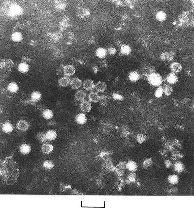
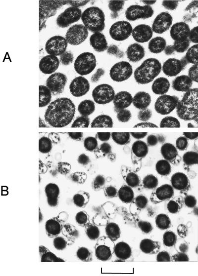
To find direct evidence for the presence of this bacteriophage, we grew the ovine C. psittaci strain MA in BGMK cells and observed the culture supernatant directly under EM after negative staining. Typical clusters of small featureless bacteriophages 25 nm in diameter were observed, some of which had characteristic stain-penetrated centers (Fig. 1). The bacteriophage (Chp2) occurred in groups and in crude cell extracts, attached to the surface of EBs (unpublished data). To investigate the morphology of the Chp2 within RBs, thin-section EM was performed on C. psittaci MA-infected BGMK cells in monolayers, which were fixed at 48 h postinfection when chlamydial inclusions were clearly visible by light microscopy. Within Chp2-infected inclusions, all RBs appeared to be darker than the uninfected counterparts (Fig. 2). Furthermore, the Chp2 particles were easily visible in association with membrane structures found in close proximity to RBs. The bacteriophages were evenly distributed within RBs and did not occur as paracrystalline arrays.
FIG. 1.
Electron micrograph showing Chp2 particles in the cell culture supernatant of C. psittaci-infected cells. The scale bar represents 50 nm.
FIG. 2.
Thin-section transmission electron micrographs of chlamydial inclusions within BGMK cells at 48 h after infection with ovine C. psittaci (without chlamydiaphage) (A) and ovine C. psittaci carrying Chp2 (B). The scale bar represents 1 μm.
Chp2 DNA.
EM evidence confirmed the presence of a bacteriophage (Chp2) infecting the ovine C. psittaci strain MA. Chp2 appeared similar to a previously described bacteriophage, Chp1, isolated from avian (duck) C. psittaci strain N352. Chp1 is a member of the Microviridae and has a genome comprising single-stranded circular DNA. Chp1 replicates through a double-stranded DNA (dsDNA) intermediate RF that is also found in EBs (26). Thus, to investigate Chp2 DNA, total DNA was extracted from purified Chp2-infected ovine C. psittaci strain MA EBs. Agarose gel electrophoresis of this DNA preparation revealed the presence of two extra DNA bands not seen in other ovine C. psittaci DNA preparations (unpublished data). The higher-molecular-weight band was extracted directly from EBs by a proprietary column binding method for purifying plasmid dsDNA. This material was digested with restriction endonucleases and had an approximate size of 4.5 kb when linearized, suggesting that it represented the RF DNA of Chp2. Thus, the dsDNA RF of Chp2 was used for subsequent cloning and sequencing studies.
The complete genome sequence of Chp2 was assembled using data obtained from sequence analysis of Chp2 RF DNA clones and overlapping PCR amplicons. Sequence analyses were performed for both strands. The double-stranded RF DNA of Chp2 is 4,567 bp with a nucleotide composition of A (28.6%), G (22.4%), T (30.4%), and C (18.6%).
Computer analysis predicted that the genome of Chp2 (Fig. 3) is organized into eight ORFs. ORFs 1 to 3, by analogy to Chp1, encode the viral structural proteins VP1, VP2, and VP3. ORFs 4 to 8 have been assigned following the ORF numbering system for the Chp1 genome (25). One or two more proteins may be encoded, depending on the usage of the second start codons within ORF 2; however, only a single protein band has been observed for this protein following SDS-PAGE (unpublished data). In Chp2, five ORFs (1 to 4 and 8) are in the same reading frame. The locations of the ORFs are shown in Fig. 3. No ORFs equivalent to Chp1 ORFs 4a, 5a, and 6 were identified. However, a new ORF (designated ORF 6), partially overlapping the start of ORF 2 and coding for a short peptide of 52 amino acids, was found in the Chp2 genome. The putative initiation codons of Chp1 ORFs 2a and 2b are conserved in Chp2. It was possible to identify proposed ribosome binding sites upstream of each ORF except ORF 2a. There are six untranslated regions (UTRs) in the Chp2 genome. The smallest UTR (between ORFs 2 and 3) is only 3 nucleotides, and the largest UTR (between ORFs 3 and 8) is 201 nucleotides. The positions of all UTRs are also shown in Fig. 3.
FIG. 3.
Comparison and diagrammatic representation of computer-predicted reading frames and genome organization of chlamydiaphages Chp1 and Chp2. Open boxes show ORFs likely to encode biologically functional proteins. Shaded boxes (ORFs 6 and 7) are small ORFs which overlap in a separate reading frame the major ORFs 1 and 2. Nomenclature of the ORFs is based on the original description of Chp1.
Database searches.
Analyses of DNA and protein sequences were performed using the FASTA program to search GenBank and SWISSPROT databases. The predicted proteins of Chp2 share homologies with the proteins of Chp1 and the honeybee pathogen Spiroplasma melliferum phage 4 (SpV4). Pairwise alignment of cognate protein sequences using the program of Pearson and Lipman (19) shown in Table 2 indicated that VP1 of Chp2 shares sequence identity with VP1 of Chp1 (48.3%) and VG1 of SpV4 (33.4%). VP2 of Chp2 shares conserved features with VP2 of Chp1 (27.3% identity) and VG4 of SpV4 (23.1% identity). The protein encoded by ORF 4 of Chp2 also has sequence identity with proteins from both Chp1 (ORF 4 protein, 19.9%) and SpV4 (VG2, 23.1%). VP3 and proteins encoded by ORFs 5, 7, and 8 of Chp2 have significant sequence identities only with their counterparts in Chp1 (Table 2).
TABLE 2.
Comparison of amino acid sequences for Chp1 and Chp2 ORFs
| ORF | No. of amino acids
|
% Identity | |
|---|---|---|---|
| Chp1 | Chp2 | ||
| 1 (VP1) | 596 | 565 | 48.3 |
| 2 (VP2) | 263 | 186 | 27.3 |
| 3 (VP3) | 145 | 148 | 26.0 |
| 4 | 399 | 336 | 19.9 |
| 5 | 96 | 84 | 27.1 |
| 6 | 82 | 52 | 11.0 |
| 7 | 31 | 32 | 43.8 |
| 8 | 36 | 44 | 54.5 |
Hybridization studies.
Chp1 and Chp2 do not cross hybridize under the standard conditions used in this study. Probing DNA from ovine strains of C. psittaci isolated in the United Kingdom revealed no evidence for the presence of related bacteriophage infection.
DISCUSSION
In this report, we present the first description of a bacteriophage (Chp2) infecting an ovine strain of C. psittaci. Chp2, although similar in morphology to Chp1, appears to have a different cytopathic effect on RBs compared with Chp1. Transmission EM shows little difference in both the size and shape of Chp2-infected RBs compared with uninfected RBs at the same time point in the developmental cycle. This is in contrast to Chp1-infected RBs, which become enlarged and in which the chlamydiaphage accumulates as paracrystalline arrays. Chp2 is found in association with membranous material, indicative of a lytic infection.
Computer analysis of the circular Chp2 genome (4,567 nucleotides) showed the presence of eight ORFs which potentially code for 8 to 10 proteins, depending on the usage of second start codons within ORF 2. The genome organization of Chp2 except for the location of ORF 6 is very similar to that of Chp1. All predicted proteins of Chp2 share amino acid sequence identities with their counterparts of Chp1 except for ORF 6 (Table 2). ORF 6 is found in a different relative location within the genome (Fig. 3). The viral coat proteins of Chp2 and Chp1 show the highest identity and are clearly related to the coat protein of SpV4. Homologies with the coat proteins of the Microviridae coliphages, φX174, G4, and α3 (11, 16, 21) are considerably lower (20%). The SpV4 and φX174 coat proteins display a major structural difference: in SpV4, a 71-amino-acid insertion loop forms “mushroom-like” protrusions at the threefold axis of symmetry (5). An equivalent insertion loop is present in both Chp1 (25) and Chp2.
The presence of this elaborate threefold related structural motif coincides with the disappearance of the prominent fivefold related spikes formed by the G proteins of the coliphages. These prominent spikes are not readily apparent in electron micrographs of Chp1 (20, 25) and Chp2 (Fig. 1) at a theoretical instrument resolution of 0.3 nm or in the cryo-EM reconstruction of SpV4 (5). However SpV4 contains one structural protein, whereas the chlamydiaphage contain at least three. Although VP2 and VP3 of Chp1 and Chp2 share no similarities with the coliphage structural proteins, it may still be possible to attribute functions to these proteins. An autoradiograph of Chp1 structural proteins separated by PAGE (26) was digitized and analyzed using NIH Image. Assuming 60 copies of VP1 per virion, there are approximately 15 copies of VP2, which is consistent with VP2 being analogous to the coliphage DNA pilot protein (protein H). In common with the H proteins, VP2 is glycine rich; in addition, a search of the protein data banks indicated some similarity with the P22 DNA pilot protein, gp16. However, it is not clear whether this protein will be located at the threefold axis or at the fivefold axes of symmetry, as seen in the E. coli phage. With the disappearance of the fivefold spikes in the chlamydiaphage and SpV4, the process of DNA injection may now be a function of the mushroom-like protrusion found at the threefold axes.
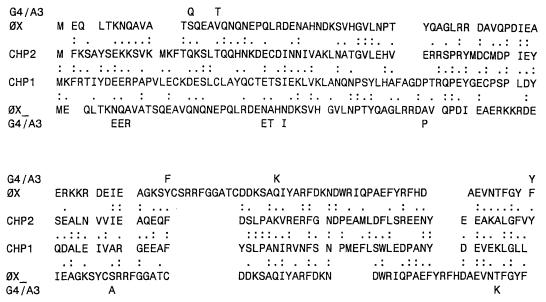
Analysis of the identity and function of VP3 may give insight into the evolution of viral scaffolding proteins. The 1:1 stoichiometry with VP1 is consistent with the stoichiometries observed for the coliphage major spike protein (protein G) or internal scaffolding protein (protein B). However, both chlamydiaphages and SpV4 appear to be spikeless. While the VP3s do not share amino acid sequence identity with the coliphage G proteins, there are intriguing homologies with the B proteins. There is approximately 20% homology in the first 120 amino acids, similar to that observed with the coliphage coat proteins (Fig. 4). Although homology is low, amino acids that mediate internal scaffolding-coat protein interactions in the atomic structure φX174 procapsid are conserved (Table 3). These data are consistent with a hypothesis that VP3 has evolved to be a structural protein in the chlamydiaphages. Similar amino acid sequence homologies also exist between the coliphage B proteins and VP3 of SpV4 although in SpV4, VP3 would remain a scaffolding protein.
FIG. 4.
Alignment of VP3 with coliphage internal scaffolding proteins. The amino acid sequence of the φX174 internal scaffolding protein was aligned to the first 120 amino acids of VP3 from Chp1 and Chp2. Amino acid variations found in the internal scaffolding proteins of bacteriophages G4 and α3 which show identity to the Chp1 and Chp2 proteins are indicated in the outer lines.
TABLE 3.
Conservation between proposed Chp2 VP3-coat protein interactions and internal scaffolding-coat protein interactions in the atomic structure of the φX174 procapsida
| φX174 procapsid interactions | Chp2 proposed interactions
|
|||
|---|---|---|---|---|
| VP3 | Coat | Nature of internal scaffolding coat interaction | ||
| D98 | R274 | D92 | R395 | Charge-charge |
| E105 | R274 | D98 | R395 | Charge-charge |
| D111 | R239 | E103 or E104 | R339 | Charge-charge |
| E113 | K166 | —b | —b | |
| E113 | R239 | E107 or E108 | R339 | Charge-charge |
| F120 | F67 | F114 or Y116 | F85 or F86 | Aromatic |
| F120 | Y134 | F114 or Y116 | Y160 | Aromatic |
| F120 | F135 | F114 or Y116 | Y160 | Aromatic |
The chlamydiaphage ORF 8 encodes small basic proteins quite similar to the coliphage DNA binding protein (protein J), which is a structural component of the virion. The ORF 8 gene products have not yet been detected within the chlamydiaphage virions. However, the J proteins are relatively small and present in only 60 copies per virion, making detection difficult. The chlamydiaphage and coliphage proteins differ in the addition of hydrophobic COOH termini in the coliphage proteins. The hydrophobic COOH termini of the coliphage B and J interact with the same binding cleft within the φX174 coat protein. Through competition, the J protein might directly mediate the extrusion of the B protein from the procapsid during DNA packaging (6–8). If VP3 has evolved into a structural protein, it follows that the ORF 8 gene products would not have hydrophobic COOH termini.
There are no proteins in Chp1, Chp2, or SpV4 that share any homologies to the coliphage external scaffolding proteins (protein D), which is the most conserved protein of the coliphages in the Microviridae. Genetic and structural analyses suggest that the primary functions of the D proteins are the mediation of twofold interactions: the placement of spike protein pentamers on the coat protein, and the organization of the coat protein at the threefold axes of symmetry (6–8). None of these functions may be required in the chlamydiaphage. First, these phage appear to be spikeless: the internal scaffolding protein also mediates second, twofold interactions, which may be analogous to the case for VP3. Second, the extensive threefold insertions, found only in the chlamydiaphage coat proteins, may circumvent the need of an external scaffolding protein to organize this region.
The functional roles assigned to many of the chlamydiaphage proteins can at this stage be only speculative. However, the discovery of a second chlamydiaphage (Chp2) showing significant sequence divergence in a host bacterial species that has an obligatory intracellular developmental cycle is both unexpected and surprising. Despite the nucleotide sequence diversity, major features of the chlamydiaphage genome remain conserved, and this information will help in developing the phage as a vector for chlamydiae.
ACKNOWLEDGMENTS
This work was supported by grant 047038 from the Wellcome Trust. Work by B. Fane is supported by the National Science Foundation.
We are grateful to Gareth Jones for supplying C. psittaci S95/3 and to Chris Storey for providing unpublished data on Chp1.
REFERENCES
- 1.Aoyama A, Hayashi M. Effects of genome size on bacteriophage phiX 174 DNA packaging in vitro. J Biol Chem. 1985;260:11033–11038. [PubMed] [Google Scholar]
- 2.Bacon E J, Richmond S J, Wood D J, Stirling P, Bevan B J, Chalmers W S K. Serological detection of phage infection in Chlamydia psittaci recovered from ducks. Vet Rec. 1986;119:618–620. [PubMed] [Google Scholar]
- 3.Bevan B J, Labram J. Laboratory transfer of a virus between isolates of Chlamydia psittaci. Vet Rec. 1983;112:280. doi: 10.1136/vr.112.12.280. [DOI] [PubMed] [Google Scholar]
- 4.Bini L, Sanchez-Campilllo M, Santucci A, Magi B, Marzocchi B, Comanducci M, Christiansen G, Birkelund S, Cevenini R, Vretou E, Ratti G, Pallini V. Mapping of Chlamydia trachomatis proteins by Immobiline-polyacrylamide two-dimensional electrophoresis: spot identification by N-terminal sequencing and immunoblotting. Electrophoresis. 1996;17:185–190. doi: 10.1002/elps.1150170130. [DOI] [PubMed] [Google Scholar]
- 5.Chipman P R, Agbandje-McKenna M, Renaudin J, Baker T S, McKenna R. Structural analysis of the spiroplasma virus, SpV4: implications for evolutionary variation to obtain host diversity among the Microviridae. Structure. 1998;6:135–145. doi: 10.1016/s0969-2126(98)00016-1. [DOI] [PMC free article] [PubMed] [Google Scholar]
- 6.Dokland T, Bernal R A, Burch A, Pletnev S, Fane B A, Rossmann M G. The role of scaffolding proteins in the assembly of the small single-stranded DNA virus φX174. J Mol Biol. 1999;288:595–608. doi: 10.1006/jmbi.1999.2699. [DOI] [PubMed] [Google Scholar]
- 7.Dokland T, McKenna R, IIag L L, Bowen B R, Incardona N L, Fane B A, Rossmann M G. Structure of a viral assembly intermediate with molecular scaffolding. Nature. 1997;389:308–313. doi: 10.1038/38537. [DOI] [PubMed] [Google Scholar]
- 8.Fane B A, Shien S, Hayashi M. Second-site suppressors of a cold sensitive external scaffolding protein of bacteriophage φX174. Genetics. 1993;134:1003–1011. doi: 10.1093/genetics/134.4.1003. [DOI] [PMC free article] [PubMed] [Google Scholar]
- 9.Francis T J, Magill T P. An unidentified virus producing acute meningitis and pneumonitis in experimental animals. J Exp Med. 1938;68:147–163. doi: 10.1084/jem.68.2.147. [DOI] [PMC free article] [PubMed] [Google Scholar]
- 10.Giannikopolou P, Bini L, Simitsek P D, Pallini V, Vretou E. Two-dimensional electrophoretic analysis of the protein family of abortifacient Chlamydia psittaci. Electrophoresis. 1997;18:2104–2108. doi: 10.1002/elps.1150181137. [DOI] [PubMed] [Google Scholar]
- 11.Godson G N, Barrell B G, Staden R, Fiddes J C. Nucleotide sequence of bacteriophage G4 DNA. Nature. 1978;276:236–247. doi: 10.1038/276236a0. [DOI] [PubMed] [Google Scholar]
- 12.Harshbanger J C, Chang S C, Otto S V. Chlamydiae (with phages), mycoplasmas, and rickettsiae in Chesapeake Bay Bivalves. Science. 1977;196:666–668. doi: 10.1126/science.193184. [DOI] [PubMed] [Google Scholar]
- 13.Hsia R-C, Ohayon H, Gounton P, Dautry A, Bavoil P M. Phage infection of Chlamydia psittaci GPIC. In: Stary A, editor. Proceedings of the Third European Society for Chlamydia Research. Bologna, Italy: Societa Editrice Esculapio; 1996. p. 48. [Google Scholar]
- 14.Hsia R, Ohayon H, Gounon P, Dautry-Varsat A, Bavoil P M. Altered development and lytic activities induced by phage infection of Chlamydia psittaci. In: Stephens R S, Byrne G I, Christiansen G, Clarke I N, Grayston J T, Rank R G, Ridgway G L, Saikku P, Schachter J, Stamm W E, editors. Chlamydial infections. 1998. pp. 131–134. Proceedings of the Ninth International Symposium on Human Chlamydial Infection. [Google Scholar]
- 15.Kalman S, Mitchell W, Marathe R, Lammel C, Fan J, Hyman R W, Olinger L, Grimwood J, Davis R W, Stephens R S. Comparative genomes of Chlamydia pneumoniae and C. trachomatis. Nat Genet. 1999;21:385–389. doi: 10.1038/7716. [DOI] [PubMed] [Google Scholar]
- 16.Kodaira K, Nakano K, Okada S, Taketo A. Nucleotide sequence of the genome of bacteriophage a3: interrelationship of the genome structure and the gene products with those of the phages φX174, G4 and φK. Biochim Biophys Acta. 1992;1130:277–288. doi: 10.1016/0167-4781(92)90440-b. [DOI] [PubMed] [Google Scholar]
- 17.McClenaghan M, Herring A J, Aitken I D. Comparison of Chlamydia psittaci isolates by DNA restriction endonuclease analysis. Infect Immun. 1984;45:384–389. doi: 10.1128/iai.45.2.384-389.1984. [DOI] [PMC free article] [PubMed] [Google Scholar]
- 18.O'Connell C M C, Maurelli A T. Introduction of foreign DNA into Chlamydia and stable expression of chloramphenical resistance. In: Stephens R S, Byrne G I, Christiansen G, Clarke I N, Grayston J T, Rank R G, Ridgway G L, Saikku P, Schachter J, Stamm W E, editors. Chlamydial infections. 1998. pp. 519–522. Proceedings of the Ninth International Symposium on Human Chlamydia Infection. [Google Scholar]
- 19.Pearson W R, Lipman D J. Improved tools for biological sequence comparison. Proc Natl Acad Sci USA. 1988;85:2444–2448. doi: 10.1073/pnas.85.8.2444. [DOI] [PMC free article] [PubMed] [Google Scholar]
- 20.Richmond S, Stirling P, Ashley C. Chlamydia have phage too. In: Mårdh P-A, Holmes K K, Oriel J D, Piot P, Schachter J, editors. Fifth International Symposium on Human Chlamydial Infection. Amsterdam, The Netherlands: Elsevier Biomedical Press; 1982. pp. 41–44. [Google Scholar]
- 21.Sanger F, Coulson A R, Friedmann C T, Air G M, Barrell B G, Brown N L, Fiddes J C, Hutchison C A, Slocombe P M, Smith M. The nucleotide sequence of bacteriophage φX174. J Mol Biol. 1978;125:225–246. doi: 10.1016/0022-2836(78)90346-7. [DOI] [PubMed] [Google Scholar]
- 22.Schachter J, Meyer K F. Lymphogranuloma venereum. II. Characterisation of some recently isolated strains. J Bacteriol. 1969;99:636–638. doi: 10.1128/jb.99.3.636-638.1969. [DOI] [PMC free article] [PubMed] [Google Scholar]
- 23.Stamp J T, McEwen A D, Watt J A A, Nisbet D I. Enzootic abortion in ewes. I. Transmission of the disease. Vet Rec. 1950;62:251–256. doi: 10.1136/vr.62.17.251. [DOI] [PubMed] [Google Scholar]
- 24.Stephens R S, Kalman S, Lammel C, Fan J, Marathe R, Aravind L, Mitchell W, Olinger L, Tatusov R L, Zhao Q, Koonin E V, Davis R W. Genome sequence of an obligate intracellular pathogen of humans: Chlamydia trachomatis. Science. 1998;282:754–759. doi: 10.1126/science.282.5389.754. [DOI] [PubMed] [Google Scholar]
- 25.Storey C C, Lusher M, Richmond S J. Analysis of the complete nucleotide sequence of Chp1, a phage which infects avian Chlamydia psittaci. J Gen Virol. 1989;70:3381–3390. doi: 10.1099/0022-1317-70-12-3381. [DOI] [PubMed] [Google Scholar]
- 26.Storey C C, Lusher M, Richmond S J, Bacon J. Further characterization of a bacteriophage recovered from an avian strain of Chlamydia psittaci. J Gen Virol. 1989;70:1321–1327. doi: 10.1099/0022-1317-70-6-1321. [DOI] [PubMed] [Google Scholar]
- 27.Tam J E, Davis C H, Wyrick P B. Expression of recombinant DNA introduced into Chlamydia trachomatis by electroporation. Can J Microbiol. 1994;40:583–591. doi: 10.1139/m94-093. [DOI] [PubMed] [Google Scholar]
- 28.Vretou E, Loutrari H, Mariani L, Costelidou K, Eliades P, Conidou G, Karamanou S, Mangana O, Siarkou V, Papadopoulus O. Diversity of abortion strains of Chlamydia psittaci demonstrated by inclusion morphology, polypeptide profiles and monoclonal antibodies. Vet Microbiol. 1996;51:275–289. doi: 10.1016/0378-1135(96)00048-x. [DOI] [PubMed] [Google Scholar]


