Abstract
Persistent inflammatory damage and suppressed immune function play a crucial role in the pathogenesis and progression of the pneumocystis jirovecii pneumonia (PjP). Therefore, we aimed to investigate the correlation between the combined immune and inflammatory indicator: the neutrophil-to-lymphocyte ratio (NLR) and prognosis of non-human immunodeficiency virus (non-HIV) PjP.
In the retrospective analysis conducted in ICUs at Beijing Chao-Yang Hospital, we examined data from 157 patients diagnosed with non-HIV PjP. Our findings reveal a concerning hospital mortality rate of 43.3%, with the 28-day mortality rate reaching 47.8%.
Through multivariable logistic and Cox regression analyses, we established a significant association between elevated NLR levels and hospital mortality (adjusted odd ratio, 1.025; 95% CI, 1.008-1.043; p = 0.004) or 28-day mortality (adjusted hazard ratio, 1.026; 95% CI, 1.008-1.045; p = 0.005). Specifically, patients with an NLR exceeding 20.3 demonstrated markedly lower overall survival rates, underscoring the biomarker's predictive value for both hospital and 28-day mortality.
In conclusion, non-HIV PjP patients in the ICU still have a high rate of mortality and a poor short-term prognosis after discharge. A high level of NLR was associated with an increased risk of hospital mortality and 28-day mortality.
Supplementary Information
The online version contains supplementary material available at 10.1186/s12890-024-03093-8.
Keywords: Pneumocystis jirovecii pneumonia, Neutrophil-to-lymphocyte ratio, Mortality
Introduction
Pneumocystis jirovecii pneumonia (PjP) constitutes an opportunistic pulmonary fungal infection primarily affecting immunocompromised individuals. Since 1979, PjP has accounted for more than a quarter of opportunistic infections in the human immunodeficiency virus (HIV) population [1] and was once considered the most common opportunistic infection and the leading cause of death among those patients [2]. The widespread use of highly active antiretroviral therapy (HAART) and trimethoprim-sulfamethoxazole (TMP-SMX) has gradually reduced the incidence and mortality rates of PjP in the HIV-infected population [3]. With the increased use of immunosuppressants or cytotoxic agents in recent years, there has been a notable rise in PjP incidence among the non-HIV immunocompromised population not receiving prophylaxis [4]. Unlike their HIV-positive counterparts, individuals with non-HIV related immunosuppression often experience an acute onset of PjP, characterized by high fever and progressive respiratory distress that typically lasts for 4 to 7 days. This acute presentation can quickly lead to respiratory failure, necessitating intensive care unit (ICU) admission for over half of these patients [5] and resulting in mortality rates between 50 and 60% [5, 6]. This divergence in clinical presentation and outcomes underscores the need for studies that specifically address the characteristics and prognostic factors in non-HIV PjP patients.
Building on this context, it's recognized that the interplay between diminished immune function and an overactive inflammatory response is a key driver of pulmonary lesions in PjP [7]. Specifically, in those with compromised immune systems, a lack of lymphocytes impairs the clearance of Pneumocystis jirovecii (Pj), triggering an influx of neutrophils and a heightened inflammatory response. This exacerbation significantly impairs lung function, leading to hypoxemia and, in severe cases, respiratory failure [8, 9].
Identifying suitable biomarkers that comprehensively reflect the inflammatory and immune balance in PjP hosts is crucial for predicting patient outcomes. Previous studies have suggested that immune indices [9], and inflammatory factors [6, 10] are closely related to adverse hospital outcomes in non-HIV immunosuppressed patients with PjP. However, focusing solely on either immune or inflammatory markers appears insufficient to reveal the extent of host immune imbalance. The neutrophil-to-lymphocyte ratio (NLR) emerges as a pivotal biomarker, shedding light on the delicate balance between systemic inflammation and immune response [11]. While NLR has been extensively utilized in the prognosis of various diseases, including cancer [12], infectious diseases [13], and cardiovascular conditions [14], its prognostic value in non-HIV PjP patients remains unexplored.
Addressing this gap, our research endeavors to elucidate the clinical profile of PjP in the non-HIV immunocompromised cohort and to investigate the prognostic relevance of NLR within this group. Our objective is to furnish novel insights into the clinical prognostics and therapeutic management of PjP, potentially enhancing patient care and outcomes for this at-risk population.
Methods
Study design and patients
Between October 1, 2011, and July 31, 2023, our investigation involved 207 patients diagnosed with PjP following their discharge from various ICUs at Beijing Chao-Yang Hospital, encompassing the respiratory ICU, emergency ICU, and surgical ICU. This study was conducted in accordance with the principles of the Declaration of Helsinki and the experimental protocol for data involving human followed the Ethical Guidelines of the Ethics Committee of Beijing Chao-Yang Hospital, under the protocol number 2021-ke-192, and the need to obtain written informed consent was waived due to the retrospective nature.
Inclusion criteria
Participants were eligible for this study if they: were 18 years of age or older; had a history of immunosuppression due to conditions such as malignancies (including hematological malignancies and solid tumors), bone marrow or solid organ transplantation, autoimmune diseases, or congenital immunodeficiency diseases; met the diagnostic criteria of acute respiratory distress syndrome [15].
Referencing prior research and a diagnostic guideline for non-HIV PjP [16], we defined the diagnosis of PjP using all of the following criteria:(a)Clinical symptoms like fever, cough, and dyspnea; (b)Chest imaging revealing bilateral diffuse ground glass opacities; (c)Positive identification of Pneumocystis cysts or trophozoites in respiratory specimens [qualified sputum samples, induced sputum, or bronchoalveolar lavage fluid (BALF)] via Grocott’s methenamine silver stain, or respiratory specimens testing positive for nucleic acid by metagenomics next-generation sequencing or polymerase chain reaction; (d)Elevated serum 1,3-β-glucan levels, with other fungal infections excluded [17].
A definitive diagnosis of PjP was established when a patient met the above clinical and diagnostic criteria (a and b, plus one of the conditions in c and d).
Exclusion criteria
Patients were excluded if they: had positive serum HIV tests; were pregnant. For those with multiple admissions, the study considered only the first admission record to avoid duplication of data.
Data collection
We systematically collected clinical data from the electronic medical record system at Beijing Chao-Yang Hospital. This comprehensive dataset included: demographic information (age, gender, and other relevant patient demographics), clinical characteristics (key features of each patient's clinical presentation and history), laboratory results (critical laboratory findings relevant to the prognosis and co-infections). The NLR was calculated based on the results of the first complete blood count performed within 24 h of patient admission. Treatment outcomes: this encompassed the length of ICU stay, the use and duration of invasive mechanical ventilation (IMV), and the use of veno-venous extracorporeal membrane oxygenation (VV-ECMO). Mortality Data: both hospital mortality and 28-day mortality rates were meticulously recorded to assess patient outcomes.
Management
When non-invasive ventilation fails due to severe hypoxemia or excessive secretions, our protocol shifts to IMV using low tidal volumes and carefully adjusted positive end-expiratory pressure to optimize gas exchange and minimize lung injury [18]. If the PaO2/FiO2 ratio (PFR) is below 50 mmHg for over 30 min or if barotrauma occurs during IMV, VV-ECMO is used. All diagnosed cases in our study were managed according to the first-line treatment which involved administering trimethoprim at 15–20 mg/kg/day and sulfamethoxazole at 75–100 mg/kg/day for 21 days. For patients who cannot tolerate TMP-SMX, the combination of clindamycin and primaquine or caspofungin is the preferred alternative treatment [19].
Statistical analysis
We performed all statistical analyses using the software R, version 4.0.2, and GraphPad Prism, version 9.0. We considered results to be statistically significant if the chance of the result occurring by random was less than 5% (p-value < 0.05).
For data that vary continuously, we described these using either the average value and the range around this average (mean and standard deviation, mean ± SD) for data that follow a normal distribution, or the middle value and the range between the lower and upper middle values [median and interquartile range, median(IQR)] for data that do not. For categories of data, we showed these as counts and percentages.
To compare groups for continuous data, we used either the One-Way ANOVA test (for data that's normally distributed) or the Mann-Whitney test (for data that's not). For categorical data, we used either the chi-squared test or Fisher’s exact test, depending on what was most suitable. We then identified factors that significantly affected the outcomes (p-value < 0.05 in univariate analysis) and important risk factors to include in a more detailed analysis using multivariate logistic and Cox regression models. We used receiver operating characteristic (ROC) curves to assess how well our predictive models could distinguish between different outcomes. To estimate survival probabilities over time for different groups, we used the Kaplan–Meier method and compared these groups using the log-rank test. We also looked into whether certain factors like age, gender, the PFR, and the use of IMV influenced the outcomes differently, performing subgroup analysis.
Results
Study population
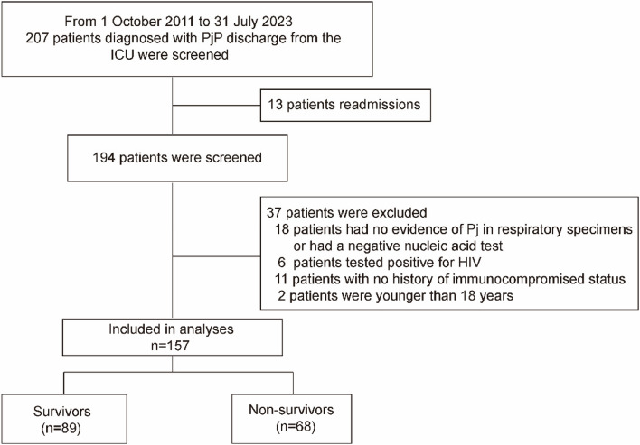
Between October 2011 and July 2023, 207 patients diagnosed with PjP and discharged from the ICUs of Beijing Chao-Yang Hospital, were screened. Among them, 13 patients had multiple admissions, 6 patients were HIV-positive, 18 patients either had no evidence of Pj in respiratory specimens or had a negative nucleic acid test, 11 patients had no history of immunocompromised status. In addition, 2 patients were under 18 years of age, which led to their exclusion. Finally, 157 non-HIV PjP patients who met the inclusion criteria were analyzed (Fig. 1).
Fig. 1.
Study flow
Baseline characteristics
Table 1 shows the patients' baseline clinical characteristics. The mean (SD) age was 54.6 ± 15.0 years, 98/157 (62.4%) were male and median (IQR) body mass index (BMI) was 23.1(20.7-25.6) kg/m2. Solid organ transplantation was the initial cause of PjP, for 49 cases (31.2%), versus 41 (26.1%) with connective diseases and 30 (19.1%) with hematological diseases. Prior to the onset of PjP, 118 (75.2%) and 93 (59.2%) of the patients had been treated with corticosteroids and immunosuppressants including tacrolimus, mycophenolate mofetil, sirolimus, cyclophosphamide, methotrexate, and leflunomide. PjP was frequently associated with other pulmonary infections, with cytomegalovirus infection being the most common (68.2%), followed by bacterial infection (40.8%).
Table 1.
Differences in baseline characteristics between survivors and non-survivors
| Baseline characteristics | Whole cohort (n = 157) |
Survivor (n = 89) |
Non-survivors (n = 68) |
P |
|---|---|---|---|---|
| Age, mean ± SD, year | 54.6 ± 15.0 | 52.3 ± 15.4 | 57.6 ± 14.1 | 0.03 |
| Male, n (%) | 98(62.4) | 55(61.8) | 43(63.2) | 0.80 |
| BMI, median (IQR), kg/m2 | 23.1(20.7-25.6) | 22.7(20.6-25.0) | 23.9(21.2-26.2) | 0.20 |
| Time form onset symptoms to ICU, median (IQR), day | 10(6-20) | 10(3-22) | 12(7-20) | 0.83 |
| Severity evaluation, median (IQR) | ||||
| APACHEII Score | 12(8-15) | 11(8-14) | 13(9-15) | 0.06 |
| SOFA score | 4(3-7) | 4(3-6) | 5(3-8) | 0.05 |
| Comorbidities, n (%) | ||||
| Hypertension | 70(44.6) | 45(50.6) | 25(36.8) | 0.08 |
| Diabetes | 31(19.7) | 19(21.3) | 12(17.6) | 0.56 |
| Coronary artery disease | 13(8.3) | 5(5.6) | 8(11.8) | 0.17 |
| Chronic pulmonary disease | 5(3.2) | 1(1.1) | 4(5.9) | 0.09 |
| Immunocompromised status, n (%) | ||||
| Solid organ transplantation | ||||
| Kidney transplantation | 36(22.9) | 24(27.0) | 12(17.6) | 0.17 |
| Liver transplantation | 13(8.3) | 10(11.2) | 3(4.4) | 0.12 |
| Cancer | ||||
| Lung cancer | 1(0.6) | 0 | 1(1.5) | 0.25 |
| Breast cancer | 3(1.9) | 3(3.4) | 0 | 0.13 |
| Esophageal cancer | 3(1.9) | 2(2.2) | 1(1.5) | 0.25 |
| Thymoma | 3(1.9) | 2(2.2) | 1(1.5) | 0.25 |
| Connective disease | ||||
| ANCA-associated vasculitis | 6(3.8) | 3(3.4) | 3(4.4) | 0.74 |
| SLE | 4(2.5) | 3(3.4) | 1(1.5) | 0.45 |
| Pemphigus | 7(4.5) | 4(4.5) | 3(4.4) | 0.98 |
| Rheumatoid arthritis | 9(5.7) | 3(3.4) | 6(8.8) | 0.15 |
| Dermatomyositis | 7(4.5) | 2(2.2) | 5(7.4) | 0.44 |
| Autoimmune hemolytic anemia | 4(2.5) | 1(1.1) | 3(4.4) | 0.20 |
| Others | 4(2.5) | 1(1.1) | 3(4.4) | 0.20 |
| Hematological disease | ||||
| Non-Hodgkin's lymphoma | 2(1.3) | 1(1.1) | 1(1.5) | 0.85 |
| Hodgkin's lymphoma | 4(2.5) | 1(1.1) | 3(4.4) | 0.20 |
| Multiple myeloma | 3(1.9) | 1(1.1) | 2(2.9) | 0.41 |
| Leukaemia | 3(1.9) | 3(3.4) | 0 | 0.13 |
| Hemophilia | 3(1.9) | 3(3.4) | 0 | 0.13 |
| Aplastic anemia | 4(2.5) | 2(2.2) | 2(2.9) | 0.79 |
| Idiopathic thrombocytopenic purpura | 11(7.0) | 7(7.9) | 4(5.9) | 0.16 |
| Idiopathic pulmonary fibrosis | 13(8.3) | 5(5.6) | 8(11.8) | 0.17 |
| Nephrotic syndrome | 18(11.5) | 13(14.6) | 5(7.4) | 0.16 |
| Immunosuppressive drugs before admission, n (%) | ||||
| Corticosteroids | 118(75.2) | 67(75.3) | 51(75.0) | 0.97 |
| Immunosuppressant | ||||
| One immunosuppressant | 45(28.6) | 26(29.2) | 19(27.9) | 0.86 |
| Two immunosuppressants | 42(26.8) | 25(28.1) | 17(25.0) | 0.67 |
| Three immunosuppressants | 6(3.8) | 4(4.5) | 2(2.9) | 0.62 |
| Vital signs, median (IQR) | ||||
| HR, rate/minute | 94(80-108) | 94(80-109) | 95(79-105) | 0.80 |
| RR, rate/minute | 25(20-29) | 24(20-28) | 26(22-30) | 0.05 |
| SBP, mmHg | 123(112-138) | 125(112-138) | 120(112-137) | 0.53 |
| DBP, mmHg | 75(63-82) | 75(65-83) | 73(61-81) | 0.21 |
| Laboratory results, median (IQR) | ||||
| WBC, median, 109/L | 8.4(5.5-10.8) | 8.0(5.4-10.8) | 8.5(5.7-10.8) | 0.50 |
| Neutrophil count, 109/L | 7.2(4.8-9.6) | 7.2(4.8-9.3) | 7.5(4.8-9.9) | 0.47 |
| Lymphocyte count, 109/L | 0.25(0.13-0.53) | 0.29(0.18-0.55) | 0.24(0.11-0.50) | 0.22 |
| NLR | 24.4(13.3-40.4) | 16.2(9.9-29.0) | 33.7(23.3-60.9) | < 0.001 |
| Monocyte, 109/L | 0.45(0.27-0.93) | 0.56(0.30-1.13) | 0.43(0.24-0.70) | 0.60 |
| Hemoglobin, g/L | 108(93-120) | 108(95-119) | 102(57-125) | 0.88 |
| Platelet, 109/L | 166(107-234) | 179(137-243) | 136(71-205) | < 0.001 |
| Albumin, g/L | 27(24-31) | 28(25-32) | 26(22-29) | < 0.001 |
| CRP, mg/L | 12.5(8.7-15.9) | 11.6(8.3-14.2) | 14.4(10.4-17.3) | < 0.001 |
| AST, U/L | 20(10-32) | 16(10-35) | 31(12-31) | 0.33 |
| ALT, U/L | 33(21-52) | 32(22-51) | 36(20-59) | 0.87 |
| BUN, mmol/L | 8.3(5.8-14.3) | 8.5(5.9-13.9) | 7.6(5.6-15.3) | 0.84 |
| Crea, umol/L | 78.3(54.4-147.8) | 83.2(54.4-145.5) | 77.2(54.1-157.0) | 0.85 |
| LDH, U/L | 494(330-731) | 406(283-551) | 580(430-879) | < 0.001 |
| TBIL, umol/L | 8.9(6.1-14.4) | 8.8(5.7-13.3) | 9.3(6.1-15.1) | 0.56 |
| IBIL, umol/L | 5.1(3.3-8.0) | 5.2(3.2-7.9) | 4.9(3.4-8.1) | 0.82 |
| Blood gas analysis | ||||
| PH | 7.43(7.40-7.47) | 7.43(7.40-7.46) | 7.45(7.40-7.47) | 0.34 |
| PaCO2, mmHg | 35.8(31.0-40.0) | 35.0(30.6-38.0) | 36.5(31.0-41.0) | 0.34 |
| PaO2, mmHg | 84(68-98) | 86(73-99) | 76(63-95) | 0.03 |
| PFR, mmHg | 150.0(101.2-242.7) | 190(120-268) | 124(92-174) | < 0.001 |
| Lymphocyte subsets | ||||
| CD3+ T cell, cell/ul | 272(143-448) | 306(162-489) | 208(116-395) | 0.02 |
| CD4+ T cell, cell/ul | 98(61-220) | 139(82-251) | 68(47-134) | < 0.001 |
| CD8+ T cell, cell/ul | 100(63-200) | 125(75-251) | 81(54-117) | < 0.001 |
| Co-infections | ||||
| Bacterial infection | 64(40.8) | 43(48.3) | 21(30.9) | 0.03 |
| Cytomegalovirus infection | 107(68.2) | 58(65.2) | 49(72.1) | 0.36 |
| Fungal infection | 58(36.9) | 29(32.6) | 29(42.6) | 0.20 |
| Outcomes | ||||
| Length of hospital | 17(10-26) | 12(8-24) | 15(10-28) | < 0.001 |
| IMV | 90(57.3) | 31(34.8) | 59(86.8) | < 0.001 |
| Duration of IMV | 10(5.8-22) | 8(5-23) | 12(6-22) | 0.62 |
| VV-ECMO | 18(11.5) | 8(9.0) | 10(14.7) | 0.27 |
Data are presented as median (interquartile range), mean (standard deviation) or n (%); Other connective diseases included retroperitoneal fibrosis, sicca syndrome, adult-onset Still's disease, ankylosing spondylitis
Abbreviations: SD Standard deviation, BMI Body mass index, IQR Interquartile range, ICU Intensive care unit, APACHEII Acute physiology and chronic health evaluation II, SOFA Sepsis-related organ failure assessment, ANCA Anti-neutrophil cytoplasmic antibodies, SLE Systemic Lupus Erythematosus, HR Heart rate, RR Respiratory rate, SBP Systolic blood pressure, DBP Diastolic blood pressure, WBC White blood cell count, NLR Neutrophil–lymphocyte ratio, CRP C-reactive protein, AST Aspartate aminotransferase, ALT Alanine aminotransferase, BUN Blood urea nitrogen, LDH Lactate dehydrogenase, TBIL Total bilirubin, IBIL Indirect bilirubin, PFR PaO2/FiO2 ratio, IMV Invasive mechanical ventilation, VV-ECMO veno-venous extracorporeal membrane oxygenation
Distribution of NLR in poor outcomes and subgroup patients
The NLR showed significant statistical differences between patients without IMV and those with IMV [18.1(9.8-28.0) vs 30.7(19.1-51.0), p < 0.001], between survivors and non-survivors [16.2(9.9-29.0) vs 33.7(23.3-60.9), p < 0.001], between 28-day mortality and non-28-day mortality [33.7(22.0-56.6) vs 16.2(10.0-28.9), p < 0.001], but no statistically significant difference between patients receiving VV-ECMO and those not receiving [34.3(15.0-64.8) vs 24.1(12.8-35.9), p = 0.153].The differences in NLR were not statistically significant in subgroups (p > 0.05) (shown in Supplementary Fig. 1, Supplementary Fig. 2 and Supplementary excel).
Influence of NLR on the risk of hospital mortality and 28-day mortality
In multivariate logistic regression analysis using significant variables, together with clinically relevant risk factors, the NLR level was associated with both hospital mortality [adjusted odd ratio (OR), 1.025; 95% confidence interval (CI), 1.008-1.043; p = 0.004] and 28-day mortality [adjusted hazard ratio (HR), 1.026; 95% CI, 1.008-1.045; p = 0.005] (Table 2).
Table 2.
Multivariable regression model results for hospital mortality and 28-day mortality
| Patient characteristic | Hospital mortality | 28-day mortality | ||
|---|---|---|---|---|
| Multivariable logistic regression | Multivariable Cox regression | |||
| OR (95% CI) | Pa | HR (95% CI) | Pa | |
| NLR | 1.025(1.008-1.043) | 0.004 | 1.026(1.008-1.045) | 0.005 |
| Age | 1.021(0.994-1.049) | 0.122 | 1.015(0.990-1.041) | 0.243 |
| BMI | 1.058(0.955-1.171) | 0.280 | 1.081(0.964-1.186) | 0.114 |
| APACHE II score | 1.006(0.926-1.093) | 0.884 | 0.996(0.922-1.075) | 0.914 |
| SOFA score | 1.160(0.997-1.349) | 0.054 | 1.130(0.978-1.305) | 0.097 |
| Platelet | 0.998(0.994-1.002) | 0.354 | 0.997(0.993-1.001) | 0.193 |
| Albumin | 0.919(0.844-1.001) | 0.052 | 0.972(0.898-1.051) | 0.476 |
| LDH | 1.001(0.999-1.002) | 0.217 | 1.000(0.999-1.001) | 0.911 |
| PFR | 0.998(0.994-1.002) | 0.154 | 0.998(0.995-1.001) | 0.188 |
| CD4+ T cell | 0.998(0.994-1.002) | 0.268 | 0.997(0.994-1.001) | 0.171 |
Abbreviations: OR Odd ratio, HR Hazard ratio, CI Confidence interval, NLR Neutrophil-lymphocyte ratio, BMI Body mass index, APACHEII Acute physiology and chronic health evaluation II, SOFA Sepsis-related organ failure assessment, LDH Lactate dehydrogenase, PFR PaO2/FiO2 ratio
aAdjusted by bacterial infection and cytomegalovirus infection
Predictive power of NLR and other biomarkers for in-hospital mortality
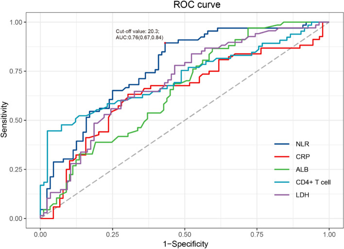
The NLR had a maximum area under the Area Under Curve (AUC) (0.76; 95%CI, 0.67-0.84; p < 0.001), compared with C-reactive proteins (CRP) (ACU,0.63; 95%CI, 0.55-0.73; p = 0.002), albumin (ALB) (ACU,0.65; 95%CI, 0.57-0.74; p = 0.001), CD4+ T cell (AUC, 0.72; 0.63-0.80; p < 0.001), lactate dehydrogenase (LDH) (ACU,0.70; 95%CI, 0.61-0.78; p < 0.001) (Fig. 2 and Supplementary Table 1).
Fig. 2.
Receiver operating characteristic (ROC) curves for hospital mortality
We established a cutoff value of 20.3 for NLR based on our analysis. Among patients with an NLR greater than or equal to 20.3, we found significantly higher proportions requiring invasive mechanical ventilation (75.4%), as well as higher in-hospital (88.5%) and 28-day mortality rates (77.6%). Kaplan-Meier analysis of survival in patients stratified for NLR < and ≥ 20.3 during 28 days (p log-rank test < 0.0001) (Supplementary Fig. 3).
Subgroup analysis
Significant interaction relationships were observed among patients received IMV in hospital mortality (OR 1.060 [95% CI 1.021-1.110]; P for interaction < 0.001) and 28-day mortality (HR 1.008 [95% CI 1.001-1.015]; P for interaction = 0.023). (Supplementary Table 2).
Differences in characteristics between the high NLR and low NLR groups
There was no statistical difference in baseline characteristics, underlying diseases, and vital signs between the two patient groups. Compared with the low NLR group, the high NLR group exhibited lower PaO2 [88.0(79.0-99.5) vs 78.0(63.5-96.0), p = 0.01]and PFR [205.0(119.3-278.9) vs 131.0(94.0-208.0), p = 0.01], and a higher proportion of IMV (37.9% vs 68.7%, p < 0.001).
Discussion
Our study's exploration of non-HIV PjP in ICU-admitted patients uncovers pivotal insights into the prognosis of this immunosuppressed cohort. The utilization of IMV in 57.3% of patients and VV-ECMO in 11.5%, alongside observed hospital and 28-day mortality rates of 43.3% and 47.8% respectively, underscores the severity of PjP in these populations. Our analysis reveals that an elevated NLR significantly correlates with increased hospital and 28-day mortality, highlighting NLR above 20.3 as a critical marker of deteriorated survival prospects.
Recently, there has been a marked increase in the incidence of PjP among immunosuppressed individuals, particularly those with malignant tumors, undergoing organ or bone marrow transplantation, or suffering from inflammatory diseases [20]. Compared to HIV-infected PjP patients, non-HIV PjP patients often have more rapid disease progression and a poorer prognosis. Roux et al. [21] found that the non-HIV PjP patients presented mainly with non-specific symptoms such as fever, cough, and dyspnea. Their disease progression was significantly more acute, with a median onset of 5 days compared to 21 days in the HIV-infected PjP patients (p < 0.05). They also had more severe hypoxemia, were more likely to require ICU admission (50% vs. 35%, p < 0.05), and were more likely to require both non-invasive ventilation (16% vs. 8%, p < 0.05) and IMV (30% vs. 11%, p < 0.05). These findings resonate with our previous research [22]. In the present study, 57.3% availed of IMV, and the observed hospital mortality rate was 43.3%. In summary, non-HIV PjP patients are at a heightened risk for unfavorable outcomes, such as mortality, and reliance on IMV or VV-ECMO. Recognizing the prognostic markers for this group is crucial for clinicians to make well-informed therapeutic decisions.
While immune markers [23] and inflammatory factors [6, 10] are associated with adverse outcomes in patients with PjP who are non-HIV immunosuppressed, the NLR offers superior prognostic accuracy for mortality. Previous studies have highlighted various biomarkers, such as LDH, CRP, and albumin, as predictors of patient outcomes in non-HIV immunosuppressed PjP patients. For instance, Schmidt et al. found that LDH levels at admission were associated with adverse outcomes during hospitalization in a mixed population of HIV-infected and non-HIV immunosuppressed PjP patients [6]. In addition, compared with HIV-infected individuals who have not undergone HAART, those who initiate HAART treatment exhibit a decrease in serum LDH levels, which may indicate a reduction in the extent of lung damage [24]. Similarly, high CRP levels have been correlated with adverse outcomes, underscoring its role in reflecting the inflammatory status [25]. It is currently believed that hypoproteinemia in non-HIV immunosuppressed PjP patients results from both acute inflammation caused by PjP and chronic inflammation due to underlying diseases. Akahane et al. found that survivors of non-HIV PjP had significantly higher albumin levels than non-survivors [26]. Despite these associations, NLR stands out by providing a more direct and potent indicator of systemic inflammation and immune response, making it a more reliable predictor of mortality in PjP patients.
In HIV patients, the HIV virus targets CD4+ T lymphocytes, leading to their progressive decline, which significantly increases the risk of PjP [27]. This reduction in CD4+ T lymphocytes is a central feature of HIV progression and a critical marker for immunosuppression in this population. In contrast, non-HIV immunosuppressed patients face a broader spectrum of immunosuppressive influences due to various medications, impacting numerous immune pathways and biological processes. Our previous study showed significant suppression of B lymphocyte immune-related gene expression with glucocorticoid-induced Pneumocystis pneumonia [28]. Rong et al. further demonstrated an increased burden of Pneumocystis carinii (Pc) and delayed clearance of Pc in the lungs of mice with a lack of B-cell function [29]. We propose that NLR might offer a more universally applicable prognostic marker across different immunocompromised states, including but not limited to HIV.
In our analysis, patients with high NLR exhibited poorer oxygenation and a greater need for IMV compared to those with low NLR, indicating that a high NLR may reflect severe lung injury. A detailed analysis [30] of the BALF from 166 non-HIV PjP patients showed that a neutrophil-dominant presence was rare in cases of mild to moderate non-HIV PjP. However, it was present in about one-third of the more severe cases. Interestingly, the proportion of neutrophils in BALF was significantly associated with 30-day (OR: 1.02, 95%CI: 1.01-1.03) and 60-day all-cause mortality (OR: 1.02, 95%CI: 1.01-1.04). At the same time, we also found that compared with patients with low NLR, patients with high NLR showed a downward trend in T cell subsets, but the difference was not statistically significant. This finding suggests that low T cell counts do not necessarily correlate with high NLR. Factors beyond simple lymphocyte depletion, such as acute phase reactions or other pathophysiological mechanisms, may influence NLR values in this specific patient cohort. Therefore, while T cell counts are critical indicators of immune status, NLR may provide a more comprehensive understanding of the complex interplay between immune response and systemic inflammatory in patients with PjP.
Although our retrospective analysis provides initial insights into the use of the NLR, comprehensive prospective studies are required to fully exploit its potential as a clinical tool. Future research should focus on monitoring changes in NLR and correlating these with patient progression, evaluating the generalizability and adaptability of NLR across various medical settings, and determining optimal thresholds to guide or escalate clinical interventions. These efforts are essential for integrating NLR into routine clinical practice effectively.
Our study has several limitations. Firstly, this study is a single-center study, and the immunosuppressed population is dominated by autoimmune diseases and solid organ transplantation, which may limit the generalization of the results; Secondly, NLR might be influenced by the diverse clinical backgrounds or medications of the patients in our cohort, and a larger cohort is needed to reduce heterogeneity; Thirdly, we evaluated the patients' primary clinical outcomes including ICU deaths, but did not examine the patients' long term survivorship.
Conclusion
In conclusion, non-HIV PjP patients in the ICU still have a high rate of mortality and a poor short-term prognosis after discharge. A high level of NLR was associated with an increased risk of hospital mortality and 28-day mortality.
Supplementary Information
Acknowledgements
Not applicable
Abbreviations
- PjP
Pneumocystis jirovecii pneumonia
- HIV
Human immunodeficiency virus
- HAART
Highly active antiretroviral therapy
- TMP-SMX
Trimethoprim-sulfamethoxazole
- ICU
Intensive care unit
- Pj
Pneumocystis jirovecii
- NLR
Neutrophil to lymphocyte ratio
- BALF
Bronchoalveolar lavage fluid
- IMV
Invasive mechanical ventilation
- VV-ECMO
Veno-venous extracorporeal membrane oxygenation
- PFR
PaO2/FiO2 ratio
- SD
Standard deviation
- IQR
Interquartile range
- ROC
Receiver operating characteristic
- BMI
Body mass index
- OR
Odd ratio
- CI
Confidence interval
- HR
Hazard ratio
- AUC
Area under curve
- CRP
C-reactive proteins
- ALB
Albumin
- LDH
Lactate dehydrogenase
- Pc
Pneumocystis carinii
Authors’ contributions
Wang Dong and Guan Lujia collected the data and wrote the first draft. Li Xuyan and Tong Zhaohui revised and finalized the final version. All authors reviewed the manuscript.
Funding
This work was supported by the National Natural Science Foundation of China (NO. 82100005).
Availability of data and materials
The datasets used and/or analyzed during the current study are available from the corresponding author on reasonable requests.
Declarations
Ethics approval and consent to participate
The study was approved by the Ethics Committee of Beijing Chao-Yang Hospital (No.2021-ke-192) and the need for informed consent was waived by the Ethics Committee of Beijing Chao-Yang Hospital.
Consent for publication
Not applicable.
Competing interests
The authors declare no competing interests.
Footnotes
Publisher’s Note
Springer Nature remains neutral with regard to jurisdictional claims in published maps and institutional affiliations.
Dong Wang and Lujia Guan contributed equally to this work.
Contributor Information
Xuyan Li, Email: 13581851048@163.com.
Zhaohui Tong, Email: tongzhaohuicy@sina.com.
References
- 1.Centers for Disease Control and Prevention (CDC). HIV and AIDS–United States, 1981–2000. MMWR Morb Mortal Wkly Rep. 2001;50(21):430–4. [PubMed]
- 2.Apostolopoulou A, Fishman JA. The Pathogenesis and Diagnosis of Pneumocystis jiroveci Pneumonia. J Fungi (Basel, Switzerland). 2022;8(11):1167. doi: 10.3390/jof8111167. [DOI] [PMC free article] [PubMed] [Google Scholar]
- 3.Buchacz K, Lau B, Jing Y, Bosch R, Abraham AG, Gill MJ, et al. Incidence of AIDS-Defining Opportunistic Infections in a Multicohort Analysis of HIV-infected Persons in the United States and Canada, 2000–2010. J Infect Dis. 2016;214(6):862–872. doi: 10.1093/infdis/jiw085. [DOI] [PMC free article] [PubMed] [Google Scholar]
- 4.Ghembaza A, Vautier M, Cacoub P, Pourcher V, Saadoun D. Risk factors and prevention of pneumocystis jirovecii pneumonia in patients with autoimmune and inflammatory diseases. Chest. 2020;158(6):2323–2332. doi: 10.1016/j.chest.2020.05.558. [DOI] [PubMed] [Google Scholar]
- 5.Cillóniz C, Dominedò C, Álvarez-Martínez MJ, Moreno A, García F, Torres A, et al. Pneumocystis pneumonia in the twenty-first century: HIV-infected versus HIV-uninfected patients. Expert Rev Anti Infect Ther. 2019;17(10):787–801. doi: 10.1080/14787210.2019.1671823. [DOI] [PubMed] [Google Scholar]
- 6.Schmidt JJ, Lueck C, Ziesing S, Stoll M, Haller H, Gottlieb J, et al. Clinical course, treatment and outcome of Pneumocystis pneumonia in immunocompromised adults: a retrospective analysis over 17 years. Critical Care (London, England) 2018;22(1):307. doi: 10.1186/s13054-018-2221-8. [DOI] [PMC free article] [PubMed] [Google Scholar]
- 7.Charpentier E, Marques C, Ménard S, Chauvin P, Guemas E, Cottrel C, et al. New Insights into Blood Circulating Lymphocytes in Human Pneumocystis Pneumonia. J Fungi (Basel, Switzerland). 2021;7(8):652. doi: 10.3390/jof7080652. [DOI] [PMC free article] [PubMed] [Google Scholar]
- 8.Swain SD, Meissner N, Han S, Harmsen A. Pneumocystis infection in an immunocompetent host can promote collateral sensitization to respiratory antigens. Infect Immun. 2011;79(5):1905–1914. doi: 10.1128/IAI.01273-10. [DOI] [PMC free article] [PubMed] [Google Scholar]
- 9.Jin F, Xie J, Wang HL. Lymphocyte subset analysis to evaluate the prognosis of HIV-negative patients with pneumocystis pneumonia. BMC Infect Dis. 2021;21(1):441. doi: 10.1186/s12879-021-06124-5. [DOI] [PMC free article] [PubMed] [Google Scholar]
- 10.Jin F, Liang H, Chen WC, Xie J, Wang HL. Development and validation of tools for predicting the risk of death and ICU admission of non-HIV-infected patients with Pneumocystis jirovecii pneumonia. Front Public Health. 2022;10:972311. doi: 10.3389/fpubh.2022.972311. [DOI] [PMC free article] [PubMed] [Google Scholar]
- 11.Song M, Graubard BI, Rabkin CS, Engels EA. Neutrophil-to-lymphocyte ratio and mortality in the United States general population. Sci Rep. 2021;11(1):464. doi: 10.1038/s41598-020-79431-7. [DOI] [PMC free article] [PubMed] [Google Scholar]
- 12.Templeton AJ, McNamara MG, Šeruga B, Vera-Badillo FE, Aneja P, Ocaña A, et al. Prognostic role of neutrophil-to-lymphocyte ratio in solid tumors: a systematic review and meta-analysis. J Natl Cancer Inst. 2014;106(6):du124. doi: 10.1093/jnci/dju124. [DOI] [PubMed] [Google Scholar]
- 13.de Jager CP, van Wijk PT, Mathoera RB, de Jongh-Leuvenink J, van der Poll T, Wever PC. Lymphocytopenia and neutrophil-lymphocyte count ratio predict bacteremia better than conventional infection markers in an emergency care unit. Critical Care (London, England) 2010;14(5):R192. doi: 10.1186/cc9309. [DOI] [PMC free article] [PubMed] [Google Scholar]
- 14.Lee MJ, Park SD, Kwon SW, Woo SI, Lee MD, Shin SH, et al. Relation between neutrophil-to-lymphocyte ratio and index of microcirculatory resistance in patients with St-segment elevation myocardial infarction undergoing primary percutaneous coronary intervention. Am J Cardiol. 2016;118(9):1323–1328. doi: 10.1016/j.amjcard.2016.07.072. [DOI] [PubMed] [Google Scholar]
- 15.Ranieri VM, Rubenfeld GD, Thompson BT, Ferguson ND, Caldwell E, Fan E, et al. Acute respiratory distress syndrome: the Berlin Definition. JAMA. 2012;307(23):2526–2533. doi: 10.1001/jama.2012.5669. [DOI] [PubMed] [Google Scholar]
- 16.Lagrou K, Chen S, Masur H, Viscoli C, Decker CF, Pagano L, et al. Pneumocystis jirovecii Disease: Basis for the Revised EORTC/MSGERC Invasive Fungal Disease Definitions in Individuals Without Human Immunodeficiency Virus. Clin Infect Dis. 2021;72(Suppl 2):S114–S120. doi: 10.1093/cid/ciaa1805. [DOI] [PMC free article] [PubMed] [Google Scholar]
- 17.Del Corpo O, Butler-Laporte G, Sheppard DC, Cheng MP, McDonald EG, Lee TC. Diagnostic accuracy of serum (1–3)-β-D-glucan for Pneumocystis jirovecii pneumonia: a systematic review and meta-analysis. Clin Microbiol Infect. 2020;26(9):1137–1143. doi: 10.1016/j.cmi.2020.05.024. [DOI] [PubMed] [Google Scholar]
- 18.Popat B, Jones AT. Invasive and non-invasive mechanical ventilation. Medicine (Abingdon) 2012;40(6):298–304. doi: 10.1016/j.mpmed.2012.03.010. [DOI] [PMC free article] [PubMed] [Google Scholar]
- 19.Weyant RB, Kabbani D, Doucette K, Lau C, Cervera C. Pneumocystis jirovecii: a review with a focus on prevention and treatment. Expert Opin Pharmacother. 2021;22(12):1579–1592. doi: 10.1080/14656566.2021.1915989. [DOI] [PubMed] [Google Scholar]
- 20.Sepkowitz KA. Opportunistic infections in patients with and patients without Acquired Immunodeficiency Syndrome. Clin Infect Dis. 2002;34(8):1098–1107. doi: 10.1086/339548. [DOI] [PubMed] [Google Scholar]
- 21.Roux A, Canet E, Valade S, Gangneux-Robert F, Hamane S, Lafabrie A, et al. Pneumocystis jirovecii pneumonia in patients with or without AIDS. France Emerg Infect Dis. 2014;20(9):1490–1497. doi: 10.3201/eid2009.131668. [DOI] [PMC free article] [PubMed] [Google Scholar]
- 22.Guo F, Chen Y, Yang SL, Xia H, Li XW, Tong ZH. Pneumocystis pneumonia in HIV-infected and immunocompromised non-HIV infected patients: a retrospective study of two centers in China. PLoS ONE. 2014;9(7):e101943. doi: 10.1371/journal.pone.0101943. [DOI] [PMC free article] [PubMed] [Google Scholar]
- 23.Tang G, Tong S, Yuan X, Lin Q, Luo Y, Song H, et al. Using routine laboratory markers and immunological indicators for predicting pneumocystis jiroveci pneumonia in immunocompromised patients. Front Immunol. 2021;12:652383. doi: 10.3389/fimmu.2021.652383. [DOI] [PMC free article] [PubMed] [Google Scholar]
- 24.Ramana KV, Rao R, Kandi S, Singh PA, Kumar VBP. Elevated activities of serum lactate dehydrogenase in human immunodeficiency virus sero-positive patients in highly active antiretroviral therapy era. J Dr YSR Univ Health Sci. 2013;2(3):162–166. doi: 10.4103/2277-8632.117180. [DOI] [Google Scholar]
- 25.Lécuyer R, Issa N, Camou F, Lavergne RA, Gabriel F, Morio F, et al. Characteristics and prognosis factors of pneumocystis jirovecii pneumonia according to underlying disease: a retrospective multicenter study. Chest. 2024;165(6):1319–1329. doi: 10.1016/j.chest.2024.01.015. [DOI] [PubMed] [Google Scholar]
- 26.Akahane J, Ushiki A, Kosaka M, Ikuyama Y, Matsuo A, Hachiya T, et al. Blood urea nitrogen-to-serum albumin ratio and A-DROP are useful in assessing the severity of Pneumocystis pneumonia in patients without human immunodeficiency virus infection. J Infect Chemother. 2021;27(5):707–714. doi: 10.1016/j.jiac.2020.12.017. [DOI] [PubMed] [Google Scholar]
- 27.Kaplan JE, Hanson D, Dworkin MS, Frederick T, Bertolli J, Lindegren ML, et al. Epidemiology of human immunodeficiency virus-associated opportunistic infections in the United States in the era of highly active antiretroviral therapy. Clin Infect Dis. 2000;30(Suppl 1):S5–14. doi: 10.1086/313843. [DOI] [PubMed] [Google Scholar]
- 28.Hu Y, Wang D, Zhai K, Tong Z. Transcriptomic analysis reveals significant b lymphocyte suppression in corticosteroid-treated hosts with pneumocystis pneumonia. Am J Respir Cell Mol Biol. 2017;56(3):322–331. doi: 10.1165/rcmb.2015-0356OC. [DOI] [PubMed] [Google Scholar]
- 29.Rong HM, Li T, Zhang C, Wang D, Hu Y, Zhai K, et al. IL-10-producing B cells regulate Th1/Th17-cell immune responses in Pneumocystis pneumonia. Am J Physiol Lung Cell Mol Physiol. 2019;316(1):L291–l301. doi: 10.1152/ajplung.00210.2018. [DOI] [PubMed] [Google Scholar]
- 30.Lee JY, Park HJ, Kim YK, Yu S, Chong YP, Kim SH, et al. Cellular profiles of bronchoalveolar lavage fluid and their prognostic significance for non-HIV-infected patients with Pneumocystis jirovecii pneumonia. J Clin Microbiol. 2015;53(4):1310–1316. doi: 10.1128/JCM.03494-14. [DOI] [PMC free article] [PubMed] [Google Scholar]
Associated Data
This section collects any data citations, data availability statements, or supplementary materials included in this article.
Supplementary Materials
Data Availability Statement
The datasets used and/or analyzed during the current study are available from the corresponding author on reasonable requests.


