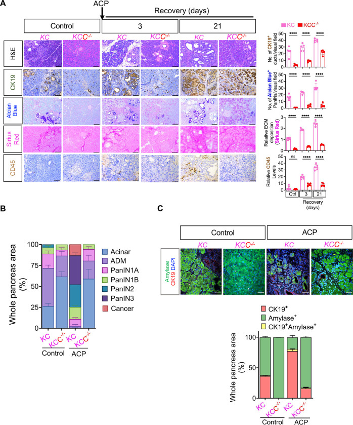
Figure 5.
Acinar specific ablation of Creb attenuates Kras* induced progression of ADM/PanINs towards pancreatic cancer with ACP. (A) Comparative histological evaluation of mouse pancreas, accompanied by representative photomicrographs, showcases H&E, CK-19, Alcian Blue, Sirius Red, and CD45+ staining within the pancreata of KC and KCC −/− mice in control and KC mice at 3- and 21-day ACP recovery period (left) and their quantification (right) (n=5 mice per group). (B) H&E-based histological quantification illustrating the presence of acinar cells, ADMs, PanINs and cancerous regions in control or within the pancreata of KC and KCC −/− mice harvested at 21 days of ACP recovery period (n=3 mice per group). (C) Representative pancreas images depicting amylase (green)/CK19 (red) and DAPI (blue) immunofluorescent labeling (top) and the corresponding quantification of CK19+ amylase+ cells (bottom) in control (KC and KCC −/−) or with ACP induction (n=3 mice per group). Scale bar, 50μm. ns nonsignificant; ****p<0.0001 by ANOVA.

