ABSTRACT
Fetus in fetu is a rare congenital anomaly in which a malformed parasitic twin is found within the body of a living child or adult. In this case report, a 1-day-old child presented with a large firm abdominal mass on the left side of the upper abdomen. Imaging studies misdiagnosed the mass as an intraperitoneal benign dermoid cyst displacing the bowel loops and internal viscera. A surgical resection was performed on 21 days of life, and pathology confirmed eight fetuses inside the cyst.
KEYWORDS: Eight, fetus in fetu, fetuses
INTRODUCTION
Fetus in fetu (FIF) is a rare anomaly of embryogenesis in which a malformed parasitic twin is found within the body of a normally developed host.[1] The exact etiology is unclear. One of the most accepted theories is the occurrence of an embryological insult occurring in diamniotic monochorionic twins, leading to the unsymmetrical division of the blastocyst mass, ending in a smaller cell mass engulfed within a mature sister embryo.[2] About 80% of cases are located in the abdomen, retroperitoneally. Other less common sites include the head, sacrum, scrotum, and chest.[3] Diagnosis is often made preoperatively with ultrasonography, plain radiography, computed tomography (CT), or magnetic resonance imaging. Histopathologic findings confirm the diagnosis, and the recommended treatment is complete excision. We report one rare case of FIF, in which eight fetuses are located intraperitoneally in a female newborn baby.
CASE REPORT
A 1-day-old female baby presented with a hard mass on the left side of the upper abdomen. On clinical examination, there was a palpable hard mass in the left hypochondrium, reaching up to the left iliac fossa, and no jaundice.
Systematic evaluation and radiological studies were done. Laboratory results were normal with respect to complete blood counts, and serum alpha-fetoprotein (AFP) was also within the normal range for newborns (47,985.20 ng/ml).
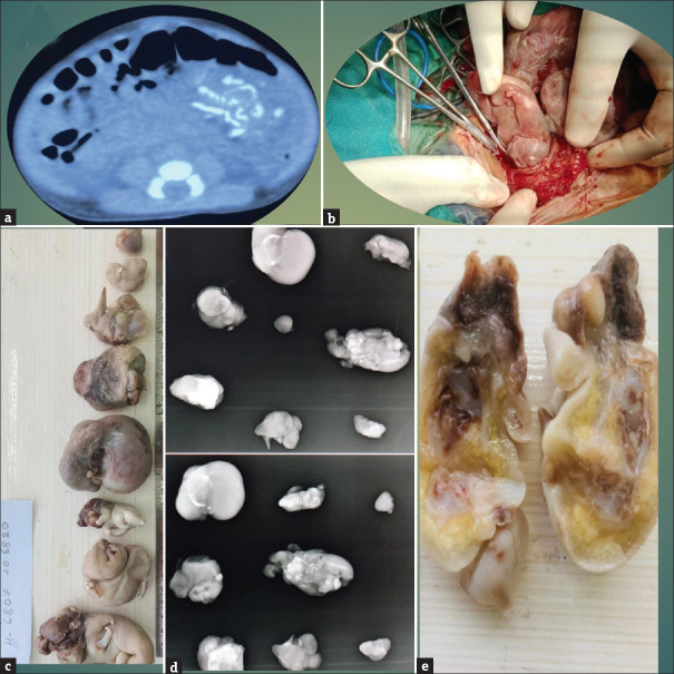
CT scans of the abdomen revealed a large well-defined cystic mass lesion with multiple thick septation within it seen in the peritoneal cavity extending from the left subdiaphragmatic area superiorly, pushing and compressing the stomach extending inferiorly till the lower abdomen level. It is laterally displacing the large bowel loop more laterally. The approximate size of the cystic lesion is 10.5 cm × 8.5 cm × 5.2 cm (AP × TR × CC). Dense calcification or ossification is seen within the cystic lesion [Figure 1a], suggestive likely of a benign dermoid cyst.
Figure 1.
(a) Contrast-enhanced computed tomography abdomen showing dense calcification or ossification seen within the cystic lesion, (b) Intraoperative picture showing fetuses being taken out, (c) Film showing all eight fetuses together, (d) Anteroposterior and lateral X-ray views of all eight fetuses, (e) Cut section of a fetus
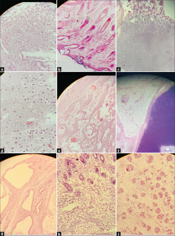
A decision was made to perform exploratory laparotomy through a left supraumbilical transverse incision that revealed a cystic mass about 10 cm × 8 cm × 5 cm just below the left dome of the diaphragm compressing the stomach and bowel loops laterally and extending below in the left iliac fossa [Figure 1b]. The cyst was opened fluid aspirated, and one fetus was found attached with the umbilical cord to the cyst wall. After removing the first fetus successively, eight fetuses were removed similarly attached with the umbilical cord to the cyst wall after ligation and division of the cord [Figure 1c]. Marsupialization of the cyst wall was done after removing the part of the cyst as the rest were adhered medially to the pancreas, stomach, and retroperitoneum. No enlarged lymph nodes were detected; closure of the abdomen in layers was done. Postoperatively, the patient started feeding on the third postoperative day and had an uneventful recovery. X-ray of all fetuses was done which showed bony structures in all of them [Figure 1d]. On histopathology all 8 fetuses have fetal parts – upper and lower limbs, vertebral bodies, and were totally covered with skin [Figure 1e]. Microscopically, there were elements from the three germ layers with no evidence of immature elements, confirming the diagnosis as a FIF [Figure 2].
Figure 2.
(a) Glandular epithelium with lamina propria (H and E, ×10), (b) Tissue lined by squamous epithelium with subepithelium showing adnexal structures which are unremarkable (H and E, ×10), (c) Choroid tissue (H and E, ×10), (d) Brain parenchyma with blood vessels (H and E, ×40) (e) Anastomosing bony trabeculae with red blood cells (H and E, ×40), (f) Chondroid tissue on the right side with adipose tissue adjacent to it (H and E, ×10), (g) Cystically dilated glandular cells with fibrous stroma in between (H and E, ×40), (h) Adnexal structures along with adipose tissue (H and E, ×10), (i) Glandular epithelium with loose stoma in between (H and E, ×10)
DISCUSSION
FIF pathology is rare and the incidence is 1 per 500,000 births. In families with a previous history of twin pregnancies, there is an increased incidence. They are usually single and genetically identical to their host.[4] Some histopathologists consider FIF as a highly differentiated teratoma and some as a separate entity. FIF is differentiated from a highly organized teratoma (with potential malignancy) prenatally by the presence of the spatially organized organs (anterior-posterior, craniocaudal, and lateral/symmetrical development) depending on the vertebral bodies, which presence indicates that notochord stage of fetal development has been reached.
FIF is genetically identical to its host; on the contrary, fetiform teratomas are homozygous. FIF is derived from totipotent inner cell mass (which can give rise to both embryonic or extra-embryonic cells), while teratomas arise from pluripotent cells without organogenesis or vertebral segmentation, both containing different tissues from one or more germinal cell layers.[2]
Spencer sets criteria for a mass to be defined as FIF:[4]
One or more of the following characteristics must be achieved:
Having a separate sac
Normal skin coverage either partially or completely
The presence of grossly recognizable anatomical parts
One or more vascular connections with its host.
The most commonly used markers are β-human choriogonadotropin, AFP, and urine homovanillic acid.[4] The role of tumor markers is to differentiate between FIF and other causes of intra-abdominal calcification, including teratoma, neuroblastoma, adrenal hemorrhage, meconium pseudocyst, and viral infection.
Due to the mass effect, FIF may exhibit its symptoms by compressing the surrounding structures leading to abdominal distension, feeding difficulties, vomiting, jaundice, renal dysfunction, and respiratory distress.[5]
In our case, the diagnosis was misleading on contrast-enhanced CT abdomen report as a benign dermoid cyst, but on laparotomy, the mass was found to be encased around a cyst like covering containing eight fetuses each attached with a separate umbilical cord to the cyst wall. All fetuses were confirmed on X-ray showing spine and histopathological examinations confirming fetuses and no immature elements. AFP levels decreased repeating 1 month after operation and were also normal for age (481.15 ng/ml).
CONCLUSION
FIF is a rare entity and requires a high degree of suspicion and proper diagnosis. FIF with a single fetus have been reported but this case is unique as we have found eight fetuses in a single fetu.
Declaration of patient consent
The authors certify that they have obtained all appropriate patient consent forms. In the form, the patient(s) has/have given his/her/their consent for his/her/their images and other clinical information to be reported in the journal. The patients understand that their names and initials will not be published and due efforts will be made to conceal their identity, but anonymity cannot be guaranteed.
Financial support and sponsorship
Nil.
Conflicts of interest
There are no conflicts of interest.
REFERENCES
- 1.Arlikar JD, Mane SB, Dhende NP, Sanghavi Y, Valand AG, Butale PR. Fetus in fetu: Two case reports and review of literature. Pediatr Surg Int. 2009;25:289–92. doi: 10.1007/s00383-009-2328-8. [DOI] [PubMed] [Google Scholar]
- 2.Taher HM, Abdellatif M, Wishahy AM, Waheeb S, Saadeldin Y, Kaddah S, et al. Fetus in fetu: Lessons learned from a large multicenter cohort study. Eur J Pediatr Surg. 2020;30:343–9. doi: 10.1055/s-0039-1698765. [DOI] [PubMed] [Google Scholar]
- 3.Barakat RM, Garzon S, Laganà AS, Franchi M, Ghezzi F. Fetus-in-fetu: A rare condition that requires common rules for its definition. Arch Gynecol Obstet. 2020;302:1541–3. doi: 10.1007/s00404-019-05211-y. [DOI] [PubMed] [Google Scholar]
- 4.Spencer R. Parasitic conjoined twins: External, internal (fetuses in fetu and teratomas), and detached (acardiacs) Clin Anat. 2001;14:428–44. doi: 10.1002/ca.1079. [DOI] [PubMed] [Google Scholar]
- 5.Ruffo G, Di Meglio L, Di Meglio L, Sica C, Resta A, Cicatiello R. Fetus-in-fetu: Two case reports. J Matern Fetal Neonatal Med. 2019;32:2812–9. doi: 10.1080/14767058.2018.1449207. [DOI] [PubMed] [Google Scholar]


