Abstract
Background
Research on biomarkers associated with the severity and adverse prognosis of COVID-19 can be beneficial for improving patient outcomes. However, there is limited research on the role of soluble TREM-1 (sTREM-1) in predicting the severity and prognosis of COVID-19 patients.
Methods
A total of 115 COVID-19 patients admitted to the emergency department of Beijing Youan Hospital from February to May 2023 were included in the study. Demographic information, laboratory measurements, and blood samples for sTREM-1 levels were collected upon admission.
Results
Our study found that sTREM-1 levels in the plasma of COVID-19 patients increased with the severity of the disease (moderate vs mild, p=0.0013; severe vs moderate, p=0.0195). sTREM-1 had good predictive value for disease severity and 28-day mortality (area under the ROC curve was 0.762 and 0.805, respectively). sTREM-1 also exhibited significant correlations with age, body temperature, respiratory rate, PaO2/FiO2, PCT, CRP, and CAR. Ultimately, through multivariate logistic regression analysis, we determined that sTREM-1 (OR 1.008, 95% CI: 1.002–1.013, p=0.005), HGB (OR 0.966, 95% CI: 0.935–0.998, p=0.036), D-dimer (OR 1.001, 95% CI: 1.000–1.001, p=0.009), and CAR (OR 1.761, 95% CI: 1.154–2.688, p=0.009) were independent predictors of 28-day mortality in COVID-19 patients. The combination of these four markers yielded a strong predictive value for 28-day mortality in COVID-19 cases with an AUC of 0.919 (95% CI: 0.857 −0.981).
Conclusion
sTREM-1 demonstrated good predictive value for disease severity and 28-day mortality, serving as an independent prognostic factor for adverse patient outcomes. In the future, we anticipate conducting large-scale multicenter studies to validate our research findings.
Keywords: COVID-19, sTREM-1, inflammation-related markers, disease severity, 28-day mortality
Introduction
COVID-19 is an infectious disease caused by the novel coronavirus SARS-CoV-2. Currently, the world is experiencing persistent outbreaks of this disease on a global scale.1–3 Although vaccines can effectively enhance the population’s resistance to the virus and reduce the mortality rate among patients, the continuous mutation of the virus leads to immune escape.4,5 As a result, even individuals who have previously been infected with COVID-19 can still experience reinfection.6 Elderly individuals or those with weakened immune systems and underlying cardiovascular diseases are still at risk of death.7–9
Timely prediction of patient prognosis and early intervention play a crucial role in saving lives during the treatment of COVID-19.10,11 Current research suggests that COVID-19 is a systemic disease in which inflammatory responses play a significant role.12,13 In the context of COVID-19, inflammatory markers have shown promise in predicting patient prognosis. Early research has highlighted the influence of various biomarkers, including C-reactive protein (CRP), procalcitonin (PCT), D-dimer, ferritin, erythrocyte sedimentation rate (ESR), serum ferritin, IL-1, IL-6, IL-8, and IL-18, on the severity of the disease.14–20
Triggering receptor expressed on myeloid cells 1 (TREM-1) is a member of the immunoglobulin superfamily, primarily expressed in bone marrow and epithelial cells.21,22 It is considered an amplifier of inflammation and, when activated, can induce the secretion of cytokines such as TNF-α, IL-6, and IL-1β in monocytes, macrophages, and dendritic cells.23,24 Research has shown that TREM-1 expression can be induced by various viral infections, including influenza A virus and hepatitis C virus, as well as bacterial infections like Staphylococcus aureus and Pseudomonas aeruginosa.25–27 Soluble TREM-1 (sTREM-1) is a 27 kDa peptide composed of the extracellular domain of TREM-1, which can be shed into the bloodstream.28,29 Multiple studies have investigated the use of sTREM-1 for predicting mortality in sepsis patients.30,31 However, there is limited research on the role of sTREM-1 in predicting disease severity and prognosis in COVID-19 patients.32
To investigate the predictive value of sTREM-1 for disease severity and prognosis in patients with COVID-19, we conducted this prospective study. We employed univariate and multivariate logistic regression analyses to explore independent predictive factors of COVID-19 and evaluate their predictive value for disease prognosis. The primary outcome of our study is the mortality status of patients within 28 days after hospital admission. This metric is commonly used in clinical studies and trials to assess the short-term outcomes and mortality rates associated with a particular condition or intervention. This study aims to provide reference for the early identification of critically ill patients in a clinical setting, thereby improving patient outcomes.
Patients and Methods
Study Design and Participants
We conducted prospective study involving 115 COVID-19 patients admitted to the emergency department of Beijing You’an Hospital between February 1, 2023, and May 31, 2023. All patients received a diagnosis based on the World Health Organization’s recommendations. Following the guidelines outlined in the National Health Commission of China’s “Diagnosis and Treatment protocol for COVID-19 patients (tentative 9 version)”,33 patients were categorized as having either mild/moderate or severe/critical COVID-19 cases. The objective of this study was to assess the predictive value of sTREM-1 and clinical parameters on the severity and prognosis of COVID-19 patients upon admission. The primary outcomes included the classification of patients as severe/critical cases and the occurrence of mortality within 28 days. The study obtained approval from the Ethics Committee of Beijing You’an Hospital, Capital Medical University, and adhered to the principles of the Helsinki Declaration (Approval No. LL-2023-006-K). Informed consent was acquired from all participating patients, and the data utilized in the study were anonymized.
Inclusion and Exclusion Criteria
The study’s inclusion criteria encompassed patients who met the requirements outlined in the Diagnosis and Treatment protocol for COVID-19 patients (tentative 9th version)33 released by the National Health Commission of China. Additionally, patients were required to provide informed consent and express their willingness to participate in the study.
Conversely, the exclusion criteria encompassed patients who declined to participate in the study. Furthermore, individuals under the age of 18, pregnant women, and patients who unfortunately passed away within 48 hours of hospital admission were also excluded. Additionally, patients for whom follow-up blood samples were either unavailable or lost were excluded from the study.
Data Collection
Demographic data, comorbidities, baseline characteristics, vital signs, arterial blood gas, laboratory data, and prognosis status were obtained from electronic medical records. The severity of illness categories were determined based on the COVID-19 treatment guidelines provided by the National Institute of Health. These categories encompass asymptomatic infection, mild illness, moderate illness, severe illness, and critical illness, taking into account various clinical manifestations (source: https://www.covid19treatmentguidelines.nih.gov/overview/clinical-spectrum/).
The study considered several comorbidities, including hypertension, diabetes mellitus (DM), coronary heart disease, chronic obstructive pulmonary disease (COPD), and malignant tumor. Vital signs assessed in the study included body temperature measured in degrees Celsius (°C), respiratory rate (RR) measured in breaths per minute, heart rate (HR) measured in beats per minute, and systolic blood pressure (SBP) measured in millimeters of mercury (mmHg). Arterial blood gas analysis parameters included pH, partial pressure of carbon dioxide (PaCO2), partial pressure of oxygen (PaO2), peripheral oxygen saturation (SpO2), and the ratio of arterial oxygen partial pressure to fractional inspired oxygen concentration (PaO2/FiO2). The study included various laboratory parameters for analysis. The complete blood count (CBC) was assessed, which included measurements of hemoglobin (HGB) levels, white blood cell (WBC) count, neutrophil count, lymphocyte count, monocyte count, and eosinophil count. Infection-related indicators: procalcitonin (PCT) levels in ng/mL, C-reactive protein (CRP) levels in mg/L, and CAR (C-reactive protein-to-albumin ratio). Coagulation-related indicators: international normalized ratio (INR), D-dimer levels in mg/L. Additionally, biochemical function tests were conducted to measure glucose levels in mmol/L, alanine transaminase (ALT) levels in U/L, aspartate transaminase (AST) levels in U/L, total bilirubin (TBIL) levels in μmol/L, direct bilirubin (DBIL) levels in μmol/L, estimated glomerular filtration rate (eGFR) in mL/min/1.73 m², and B-type natriuretic peptide (BNP) levels in pg/mL. Derived scores for inflammatory markers were calculated by combining two or more laboratory parameters. These parameters included the NLR (Neutrophil-to-lymphocyte ratio), MLR (Monocyte-to-lymphocyte ratio), PLR (Platelet-to-lymphocyte ratio), LCR (Lymphocyte-to-monocyte ratio), SIRI (Systemic inflammation response index), and SII (Systemic inflammation index). The SIRI, which represents the systemic inflammation response index, was calculated using the formula: SIRI = (Neutrophil count × Monocyte count) / Lymphocyte count. Similarly, the SII, known as the systemic inflammation index, was calculated using the formula: SII = (Neutrophil count × Platelet count) / Lymphocyte count. These derived scores play a crucial role in evaluating the severity of inflammation and providing valuable insights into the inflammatory response associated with the condition under study. These parameters are summarized in Table 1.
Table 1.
Baseline Characteristics and Clinical Data After Hospitalization of Study Population
| Variables | Total (n=115) | 28-day Survival (n=100) | 28-day Mortality (n=15) | P-value |
|---|---|---|---|---|
| Demographic data | ||||
| Sex, male, n (%) | 69 (60%) | 62 (62%) | 7 (47%) | 0.302 |
| Age (years) | 69.0 (59.5, 79.5) | 68.5 (56.8, 78.3) | 83.0 (69.0, 88.5) | 0.014* |
| Co-morbidities | ||||
| Hypertension, n (%) | 51 (44%) | 41 (41%) | 10 (67%) | 0.062 |
| Diabetes mellitus, n (%) | 26 (23%) | 20 (20%) | 6 (40%) | 0.104 |
| Coronary heart disease, n (%) | 22 (19%) | 19 (19%) | 3 (20%) | 0.912 |
| COPD, n (%) | 15 (13%) | 12 (12%) | 3 (20%) | 0.424 |
| Malignant tumor, n (%) | 20 (17%) | 17 (17%) | 3 (20%) | 0.762 |
| Vital signs | ||||
| Body temperature, °C | 36.7 (36.3,37.2) | 36.6 (36.3,37.0) | 37.4 (36.9,38.0) | <0.001* |
| RR, breaths/min | 20 (20,23) | 20 (20,22) | 25 (22,31) | 0.017* |
| HR, beats/min | 88 (78,100) | 88 (78,99) | 98 (85,104) | 0.090 |
| SBP, mmHg | 130 (120,142) | 130 (120,141) | 127 (114,140) | 0.516 |
| Arterial blood gas | ||||
| PH | 7.420 (7.400,7.453) | 7.421 (7.400,7.450) | 7.405 (7.322,7.480) | 0.469 |
| PaCO2, mmHg | 36.7 (32.1,39.8) | 36.6 (32.5,39.7) | 37.5 (24.9,39.9) | 0.624 |
| PaO2, mmHg | 93.1 (76.8,108.0) | 95.0 (80.1,108.0) | 75.5 (58.0,116.4) | 0.114 |
| SpO2, % | 98.0 (96.1,99.0) | 98.1 (96.5,99.0) | 95.4 (92.0,99.2) | 0.127 |
| PaO2/FiO2, mmHg | 272 (202,334) | 285 (224,341) | 178 (57,222) | <0.001* |
| COVID-19 severity class, n (%) | <0.001* | |||
| Mild illness | 34 (30%) | 34 (34%) | 0 (0%) | |
| Moderate illness | 42 (37%) | 42 (42%) | 0 (0%) | |
| Severe/critical illness | 39 (34%) | 24 (24%) | 15 (100%) | |
| Laboratory parameters | ||||
| sTREM-1, pg/mL | 180 (79,480) | 161 (68,300) | 680 (635,703) | <0.001* |
| PCT, ng/mL | 0.08 (0.05,0.23) | 0.07 (0.05,0.178) | 0.23 (0.115,1.68) | 0.009* |
| CRP, mg/L | 21.2 (7.28,56.6) | 16.4 (5.9,49.5) | 72.7 (48.9,109.2) | <0.001* |
| HGB, g/L | 126 (107,138) | 129 (110,138) | 115 (91,130) | 0.041* |
| Platelets count, ×10^9/L | 148 (113,198) | 151 (114,198) | 148 (110,210) | 0.960 |
| WBC count, ×10^9/L | 5.16 (3.49,7.21) | 5.19 (3.46,7.16) | 5.16 (4.10,7.92) | 0.787 |
| Neutrophils count, ×10^9/L | 3.53 (2.18,5.82) | 3.50 (2.18,5.65) | 3.85 (2.66,6.87) | 0.642 |
| Lymphocytes count, ×10^9/L | 0.92 (0.63,1.27) | 0.93 (0.63,1.29) | 0.77 (0.63,1.12) | 0.313 |
| Monocytes count, ×10^9/L | 0.39 (0.26,0.58) | 0.39 (0.26,0.57) | 0.45 (0.27,0.64) | 0.609 |
| Eosinophils count, ×10^9/L | 0.010 (0.00,0.045) | 0.015 (0.00,0.050) | 0.010 (0.00,0.020) | 0.120 |
| Glucose, mmol/L | 6.80 (5.94,7.90) | 6.70 (5.92,7.80) | 8.19 (6.62,9.06) | 0.051 |
| ALT, U/L | 20 (13,30) | 19 (13,31) | 23 (18,27) | 0.706 |
| AST, U/L | 26 (19,36) | 25 (18,33) | 35 (22,52) | 0.079 |
| Albumin, g/L | 35.8 (31.6,38.3) | 36.2 (32.6,38.5) | 30.8 (29.8, 35.9) | 0.006* |
| TBIL, μ mol/L | 11.15 (8.55,16.13) | 11.20 (8.50,16.05) | 10.80 (9.35,15.05) | 0.970 |
| DBIL, μ mol/L | 4.5 (3.0,6.9) | 4.2 (2.9,6.9) | 5.8 (3.4,6.7) | 0.456 |
| Prothrombin time activity (%) | 89 (78,97) | 91 (79,97) | 86 (73,94) | 0.186 |
| INR | 1.05 (1.00,1.11) | 1.04 (1.00,1.11) | 1.08 (1.02,1.24) | 0.190 |
| D-dimer, mg/L | 171 (0.775,588) | 148 (0.600,473) | 646 (5.00,2046) | 0.021* |
| eGFR, mL/min/1.73 m² | 87 (69,100) | 89 (72,101) | 69 (57,85) | 0.022* |
| BNP, pg/mL | 79 (29,282) | 76 (25,281) | 83 (70,351) | 0.132 |
| CAR | 0.668 (0.181,1.735) | 0.456 (0.149,1.428) | 2.315 (1.581,3.653) | <0.001* |
| BCDIMs | ||||
| NLR | 3.80 (2.31,7.06) | 3.63 (2.31,6.99) | 5.36 (2.74,8.11) | 0.368 |
| MLR | 0.40 (0.24,0.70) | 0.39 (0.24,0.67) | 0.49 (0.32,0.78) | 0.325 |
| PLR | 168 (119,230) | 167 (115,230) | 171 (140,257) | 0.457 |
| LCR | 0.040 (0.013,0.143) | 0.053 (0.017,0.181) | 0.011 (0.005,0.018) | <0.001* |
| SIRI | 1.642 (0.609,3.356) | 1.598 (0.607,3.268) | 1.879 (1.148,4.471) | 0.385 |
| SII | 522 (304,1203) | 492 (294,1177) | 812 (463,1203) | 0.342 |
Notes: Normally distributed continuous variables are displayed as mean ± standard deviation (SD) and were compared using the independent-samples Student’s t-test. Non-normally distributed continuous variables are displayed as a median with interquartile range (IQR) and were compared using the Mann–Whitney U-test. Categorical variables are expressed as counts with percentages and were compared using Pearson’s chi-square or Fisher’s exact test. SIRI = (Neutrophil count × Monocyte count) / Lymphocyte count; SII = (Neutrophil count × Platelet count) / Lymphocyte count. *p-value <0.05 was considered significant.
Abbreviations: COPD, Chronic Obstructive Pulmonary Disease; RR, respiratory rate; HR, heart rate; SBP, systolic blood pressure; PaCO2, arterial carbon dioxide tension; PaO2, oxygen tension; SpO2, peripheral oxygen saturation; FiO2, fraction of inspired oxygen; sTREM, soluble Triggering Receptors Expressed on Myeloid Cell; PCT, Procalcitonin; CRP, C-reactive protein; HGB, Hemoglobin; WBC, White blood cell; ALT, Alanine aminotransferase; AST, Aspartate aminotransferase; TBIL, Total bilirubin; DBIL, Direct bilirubin; INR, International normalized ratio; eGFR, estimated glomerular filtration rate; BNP, B-type natriuretic peptide; CAR, C-reactive protein-to-albumin ratio; BCDIMs, blood count-derived inflammatory markers; NLR, Neutrophil-to-lymphocyte ratio; MLR, Monocyte-to-lymphocyte ratio; PLR, Platelet-to-lymphocyte ratio; LCR, Lymphocyte-to- C-reactive protein ratio; SIRI, Systemic inflammation response index; SII, Systemic inflammation index.
Blood Sample Collection and Testing
At the time of admission, blood sample collection and testing were carried out simultaneously. The blood samples were collected using anticoagulant tubes containing ethylenediaminetetraacetic acid (EDTA). To obtain plasma, the collected whole blood was centrifuged at 1350g for 12 minutes. The concentration of plasma sTREM-1 was determined using the human sTREM-1 ELISA Kit (Keshun Biotechnology, Shanghai, China; REF: KS18244, Lot: 202305), following the instructions provided in the kit manual.
Statistical Analysis
The normality of continuous variables was assessed using the Shapiro–Wilk test. Continuous variables that were normally distributed are presented as mean ± standard deviation (SD) and were compared using independent-samples Student’s t-test. Non-normally distributed continuous variables are presented as median with interquartile range (IQR) and were compared using the Mann–Whitney U-test. Categorical variables are reported as counts with percentages and were compared using Pearson’s chi-square or Fisher’s exact test. Multiple samples were compared using the non-parametric Kruskal–Wallis test. Variables with P values < 0.05 were considered statistically significant. The Receiver Operating Characteristic (ROC) curve was employed to assess the predictive performance of parameters for disease severity and 28-day mortality in COVID-19 patients. Kaplan-Meier curves were used to evaluate the risk prediction of parameters for 28-day mortality in COVID-19 patients. Spearman’s rank correlation is used to analyze the correlation between sTREM-1 and age, vital signs, and laboratory markers of inflammation. The results are visualized using a heat map. Univariable logistic regression analysis is employed to identify independent predictive factors for 28-day mortality (P<0.05). Parameters showing statistical differences are included in the multivariable logistic regression analysis. Data were analyzed using SPSS software (version 22.0; IBM Corp) and R language (version 4.2.1; R Foundation for Statistical Computing) and illustrated using GraphPad Prism 9 (GraphPad Software Inc).
Results
Patient Characteristics and Clinical Parameters Upon Admission
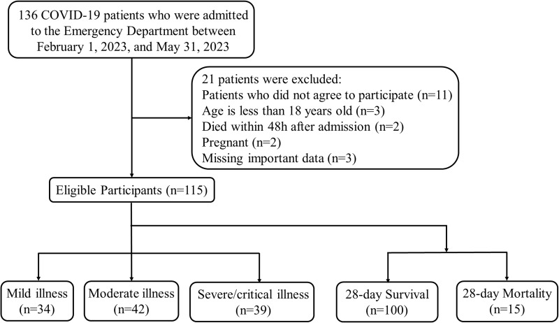
Out of the 136 patients admitted to the emergency department, 115 patients were eligible for further analysis. Figure 1 illustrates the process of patient enrollment. Among them, 34 cases (30.0%) were categorized as mild, 42 cases (37.0%) as moderate, and 39 cases (34%) as severe or critically ill. Finally, within 28 days of admission, 15 cases (13.0%) resulted in mortality.
Figure 1.
Flow diagram of patients enrollment.
Table 1 provides an overview of the patients’ baseline characteristics and clinical parameters. Of the patients, 69 (60%) were male, with a median age of 69 years. The most prevalent comorbidities in this group included hypertension (51/115, 44%) and diabetes (26/115, 23%).
When comparing the patients who survived for 28 days to those who passed away within that period, several notable differences emerged. The deceased patients were generally older, had higher body temperature upon admission, and exhibited faster respiratory rates and lower oxygenation index (all p-values < 0.05). In terms of laboratory parameters, the 28-day mortality group exhibited elevated levels of sTREM-1, PCT, CRP, D-dimer and CAR, while experiencing decreased levels of Albumin, HGB, and eGFR (all p-values < 0.05). Furthermore, the level of LCR, which is a blood count-derived inflammatory marker, differed significantly between the two groups (p-value < 0.05).
The Predictive Value of sTREM-1 for the Severity and Prognosis of COVID-19 Patients
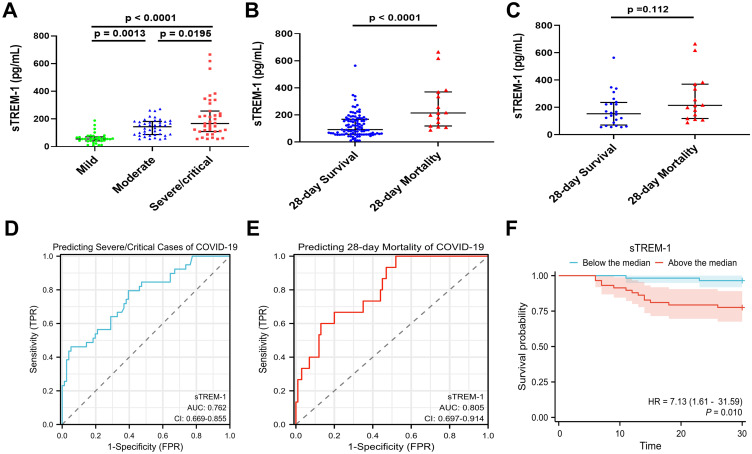
Similar to previous reports, the study uncovered a notable association between sTREM-1 levels and the severity of illness in COVID-19 patients. Patients with more severe conditions exhibited higher levels of sTREM-1 (Figure 2A). Additionally, patients who passed away within 28 days demonstrated elevated sTREM-1 levels (Figure 2B). We also compared sTREM-1 levels between the death and survival groups in critically ill patients. There was a trend, but currently no statistically significant difference between the two groups (p=0.112) (Figure 2C). sTREM-1 displayed commendable predictive value for assessing disease severity and 28-day mortality, as indicated by the area under the ROC curve of 0.762 (95% confidence interval [CI]: 0.669–0.855) and an area under the ROC curve (AUC) of 0.805 (95% CI: 0.697–0.914) respectively (Figure 2D and E). Furthermore, when patients were categorized based on the median value, those above the median exhibited a higher risk of mortality within 28 days compared to those below the median (p-value < 0.05) (Figure 2F).
Figure 2.
The predictive value of sTREM-1 for the severity of illness and 28-day mortality in COVID-19 patients. (A) Comparison of sTREM-1 levels among different severity groups of COVID-19 patients. (B) Comparison of sTREM-1 levels between the 28-day survival and death groups. (C) Comparison of sTREM-1 levels between the 28-day survival and death groups in critically ill patients. Data are displayed as a median with interquartile range (IQR) and were compared using the Mann–Whitney U-test. Multiple samples were compared using the non-parametric Kruskal–Wallis test. (D) Receiver Operating Characteristic (ROC) curve of sTREM-1 for predicting severity in COVID-19 patients, with an area under the ROC curve (AUC) of 0.762 (95% confidence interval [CI]: 0.669–0.855). (E) ROC curve of sTREM-1 for predicting 28-day mortality in COVID-19 patients, with an area under the ROC curve (AUC) of 0.805 (95% [CI]: 0.697–0.914). (F) Kaplan-Meier curve for patients divided into two groups based on the median sTREM-1 level: above-median group and below-median group, for 28-day survival. A p-value <0.05 was considered significant.
The Correlations Between sTREM-1 and Age, Vital Signs, and Laboratory Inflammatory Markers
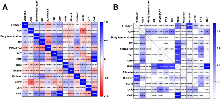
The heatmap in Figure 3 provides a visual representation of the correlations and corresponding p-values among sTREM-1, age, clinical scoring systems, and laboratory inflammatory markers, involving a total of 12 parameters (Figure 3A and B). Notably, sTREM-1 exhibited a significant correlation with age (r=0.246, p=0.0081), body temperature (r=0.202, p=0.031), respiratory rate (r=0.220, p=0.018), and PaO2/FiO2 (r= - 0.325, p=0.0004). Furthermore, significant correlations were observed between sTREM-1 and the following laboratory inflammatory markers: PCT (r=0.190, p=0.044), CRP (r=0.299, p=0.001), and CAR (r=0.273, p=0.004).
Figure 3.
Heatmap depicting the correlation between sTREM-1 and age, Vital signs, and laboratory inflammatory markers. (A) The values are presented as Spearman‘s correlation coefficient (r) for a sample of 115 runners regarding sTREM-1. The colormap ranges from 1 to −1, with blue indicating the highest value and red indicating the lowest value. (B) The Heatmap of corresponding p-values. The colormap ranges from 0 to 1, with blue representing the largest value and white representing the smallest value. White cells without numerical values indicate that the p-value is smaller than 0.00001, indicating a highly significant correlation.
Abbreviations: RR, respiratory rate; PaO2, oxygen tension; FiO2, fraction of inspired oxygen; sTREM, soluble Triggering Receptors Expressed on Myeloid Cell; PCT, Procalcitonin; CRP, C-reactive protein; HGB, Hemoglobin; eGFR, estimated glomerular filtration rate; LCR, Lymphocyte-to- C-reactive protein ratio; CAR, C-reactive protein-to-albumin ratio.
The Expression of sTREM-1 Demonstrates Equal or Better Predictive Value for Disease Severity and Prognosis in COVID-19 Patients Compared to Some Clinical Indicators
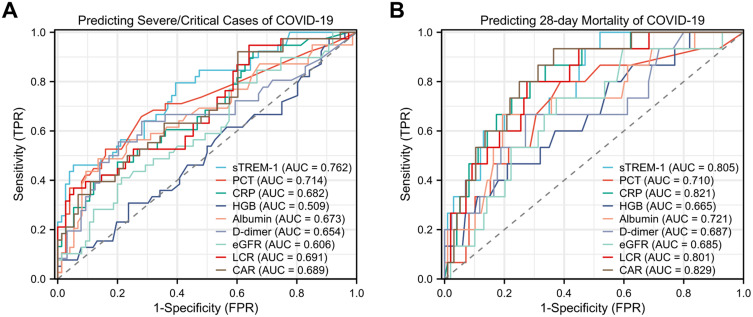
The receiver operating characteristic (ROC) curve was used to evaluate and compare the predictive value of sTREM-1 and laboratory indicators for assessing the disease severity and prognosis of COVID-19 patients. For predicting disease severity (Figure 4A and Table 2), sTREM-1 exhibited the highest predictive value among the evaluated parameters, with an area under the ROC curve of 0.762 (95% confidence interval [CI]: 0.669–0.855). PCT also showed good predictive value, with an AUC of 0.714 (95% CI: 0.607–0.822). CRP, Albumin, D-dimer, LCR, and CAR demonstrated moderate predictive values, with AUCs ranging from 0.654 to 0.691. On the other hand, HGB and eGFR showed relatively lower predictive values, with AUCs of 0.509 and 0.606, respectively. For predicting prognosis (Figure 4B and Table 2), CAR exhibited the highest predictive value with an AUC of 0.829 (95% CI: 0.732–0.927). sTREM-1 had an AUC of 0.805 (95% CI: 0.697–0.914), indicating significant predictive value. CRP and LCR also showed high predictive value, with AUCs of 0.821 and 0.801, respectively. Also, HGB had a relatively lower predictive value, with an AUC of 0.665.
Figure 4.
ROC curves of sTREM-1 and main clinical parameters for severity and prognosis of COVID-19 patients. (A) Predicting severity of COVID-19 patients. The area under the curve (AUC) for sTREM-1 was 0.762 (95% confidence interval [CI]: 0.669–0.855); PCT, AUC was 0.714 (95% CI: 0.607–0.822); CRP, 0.682 (95% CI: 0.577–0.787); HGB, AUC was 0.509 (95% CI: 0.395–0.623); Albumin, 0.673 (95% CI: 0.562–0.784); D-dimer, AUC was 0.654 (95% CI: 0.532–0.776); eGFR, AUC was 0.606 (95% CI: 0.496–0.717); LCR, AUC was 0.691 (95% CI: 0.585–0.796); CAR, AUC was 0.689 (95% CI: 0.584–0.793). (B) Predicting prognosis of COVID-19 patients. sTREM-1, AUC 0.805 (95% CI: 0.697–0.914); PCT, AUC 0.710 (95% CI: 0.565–0.855); CRP, AUC 0.821 (95% CI: 0.719–0.924); HGB, AUC 0.665 (95% CI: 0.515–0.814); Albumin, AUC 0.721 (95% CI: 0.575–0.867); D-dimer, AUC 0.687 (95% CI: 0.529–0.846); eGFR, AUC 0.685 (95% CI: 0.545–0.824); LCR, AUC 0.801 (95% CI: 0.691–0.912); CAR, AUC 0.829 (95% CI: 0.732–0.927).
Abbreviations: TPR, true positive rate; FPR, false positive rate; sTREM, soluble Triggering Receptors Expressed on Myeloid Cell; PCT, Procalcitonin; CRP, C-reactive protein; HGB, Hemoglobin; eGFR, estimated glomerular filtration rate; LCR, Lymphocyte-to- C-reactive protein ratio; CAR, C-reactive protein-to-albumin ratio.
Table 2.
Predicted Value Information of Different Variable Parameters for Disease Severity and Prognosis in COVID-19 Patients
| Variables | Cut off value | Sensitivity | Specificity | PPV | NPV | Accuracy | Youden index |
|---|---|---|---|---|---|---|---|
| Predicting disease severity | |||||||
| sTREM-1, pg/mL | 105 | 0.79 | 0.61 | 0.51 | 0.85 | 0.74 | 0.40 |
| PCT, ng/mL | 0.115 | 0.66 | 0.72 | 0.54 | 0.81 | 0.70 | 0.38 |
| CRP, mg/L | 65.9 | 0.39 | 0.88 | 0.63 | 0.74 | 0.72 | 0.27 |
| HGB, g/L | 108 | 0.31 | 0.76 | 0.40 | 0.68 | 0.61 | 0.07 |
| Albumin, g/L | 31.7 | 0.49 | 0.86 | 0.66 | 0.76 | 0.73 | 0.35 |
| D-dimer, mg/L | 248 | 0.64 | 0.72 | 0.56 | 0.78 | 0.69 | 0.36 |
| eGFR, mL/min/1.73 m² | 93.2 | 0.79 | 0.41 | 0.41 | 0.79 | 0.54 | 0.21 |
| LCR | 0.00879 | 0.37 | 0.95 | 0.78 | 0.75 | 0.75 | 0.32 |
| CAR | 0.224 | 0.92 | 0.40 | 0.44 | 0.91 | 0.58 | 0.32 |
| Predicting prognosis | |||||||
| sTREM-1, pg/mL | 198 | 0.60 | 0.87 | 0.41 | 0.94 | 0.86 | 0.47 |
| PCT, ng/mL | 0.105 | 0.8 | 0.61 | 0.24 | 0.95 | 0.63 | 0.41 |
| CRP, mg/L | 35.7 | 0.87 | 0.66 | 0.28 | 0.97 | 0.69 | 0.53 |
| HGB, g/L | 107 | 0.47 | 0.79 | 0.25 | 0.91 | 0.75 | 0.26 |
| Albumin, g/L | 31.7 | 0.67 | 0.81 | 0.34 | 0.94 | 0.79 | 0.47 |
| D-dimer, mg/L | 425 | 0.67 | 0.73 | 0.30 | 0.93 | 0.72 | 0.40 |
| eGFR, mL/min/1.73 m² | 79.9 | 0.73 | 0.63 | 0.23 | 0.94 | 0.64 | 0.36 |
| LCR | 0.0187 | 0.8 | 0.72 | 0.31 | 0.96 | 0.73 | 0.52 |
| CAR | 0.86 | 0.93 | 0.64 | 0.29 | 0.98 | 0.68 | 0.57 |
| Combined Model | 0.0951 | 0.93 | 0.77 | 0.42 | 0.98 | 0.80 | 0.70 |
Abbreviations: PPV, Positive predictive value; NPV, Negative predictive value; sTREM, soluble Triggering Receptors Expressed on Myeloid Cell; PCT, Procalcitonin; CRP, C-reactive protein; HGB, Hemoglobin; eGFR, estimated glomerular filtration rate; LCR, Lymphocyte-to- C-reactive protein ratio; CAR, C-reactive protein-to-albumin ratio.
sTREM-1 is an Independent Predictor of 28-Day Mortality in COVID-19 Patients
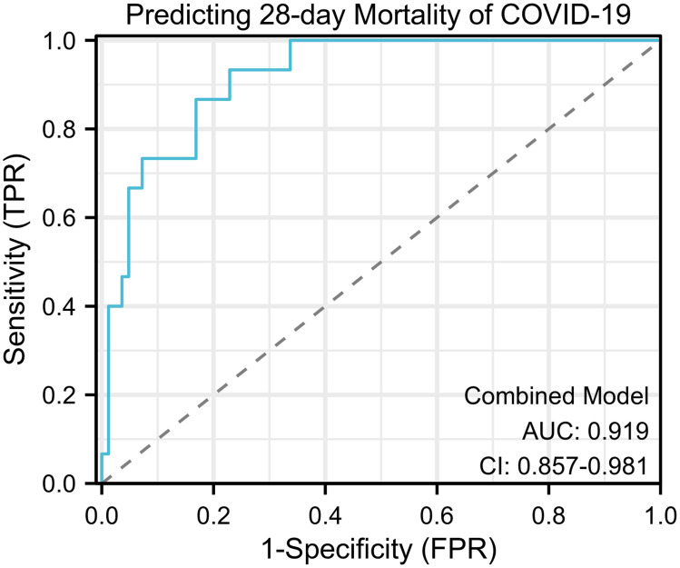
We conducted univariate logistic regression analysis on indicators that showed statistically significant differences (p<0.05) between the 28-day survival group and the deceased group. Among the 12 indicators, all except PCT demonstrated statistically significant differences (p<0.05). These variables with statistically significant differences were included in a multivariate logistic regression analysis. Finally, we found that sTREM-1 (OR 1.008, 95% CI: 1.002–1.013, p=0.005), HGB (OR 0.966, 95% CI: 0.935–0.998, p=0.036), D-dimer (OR 1.001, 95% CI: 1.000–1.001, p=0.009), and CAR (OR 1.761, 95% CI: 1.154–2.688, p=0.009) were independent predictors of 28-day mortality in COVID-19 patients (Table 3). The prognostic model for COVID-19 patients incorporating these four markers showed the highest predictive value for 28-day mortality, with an AUC of 0.919 (95% CI: 0.857–0.981) (Figure 5).
Table 3.
Univariable and Multivariable Logistic Regression Analysis for the Predictors of 28-Day Mortality in COVID-19 Patients
| Variables | UV | MV | ||||
|---|---|---|---|---|---|---|
| Wald | OR (95% CI) | P-value | Wald | OR (95% CI) | P-value | |
| Age (years) | 4.606 | 1.047 (1.004–1.092) | 0.032* | |||
| Body temperature, °C | 12.637 | 4.370 (1.938–9.854) | 0.001* | |||
| RR, breaths/min | 10.179 | 1.151 (1.056–1.256) | 0.001* | |||
| PaO2/FiO2, mmHg | 9.749 | 0.991 (0.986–0.997) | 0.002* | |||
| sTREM-1, pg/mL | 11.793 | 1.009 (1.004–1.014) | 0.001* | 7.975 | 1.008 (1.002–1.013) | 0.005* |
| PCT, ng/mL | 2.348 | 1.045 (0.988–1.106) | 0.125 | |||
| CRP, mg/L | 10.295 | 1.017 (1.007–1.028) | 0.001* | |||
| HGB, g/L | 5.170 | 0.973 (0.950–0.996) | 0.023* | 4.382 | 0.966 (0.935–0.998) | 0.036* |
| Albumin, g/L | 8.322 | 0.836 (0.741–0.944) | 0.004* | |||
| D-dimer, mg/L | 7.466 | 1.001 (1.000–1.001) | 0.006* | 6.890 | 1.001 (1.000–1.001) | 0.009* |
| eGFR, mL/min/1.73 m² | 4.595 | 0.979 (0.960–1.116) | 0.032* | |||
| LCR | 4.221 | 0.000 (0.000–0.341) | 0.040* | |||
| CAR | 10.671 | 1.747 (1.250–2.442) | 0.001* | 6.884 | 1.761 (1.154–2.688) | 0.009* |
Notes: *p-value <0.05 was considered significant.
Abbreviations: RR, respiratory rate; PaO2, oxygen tension; FiO2, fraction of inspired oxygen; sTREM, soluble Triggering Receptors Expressed on Myeloid Cell; PCT, Procalcitonin; CRP, C-reactive protein; HGB, Hemoglobin; eGFR, estimated glomerular filtration rate; LCR, Lymphocyte-to- C-reactive protein ratio; CAR, C-reactive protein-to-albumin ratio.
Figure 5.
ROC curves for the combined model for the prognosis of COVID-19 patients. Combined model: sTREM-1, CAR, HGB and D-dimer; AUC 0.919 (95% CI: 0.857–0.981).
Abbreviations: TPR, true positive rate; FPR, false positive rate; sTREM, soluble Triggering Receptors Expressed on Myeloid Cell; HGB, Hemoglobin; CAR, C-reactive protein-to-albumin ratio.
Discussion
Timely identification and intervention in critically ill patients play a pivotal role in reducing mortality among individuals with COVID-19. Our research elucidated a correlation between sTREM-1 levels in the plasma of COVID-19 patients and the severity of the disease. Moreover, sTREM-1 exhibited promising predictive capabilities for assessing the prognosis of COVID-19 patients. Additionally, our study unveiled associations between plasma sTREM-1 levels and various factors, including age, body temperature, respiratory rate, PaO2/FiO2, and laboratory inflammatory markers such as PCT, CRP, and CAR, in COVID-19 patients. Notably, the expression of sTREM-1 demonstrated comparable or superior predictive value for disease severity and prognosis in COVID-19 patients when compared to certain clinical indicators. Ultimately, through multivariate logistic regression analysis, we determined that sTREM-1 and CAR, along with HGB and D-dimer, collectively serve as independent predictive factors for 28-day mortality in COVID-19 patients. The combination of these four markers yielded a strong predictive value for 28-day mortality in COVID-19 cases.
TREM-1 is a receptor belonging to the immunoglobulin superfamily, primarily expressed on neutrophils and mature monocytes/macrophages in the bloodstream.21,22 It is considered an amplifier of inflammation, capable of inducing the release of pro-inflammatory mediators such as IL-6, TNF-α, and IL-1β. It may be associated with the cytokine storm observed in COVID-19 patients.32,34 sTREM-1 is the soluble counterpart of TREM-1, which can be proteolytically cleaved from the cell membrane and enter the plasma following stimulation by pro-inflammatory mediators.24 Therefore, it is reasonable to associate sTREM-1 with disease severity and its predictive value for patient prognosis in COVID-19.
Current research shows some discrepancies in the predictive value of sTREM-1 for patient prognosis, particularly regarding the optimal cut-off value for prediction. In a recent study, the area under the ROC curve (AUC) for sTREM-1 in predicting severe COVID-19 was reported as 0.656, but the optimal cut-off value and its predictive value for prognosis were not specified.35 However, a study has indicated that sTREM-1 has high predictive value for severe COVID-19, with an AUC of 0.96 (95% CI: 0.94–0.98).36 Regarding prognosis, an earlier study involving 76 COVID-19 patients found that sTREM-1 had an AUC of 0.86 (95% CI: 0.77–0.95) for predicting intubation/death at 30 days, with an optimal cut-off value of 689 pg/mL.34 Another study reported that sTREM-1 had an AUC of 0.73 (95% CI: 0.62–0.83) for predicting mortality, with an optimal cut-off value of 315 pg/mL.32 Our study found that sTREM-1 had AUCs of 0.762 and 0.805 for predicting severe COVID-19 and 28-day mortality, respectively, which align with the current research. These studies compared the predictive value of sTREM-1 with conventional laboratory markers such as PCT, CRP, and others for disease severity and prognosis in patients. In all cases, it was found that sTREM-1 exhibited comparable or even superior predictive performance. Due to the availability of mature commercial ELISA test kits, detecting sTREM-1 in routine clinical practice is feasible. As the number of patients increases and the sample size for testing becomes sufficient, the cost of testing will become relatively lower. Dynamic monitoring of sTREM-1 levels is more meaningful and feasible for monitoring patients’ conditions. It is important to note that the reference ranges for sTREM-1 levels used to monitor the severity of patients’ diseases and prognosis may vary, especially among different ethnic groups, which requires further research to determine.
Several studies have compared the correlation between sTREM-1 and clinical parameters and laboratory inflammatory markers.37,38 Our study also found correlations (p<0.05) between sTREM-1 and age, body temperature, respiratory rate, PaO2/FiO2, as well as laboratory inflammatory markers such as PCT, CRP, and CAR. For clinical parameters like age and body temperature, this may be due to their association with disease severity in patients. As for respiratory rate and PaO2/FiO2, these parameters directly reflect the severity of the patient’s condition, and higher levels of sTREM-1 may be observed as a result of the increased inflammatory response associated with more severe disease. Interestingly, when meaningful indicators (p<0.05) from univariate logistic regression analysis were included in the multivariable regression analysis, it was observed that, apart from sTREM-1, the other three independent predictive factors included were all laboratory markers. These three laboratory markers were HGB, D-dimer, and CAR. The measurement Results of HGB are commonly used to assess anemia, monitor blood status, and evaluate oxygen delivery and transport capacity. D-dimer is used to evaluate the activity of blood coagulation and the fibrinolytic system. CAR is a good indicator of clinical inflammatory response. These three markers have all been reported to be associated with the severity of COVID-19.39–44 In our study, apart from CAR, we did not find a correlation between sTREM-1 and the severity of the disease. Therefore, the combination of CAR, HGB, and D-dimer yielded a strong predictive value for 28-day mortality in COVID-19 cases, with an AUC of 0.919 (95% CI: 0.857–0.981).
Our study has several limitations. Firstly, it was a single-center prospective study, and the representativeness of the patient population needs to be validated. Secondly, the sample size was relatively small, mainly due to challenges in obtaining blood samples from enrolled patients. Thirdly, the primary outcome of our study focused on the 28-day mortality rate, and further investigation is needed to assess long-term survival outcomes. Fourthly, we did not conduct continuous monitoring and tracking of sTREM-1 levels in patients. Incorporating dynamic monitoring of sTREM-1 levels alongside changes in clinical conditions could offer more meaningful insights into disease progression. However, this is an aspect we intend to address in future research endeavors. We eagerly anticipate larger-scale, multicenter studies in the future to validate and expand upon our findings.
Conclusion
This study identified a correlation between sTREM-1 levels and the severity of COVID-19 in patients. sTREM-1 demonstrated good predictive value for disease severity and 28-day mortality, serving as an independent prognostic factor for adverse patient outcomes. In the future, we anticipate conducting large-scale multicenter studies to validate our research findings.
Acknowledgment
We would like to express our gratitude to Beijing You’an Hospital for granting access to the clinical data of the patients. We would also like to extend our appreciation to Dr. Qingkun Song and the team at the Biomedical Informatics Sample Center for providing the specimens.
Funding Statement
This work was supported by National Key Research and Development Program of China [Grant No. 2022YFC2305002], Beijing municipal medical research institute public welfare development and reform pilot project [Grant No. jingyiyan2021-10], COVID-19 Special Project of Beijing You’an Hospital [Grant No. 2023-6], Middle-aged and Young Talent Incubation Programs (Clinical Research) of Beijing You’an Hospital [Grant No. BJYAYY-YN2022-12, BJYAYY-YN2022-13], Beijing research center for respiratory infectious diseases project [Grant No. BJRID2024-001], Beijing Natural Science Foundation [Grant No. M22030] and Beijing Natural Science Foundation [Grant No. 7232079].
Abbreviations
COPD, Chronic Obstructive Pulmonary Disease; RR, respiratory rate; HR, heart rate; SBP, systolic blood pressure; PaCO2, arterial carbon dioxide tension; PaO2, oxygen tension; SpO2, peripheral oxygen saturation; FiO2, fraction of inspired oxygen; sTREM, soluble Triggering Receptors Expressed on Myeloid Cell; PCT, Procalcitonin; CRP, C-reactive protein; HGB, Hemoglobin; WBC, White blood cell; ALT, Alanine aminotransferase; AST, Aspartate aminotransferase; TBIL, Total bilirubin; DBIL, Direct bilirubin; INR, International normalized ratio; eGFR, estimated glomerular filtration rate; BNP, B-type natriuretic peptide; CAR, C-reactive protein-to-albumin ratio; BCDIMs, blood count-derived inflammatory markers; NLR, Neutrophil-to-lymphocyte ratio; MLR, Monocyte-to-lymphocyte ratio; PLR, Platelet-to-lymphocyte ratio; LCR, Lymphocyte-to-C-reactive protein ratio; SIRI, Systemic inflammation response index; SII: Systemic inflammation index. SIRI = (Neutrophil count × Monocyte count) / Lymphocyte count; SII = (Neutrophil count × Platelet count) / Lymphocyte count. SD, standard deviation; IQR, interquartile range; ROC, Receiver Operating Characteristic.
Data Sharing Statement
The datasets used and/or analyzed during the current study are available from the corresponding author on reasonable request.
Ethics Approval and Consent to Participate
This study was approved by the Ethical Committee of Beijing Youan Hospital (Approval No. LL-2023-006-K). All participating patients provided informed consent, and the data used in the study were anonymized.
Author Contributions
All authors made a significant contribution to the work reported, whether that is in the conception, study design, execution, acquisition of data, analysis and interpretation, or in all these areas; took part in drafting, revising or critically reviewing the article; gave final approval of the version to be published; have agreed on the journal to which the article has been submitted; and agree to be accountable for all aspects of the work.
Disclosure
The authors declare that they have no competing interests.
References
- 1.Huang C, Wang Y, Li X, et al. Clinical features of patients infected with 2019 novel coronavirus in Wuhan, China. Lancet. 2020;395(10223):497–506. doi: 10.1016/S0140-6736(20)30183-5 [DOI] [PMC free article] [PubMed] [Google Scholar]
- 2.Sachs JD, Karim SSA, Aknin L, et al. The Lancet Commission on lessons for the future from the COVID-19 pandemic. Lancet. 2022;400(10359):1224–1280. doi: 10.1016/S0140-6736(22)01585-9 [DOI] [PMC free article] [PubMed] [Google Scholar]
- 3.Flahault A, Calmy A, Costagliola D, et al. No time for complacency on COVID-19 in Europe. Lancet. 2023;401(10392):1909–1912. doi: 10.1016/S0140-6736(23)01012-7 [DOI] [PMC free article] [PubMed] [Google Scholar]
- 4.Singh P, Anand A, Rana S, et al. Impact of COVID-19 vaccination: a global perspective. Front Public Health. 2023;11:1272961. doi: 10.3389/fpubh.2023.1272961 [DOI] [PMC free article] [PubMed] [Google Scholar]
- 5.Wu N, Joyal-Desmarais K, Ribeiro P, et al. Long-term effectiveness of COVID-19 vaccines against infections, hospitalisations, and mortality in adults: findings from a rapid living systematic evidence synthesis and meta-analysis up to December, 2022. Lancet Respir Med. 2023;11(5):439–452. doi: 10.1016/S2213-2600(23)00015-2 [DOI] [PMC free article] [PubMed] [Google Scholar]
- 6.Stein C, Nassereldine H, Sorensen RJD; Team C-F. Past SARS-CoV-2 infection protection against re-infection: a systematic review and meta-analysis. Lancet. 2023;401(10379):833–842. doi: 10.1016/S0140-6736(22)02465-5 [DOI] [PMC free article] [PubMed] [Google Scholar]
- 7.Evans RA, Dube S, Lu Y, et al. Impact of COVID-19 on immunocompromised populations during the Omicron era: insights from the observational population-based INFORM study. Lancet Reg Health Eur. 2023;35:100747. doi: 10.1016/j.lanepe.2023.100747 [DOI] [PMC free article] [PubMed] [Google Scholar]
- 8.Termorshuizen F, Dongelmans DA, Brinkman S, et al. Characteristics and outcome of COVID-19 patients admitted to the ICU: a nationwide cohort study on the comparison between the consecutive stages of the COVID-19 pandemic in the Netherlands, an update. Ann Intensive Care. 2024;14(1):11. doi: 10.1186/s13613-023-01238-2 [DOI] [PMC free article] [PubMed] [Google Scholar]
- 9.Hippisley-Cox J, Khunti K, Sheikh A, et al. Risk prediction of covid-19 related death or hospital admission in adults testing positive for SARS-CoV-2 infection during the omicron wave in England (QCOVID4): cohort study. BMJ. 2023;381:e072976. doi: 10.1136/bmj-2022-072976 [DOI] [PMC free article] [PubMed] [Google Scholar]
- 10.Meyerowitz EA, Scott J, Richterman A, et al. Clinical course and management of COVID-19 in the era of widespread population immunity. Nat Rev Microbiol. 2024;22(2):75–88. doi: 10.1038/s41579-023-01001-1 [DOI] [PubMed] [Google Scholar]
- 11.Buttia C, Llanaj E, Raeisi-Dehkordi H, et al. Prognostic models in COVID-19 infection that predict severity: a systematic review. Eur J Epidemiol. 2023;38(4):355–372. doi: 10.1007/s10654-023-00973-x [DOI] [PMC free article] [PubMed] [Google Scholar]
- 12.Chen R, Lan Z, Ye J, et al. Cytokine Storm: the Primary Determinant for the Pathophysiological Evolution of COVID-19 Deterioration. Front Immunol. 2021;12:589095. doi: 10.3389/fimmu.2021.589095 [DOI] [PMC free article] [PubMed] [Google Scholar]
- 13.Fouladseresht H, Doroudchi M, Rokhtabnak N, et al. Predictive monitoring and therapeutic immune biomarkers in the management of clinical complications of COVID-19. Cytokine Growth Factor Rev. 2021;58:32–48. doi: 10.1016/j.cytogfr.2020.10.002 [DOI] [PMC free article] [PubMed] [Google Scholar]
- 14.Tjendra Y, Al Mana AF, Espejo AP, et al. Predicting Disease Severity and Outcome in COVID-19 Patients: a Review of Multiple Biomarkers. Arch Pathol Lab Med. 2020;144(12):1465–1474. doi: 10.5858/arpa.2020-0471-SA [DOI] [PubMed] [Google Scholar]
- 15.Xue G, Gan X, Wu Z, et al. Novel serological biomarkers for inflammation in predicting disease severity in patients with COVID-19. Int Immunopharmacol. 2020;89:107065. doi: 10.1016/j.intimp.2020.107065 [DOI] [PMC free article] [PubMed] [Google Scholar]
- 16.Zeng F, Huang Y, Guo Y, et al. Association of inflammatory markers with the severity of COVID-19: a meta-analysis. Int J Infect Dis. 2020;96:467–474. doi: 10.1016/j.ijid.2020.05.055 [DOI] [PMC free article] [PubMed] [Google Scholar]
- 17.Capra AP, Crupi L, Panto G, et al. Serum Pentraxin 3 as Promising Biomarker for the Long-Lasting Inflammatory Response of COVID-19. Int J Mol Sci. 2023;24(18):14195. doi: 10.3390/ijms241814195 [DOI] [PMC free article] [PubMed] [Google Scholar]
- 18.Ben Jemaa A, Salhi N, Ben Othmen M, et al. Evaluation of individual and combined NLR, LMR and CLR ratio for prognosis disease severity and outcomes in patients with COVID-19. Int Immunopharmacol. 2022;109:108781. doi: 10.1016/j.intimp.2022.108781 [DOI] [PMC free article] [PubMed] [Google Scholar]
- 19.Wilson JG, Simpson LJ, Ferreira AM, et al. Cytokine profile in plasma of severe COVID-19 does not differ from ARDS and sepsis. JCI Insight. 2020;5(17). doi: 10.1172/jci.insight.140289 [DOI] [PMC free article] [PubMed] [Google Scholar]
- 20.Duan J, Li H, Ma X, et al. Predicting SARS-CoV-2 infection among hemodialysis patients using multimodal data. Front Nephrol. 2023;3:1179342. doi: 10.3389/fneph.2023.1179342 [DOI] [PMC free article] [PubMed] [Google Scholar]
- 21.Colonna M. The biology of TREM receptors. Nat Rev Immunol. 2023;23(9):580–594. doi: 10.1038/s41577-023-00837-1 [DOI] [PMC free article] [PubMed] [Google Scholar]
- 22.Klesney-Tait J, Turnbull IR, Colonna M. The TREM receptor family and signal integration. Nat Immunol. 2006;7(12):1266–1273. doi: 10.1038/ni1411 [DOI] [PubMed] [Google Scholar]
- 23.Matos AO, Dantas P, Silva-Sales M, et al. TREM-1 isoforms in bacterial infections: to immune modulation and beyond. Crit Rev Microbiol. 2021;47(3):290–306. doi: 10.1080/1040841X.2021.1878106 [DOI] [PubMed] [Google Scholar]
- 24.Arts RJ, Joosten LA, Van Der Meer JW, et al. TREM-1: intracellular signaling pathways and interaction with pattern recognition receptors. J Leukoc Biol. 2013;93(2):209–215. doi: 10.1189/jlb.0312145 [DOI] [PubMed] [Google Scholar]
- 25.Wang X, Jiang J, Wei C, et al. Utility of Strem-1 Biomarker and Hcp Gene for Identification of Acinetobacter Baumannii Colonization and Infection in Lung. Shock. 2023;60(3):354–361. doi: 10.1097/SHK.0000000000002175 [DOI] [PMC free article] [PubMed] [Google Scholar]
- 26.Feng JY, Su WJ, Pan SW, et al. Role of TREM-1 in pulmonary tuberculosis patients- analysis of serum soluble TREM-1 levels. Sci Rep. 2018;8(1):8223. doi: 10.1038/s41598-018-26478-2 [DOI] [PMC free article] [PubMed] [Google Scholar]
- 27.De Oliveira Matos A, Dos Santos Dantas PH, Figueira Marques Silva-Sales M, et al. The role of the triggering receptor expressed on myeloid cells-1 (TREM-1) in non-bacterial infections. Crit Rev Microbiol. 2020;46(3):237–252. doi: 10.1080/1040841X.2020.1751060 [DOI] [PubMed] [Google Scholar]
- 28.Dantas P, Matos AO, Da Silva Filho E, et al. Triggering receptor expressed on myeloid cells-1 (TREM-1) as a therapeutic target in infectious and noninfectious disease: a critical review. Int Rev Immunol. 2020;39(4):188–202. doi: 10.1080/08830185.2020.1762597 [DOI] [PubMed] [Google Scholar]
- 29.Weber B, Schuster S, Zysset D, et al. TREM-1 deficiency can attenuate disease severity without affecting pathogen clearance. PLoS Pathog. 2014;10(1):e1003900. doi: 10.1371/journal.ppat.1003900 [DOI] [PMC free article] [PubMed] [Google Scholar]
- 30.Jedynak M, Siemiatkowski A, Mroczko B, et al. Soluble TREM-1 Serum Level can Early Predict Mortality of Patients with Sepsis, Severe Sepsis and Septic Shock. Arch Immunol Ther Exp. 2018;66(4):299–306. doi: 10.1007/s00005-017-0499-x [DOI] [PMC free article] [PubMed] [Google Scholar]
- 31.Su L, Liu D, Chai W, et al. Role of sTREM-1 in predicting mortality of infection: a systematic review and meta-analysis. BMJ Open. 2016;6(5):e010314. doi: 10.1136/bmjopen-2015-010314 [DOI] [PMC free article] [PubMed] [Google Scholar]
- 32.De Nooijer AH, Grondman I, Lambden S, et al. Increased sTREM-1 plasma concentrations are associated with poor clinical outcomes in patients with COVID-19. Biosci Rep. 2021;41(7). doi: 10.1042/BSR20210940 [DOI] [PMC free article] [PubMed] [Google Scholar]
- 33.The General Office of the National Health Commission T O O T S a O T C M. Diagnosis and Treatment protocol for COVID-19 patients (tentative 9 version). Available from: https://www.gov.cn/zhengce/zhengceku/2022-03/15/content_5679257.htm. Accessed March 20, 2022.
- 34.Van Singer M, Brahier T, Ngai M, et al. COVID-19 risk stratification algorithms based on sTREM-1 and IL-6 in emergency department. J Allergy Clin Immunol. 2021;147(1):99–106 e4. doi: 10.1016/j.jaci.2020.10.001 [DOI] [PMC free article] [PubMed] [Google Scholar]
- 35.Fan R, Cheng Z, Huang Z, et al. TREM-1, TREM-2 and their association with disease severity in patients with COVID-19. Ann Med. 2023;55(2):2269558. doi: 10.1080/07853890.2023.2269558 [DOI] [PMC free article] [PubMed] [Google Scholar]
- 36.Da Silva-Neto PV, De Carvalho JCS, Pimentel VE, et al. sTREM-1 Predicts Disease Severity and Mortality in COVID-19 Patients: involvement of Peripheral Blood Leukocytes and MMP-8 Activity. Viruses. 2021;13(12):2521. doi: 10.3390/v13122521 [DOI] [PMC free article] [PubMed] [Google Scholar]
- 37.Wang Y, Zhang S, Li L, et al. The usefulness of serum procalcitonin, C-reactive protein, soluble triggering receptor expressed on myeloid cells 1 and Clinical Pulmonary Infection Score for evaluation of severity and prognosis of community-acquired pneumonia in elderly patients. Arch Gerontol Geriatr. 2019;80:53–57. doi: 10.1016/j.archger.2018.10.005 [DOI] [PubMed] [Google Scholar]
- 38.Oudhuis GJ, Beuving J, Bergmans D, et al. Soluble Triggering Receptor Expressed on Myeloid cells-1 in bronchoalveolar lavage fluid is not predictive for ventilator-associated pneumonia. Intensive Care Med. 2009;35(7):1265–1270. doi: 10.1007/s00134-009-1463-y [DOI] [PMC free article] [PubMed] [Google Scholar]
- 39.Kovacs A, Hantosi D, Szabo N, et al. D-dimer levels to exclude pulmonary embolism and reduce the need for CT angiography in COVID-19 in an outpatient population. PLoS One. 2024;19(1):e0297023. doi: 10.1371/journal.pone.0297023 [DOI] [PMC free article] [PubMed] [Google Scholar]
- 40.Uzum Y, Turkkan E. Predictivity of CRP, Albumin, and CRP to Albumin Ratio on the Development of Intensive Care Requirement, Mortality, and Disease Severity in COVID-19. Cureus. 2023;15(1):e33600. doi: 10.7759/cureus.33600 [DOI] [PMC free article] [PubMed] [Google Scholar]
- 41.Scalambrino E, Clerici M, Scardo S, et al. COVID-19. Comparison of D-dimer levels measured with 3 commercial platforms. Res Pract Thromb Haemost. 2023;7(8):102247. doi: 10.1016/j.rpth.2023.102247 [DOI] [PMC free article] [PubMed] [Google Scholar]
- 42.Kurtipek E, Mermer M, Yildirim B, et al. Factors Affecting Duration of Hospital Stay in Deceased COVID-19 Patients. Int J Gen Med. 2023;16:929–936. doi: 10.2147/IJGM.S406021 [DOI] [PMC free article] [PubMed] [Google Scholar]
- 43.Garcia-Larragoiti N, Cano-Mendez A, Jimenez-Vega Y, et al. Inflammatory and Prothrombotic Biomarkers Contribute to the Persistence of Sequelae in Recovered COVID-19 Patients. Int J Mol Sci. 2023;24(24):17468. doi: 10.3390/ijms242417468 [DOI] [PMC free article] [PubMed] [Google Scholar]
- 44.Djakpo DK, Wang Z, Zhang R, et al. Blood routine test in mild and common 2019 coronavirus (COVID-19) patients. Biosci Rep. 2020;40(8). doi: 10.1042/BSR20200817 [DOI] [PMC free article] [PubMed] [Google Scholar]
Associated Data
This section collects any data citations, data availability statements, or supplementary materials included in this article.
Data Availability Statement
The datasets used and/or analyzed during the current study are available from the corresponding author on reasonable request.


