Abstract
S.A.AR86, a member of the Sindbis group of alphaviruses, is neurovirulent in adult mice and has a unique threonine at position 538 of nsP1; nonneurovirulent members of this group of alphaviruses encode isoleucine. Isoleucine was introduced at position 538 in the wild-type S.A.AR86 infectious clone, ps55, and virus derived from this mutant clone, ps51, was significantly attenuated for neurovirulence compared to that derived from ps55. Intracranial (i.c.) s55 infection resulted in severe disease, including hind limb paresis, conjunctivitis, weight loss, and death in 89% of animals. In contrast, s51 caused fewer clinical signs and no mortality. Nevertheless, comparison of the virus derived from the mutant (ps51) and wild-type (ps55) S.A.AR86 molecular clones demonstrated that s51 grew as well as or better than the wild-type s55 virus in tissue culture and that viral titers in the brain following i.c. infection with s51 were equivalent to those of wild-type s55 virus. Analysis of viral replication within the brain by in situ hybridization revealed that both viruses established infection in similar regions of the brain at early times postinfection (12 to 72 h). However, at late times postinfection, the wild-type s55 virus had spread throughout large areas of the brain, while the s51 mutant exhibited a restricted pattern of replication. This suggests that s51 is either defective in spreading throughout the brain at late times postinfection or is cleared more rapidly than s55. Further evidence for the contribution of nsP1 Thr 538 to S.A.AR86 neurovirulence was provided by experiments in which a threonine residue was introduced at nsP1 position 538 of Sindbis virus strain TR339, which is nonneurovirulent in weanling mice. The resulting virus, 39ns1, demonstrated significantly increased neurovirulence and morbidity, including weight loss and hind limb paresis. These results demonstrate a role for alphavirus nonstructural protein genes in adult mouse neurovirulence.
The outcome of experimental Sindbis virus infection is dependent on both the genetics of the virus and the age of the rodent host. In suckling mice, Sindbis virus infection results in a shock-like disease with high levels of proinflammatory cytokines and 100% mortality in the absence of encephalitis (37, 38). Introduction of attenuating mutations into the Sindbis virus genome abrogates the shock-like disease, extends the average survival time (AST), and, depending on the degree of attenuation, decreases mortality. With the less virulent Sindbis strains, an encephalitis is evident (15). In contrast to infection of suckling mice, animals greater than 10 to 14 days of age are completely resistant to Sindbis virus and the group of alphaviruses most closely related to it, even when virus is administered by the intracranial (i.c.) route (reviewed in reference 12). S.A.AR86 is a notable exception in that it remains neurovirulent in adult animals (33).
Sequence comparisons have placed the Sindbis-group viruses into two categories (28). One subgroup contains the AR339 strain of Sindbis, which was originally isolated in Egypt (36); while the second subgroup includes S.A.AR86 and GirdwoodS.A., which were isolated in South Africa (25, 33, 42); and Ockelbo, which was identified in Sweden (34). Of the viruses within the second subgroup, only S.A.AR86 is neurovirulent in weanling and adult mice, causing 90 to 100% mortality in mice of any age (33, 42). The existence of adult neurovirulent and nonneurovirulent strains makes Sindbis-group viruses particularly useful for studying alphavirus neurovirulence. The complete nucleotide sequence has been obtained for the nonneurovirulent viruses AR339 (29), Ockelbo (32), and GirdwoodS.A. (33), as well as S.A.AR86 (33). This allows identification of changes which are unique to the neurovirulent virus by direct comparison of the nucleotide and amino acid sequences. Furthermore, full-length infectious cDNA clones exist for S.A.AR86, as well as nonneurovirulent strains, such as TR339 (14). Therefore, the contribution of single-amino-acid loci to the adult neurovirulence phenotype can be evaluated by substituting specific codons from the nonneurovirulent virus into the neurovirulent virus and vice versa.
Previous studies of adult neurovirulence of Sindbis-group viruses have utilized NSV, a strain of Sindbis isolated after alternating i.c. passage of AR339 in suckling and weanling mice (7). The i.c. inoculation of 3- to 4-week-old mice with NSV results in 100% mortality, but unlike S.A.AR86, NSV causes decreased mortality in older mice (7). Studies with NSV found that the E1 and E2 glycoproteins play an important role in adult neurovirulence (24). Of particular importance is a histidine at position 55 of the E2 glycoprotein, which is essential for NSV neurovirulence (24). The mechanism by which E2 His 55 and other determinants within the glycoproteins contribute to neurovirulence has not been completely determined, although viruses containing E2 His 55 exhibit increased replication in cultured cells and are able to overcome bcl2 expression to kill mature neurons (11, 21, 22, 39). Though the viral glycoproteins certainly play an important role in adult neurovirulence, it is clear that other regions of the viral genome also contribute to this phenotype. The viral 5′ untranslated region modulates the neurovirulence of the SVNI strain of Sindbis virus in adult rats (16). Chimeric viruses, in which the glycoproteins are derived from the NSV strain and the nonstructural proteins are derived from a nonneurovirulent strain, exhibit only 44% mortality in 4-week-old mice, while NSV causes 100% mortality (24). Furthermore, S.A.AR86, which is fully neurovirulent in adult mice regardless of the animal's age, does not encode a histidine at position 55 of the E2 glycoprotein, suggesting that determinants other than E2 His 55 can affect the neurovirulence phenotype (33).
The contribution of the viral nonstructural proteins to neurovirulence has largely gone unstudied. Alphaviruses encode four nonstructural proteins, which are rapidly expressed after infection and comprise the viral replicase (reviewed in reference 35). With one exception, the nonstructural proteins of the Sindbis-group viruses are translated from the genomic RNA as two polyproteins, P123 and P1234. Termination at an opal termination codon between nsP3 and nsP4 results in production of P123, while translational readthrough to nsP4 results in P1234 (reviewed in reference 35). The exception to this rule is S.A.AR86, which encodes a cysteine rather than the opal termination codon (33) and produces only the P1234 polyprotein. These polyproteins are then processed into intermediate cleavage products and the individual nonstructural proteins by a papain-like thiol protease, which is located in the C-terminal half of nsP2 (reviewed in reference 35). Each of the nonstructural proteins is required for efficient RNA synthesis. Specific functions have been assigned to nsP1, nsP2, and nsP4. nsP1 acts as both a methyltransferase and guanyltransferase (27, 31), while nsP2 is thought to act as an RNA helicase and encodes the protease responsible for processing of the nonstructural polyprotein (4, 6, 10, 35). nsP4 acts as the core RNA polymerase (9, 13, 35). No specific function has been assigned to nsP3 (reviewed in reference 35), although the finding that nsP3 is a phosphoprotein (23) suggests a possible regulatory role. In addition to the individual nonstructural proteins, the polyprotein precursor, P123, as well as the intermediate cleavage products, plays a role in regulating viral RNA synthesis (reviewed in reference 35).
In this study, we report the identification of a single amino acid at position 538 within the nsP1 protein that plays an important role in S.A.AR86 neurovirulence for adult mice. S.A.AR86 contains a unique threonine at nsP1 538, while the nonneurovirulent Sindbis-group viruses encode an isoleucine. Conversion of the threonine to isoleucine in S.A.AR86 resulted in attenuation of neurovirulence in adult mice. The mutant S.A.AR86 clone grew as well as or better than the wild-type virus both in vivo and in vitro. However, the mutant displayed a defect in spreading throughout the brain at late times during infection. Further evidence for the contribution of nsP1 Thr 538 to neurovirulence was provided by the introduction of a threonine codon at nsP1 538 of a nonneurovirulent Sindbis-group virus. This resulted in a partially neurovirulent virus which caused significantly more disease in 18- to 21-day-old mice.
MATERIALS AND METHODS
Viruses and virus stocks.
Names of plasmids containing viral cDNA have the prefix “p,” while virus derived from the clone does not have the “p” designation; i.e., s55 denotes virus derived from the plasmid ps55. Clone ps55 represents a fully virulent infectious clone of S.A.AR86, which has been described previously (33). Virus derived from this clone exhibits all of the biologic characteristics of the natural S.A.AR86 isolate (33, 42). Clone ps51 represents an intermediate sequence used in the construction of the fully neurovirulent S.A.AR86 clone ps55 (33). ps51 differs from clone ps55 at a single nucleotide (position 1672), where ps55 has cytidine and ps51 has thymidine. This nucleotide difference results in a single amino acid change at position 538 of nsP1, where ps51 encodes an isoleucine and ps55 encodes a threonine found in natural S.A.AR86 isolates (33). Clone pTR339 represents a consensus sequence clone based on the original Sindbis virus AR339 strain, in which cell culture-derived mutations have been corrected (14, 26). Clone p39ns1 was constructed by replacing the thymidine at position 1672 of pTR339 with cytidine by PCR megaprimer mutagenesis (30). This resulted in p39ns1 encoding a threonine at nsP1 position 538, rather than the isoleucine found in pTR339. Introduction of the mutation was confirmed by sequencing at the University of North Carolina at Chapel Hill Automated DNA Sequencing Facility with a model 373A DNA sequencer (Applied Biosystems).
Virus stocks were made in the following manner. Plasmids ps55 and ps51 were linearized with XbaI (New England Biolabs), while pTR339 and p39ns1 were linearized with XhoI (New England Biolabs). Full-length transcripts were produced with SP6 specific Maxiscript in vitro transcription kits (Ambion). Transcripts were electroporated into BHK-21 cells grown in minimal essential medium (10% fetal calf serum [Gibco], 10% tryptose phosphate broth [Sigma], 1% glutamine [Biofluids]). Supernatants were harvested and aliquoted 24 to 27 h after electroporation, and virus was titrated on BHK-21 cells as previously described (33).
Animal studies.
Specific-pathogen-free female CD-1 mice were obtained from Charles River Breeding Laboratories (Raleigh, N.C.). Animal housing and care were in accordance with all University of North Carolina at Chapel Hill and Institutional Animal Care and Use Committee guidelines. Six-week-old mice were anesthetized with Metofane (Schering-Plough) prior to i.c. inoculation with 500 or 103 PFU of s55 or s51 diluted in phosphate-buffered saline (PBS [pH 7.4]), supplemented with 1% donor calf serum (DCS) (Gibco). Similar results were obtained with either dose of virus. Mock-infected mice received diluent alone. For experiments with TR339 and 39ns1, 18- to 21-day-old mice were used. Mice were scored daily for clinical signs by visual inspection and body weight. Severely ill animals were euthanized. In separate experiments, mice were anesthetized with Metofane at various times postinfection, sacrificed by exsanguination, and perfused with PBS. The left hemisphere of the brain was then removed for titration of virus load by plaque assay. Alternatively, following exsanguination, mice were perfused with 4% paraformaldehyde, and the entire brain was removed for in situ hybridization studies, as previously described (2).
Cell culture.
BHK-21 cells were maintained at 37°C in alpha minimal essential medium (Gibco) supplemented with 10% DCS, 10% tryptose phosphate broth, and 0.29 mg of l-glutamine per ml. Plaque assays of virus stocks and in vitro growth curve experiments were performed as previously described (33). For in vivo growth curves, mice were sacrificed at various times postinfection and perfused with PBS (pH 7.4), and the left hemisphere of the brain was suspended as a 25% homogenate in PBS (pH 7.4) supplemented with 1% DCS and Ca2+-Mg2+ Plaque assays were then performed as described previously (33).
In vitro growth curves were determined as follows. BHK-21 cells were plated at 105 cells/well in 24-well plates (Sarstedt) for 14 to 16 h at 37°C. Medium was removed, and wells were infected with virus in triplicate at a multiplicity of infection (MOI) of 5.0. Cells were incubated at 37°C for 1 h. Wells were then washed three times with 0.5 to 1 ml of room temperature PBS supplemented with 1% DCS and Ca2+-Mg2+. One milliliter of growth medium was then added to each well, and cells were incubated at 37°C. Samples of supernatant were removed at various time points, with an equal volume of fresh medium added to maintain a constant volume within each well. Samples were frozen at −80°C until analysis by plaque assay.
In situ hybridization.
Hybridizations were performed with a 35S-UTP-labeled S.A.AR86-specific riboprobe derived from pDS-45. Clone pDS-45 was constructed by amplifying a 707-bp fragment from ps55 by PCR with primers 7241 (5′CTGCGGCGGATTCATCTTGC-3′) and SC-3 (5′-CTCCAACTTAAGTG-3′). The resulting 707-bp fragment was purified by using Gene Clean (Bio 101, Inc., La Jolla, Calif.), digested with HhaI, and cloned into the SmaI site of pSP72 (Promega). Clone pDS-45 was linearized with EcoRV and transcribed in vitro with a Maxiscript SP6 transcription kit (Ambion) in the presence of 35S-UTP to yield a riboprobe approximately 500 nucleotides in length, of which 445 nucleotides were complementary to S.A.AR86 nucleotides 7371 to 7816. This includes the last 187 nucleotides of the nsP4 gene, the 26S promoter region, and the first 209 nucleotides of the capsid gene. A riboprobe specific for the influenza virus strain PR-8 hemagglutinin gene was used as a control probe for nonspecific binding (3). The S.A.AR86 riboprobe was hybridized to tissues from PBS-inoculated mice as an additional control for nonspecific binding. The in situ hybridizations were performed according to the method of Charles et al. (2) by using 25 μl of probe/slide at 5 × 10⁴ cpm/μl.
RESULTS
A single amino acid change in nsP1 attenuates a S.A.AR86 infectious clone for neurovirulence in adult mice.
It is well established that changes within the 5′ untranslated region and glycoprotein genes can affect alphavirus virulence (5, 16, 17, 24, 26). However, the role of alphavirus nonstructural proteins in neurovirulence has not been evaluated. Identification of molecular determinants of S.A.AR86 neurovirulence in adult mice was facilitated by the existence of molecular clones derived from a natural isolate of S.A.AR86 (33). One clone, ps55, is identical in coding sequence to the natural S.A.AR86 isolate. Furthermore, virus derived from ps55 exhibits all of the phenotypic characteristics of natural S.A.AR86 isolates, including neurovirulence in adult mice (33). Additional S.A.AR86 clones, containing mutations at various sites within the nonstructural genes, were made during the construction of the wild-type ps55 clone (D. A. Simpson and R. E. Johnston, unpublished observations). One mutant, clone ps51, was of particular interest. Clone ps51 is isogenic with ps55, except for a single amino acid change of threonine to isoleucine at position 538 of nsP1. This change was striking, since isoleucine at nsP1 538 is found in the nonneurovirulent Sindbis-group viruses (29, 32, 33), including GirdwoodS.A. and Ockelbo, which are closely related to S.A.AR86, but are not neurovirulent in adult animals (25, 32, 33; D. A. Simpson and R. E. Johnston, unpublished observations). Furthermore, the threonine at position 538 represents the only amino acid change within nsP1 that is unique to S.A.AR86 as compared to the nonneurovirulent Sindbis-group viruses.
Neurovirulence of s51 and s55 was evaluated by i.c. infection of 6-week-old female CD-1 mice. An i.c. infection with 500 PFU of s55 caused severe disease, including loss of 30 to 40% of body weight, moderate to severe hind limb paresis, conjunctivitis, and limbic disorders, such as loss of balance and tremors (Fig. 1 and Table 1). Similar results were found when mice were infected with 1,000 PFU of s55. Furthermore, 89% of mice infected with either 500 or 1,000 PFU of s55 died, with an AST of 7.0 ± 2.0 days (Table 1). In contrast, mice infected with either 500 or 1,000 PFU of s51 exhibited less severe disease signs and no mortality (Fig. 1 and Table 1). s51 caused a 20 to 25% drop in body weight, conjunctivitis in only 1 of 15 animals, no loss of motor control, and a milder degree of paresis than s55 (Fig. 1 and Table 1). s51-infected mice eventually recovered the lost body weight, although paresis persisted in some animals for up to 3 months (data not shown). Therefore, changing the threonine found in wild-type S.A.AR86 (clone s55) to isoleucine found in the nonneurovirulent Sindbis-group viruses resulted in attenuation of S.A.AR86 neurovirulence.
FIG. 1.
Virus derived from S.A.AR86 clone ps51 causes less morbidity and mortality than virus derived from the wild-type clone ps55. Six-week-old female CD-1 mice were infected i.c. with 500 PFU of the wild-type s55 or the mutant s51. (A) Mice were monitored for weight loss as an indicator of morbidity. Mouse weight is plotted as percentage of starting body weight over time. Data represent the mean percentage from five animals per group (s55 and s51) or two animals (mock). ○, s51-infected mice; ▴, s55-infected mice; +, mock-infected mice. Error bars represent the standard error. The data shown represent results from one of three comparable experiments. (B) Percentage of mice surviving over time after s51 or s55 infection. There were 7 mock-infected mice, 15 s51-infected mice, and 19 s55-infected mice. The data shown are pooled from three separate experiments.
TABLE 1.
Percentage of mice with clinical signs following s55 or s51 infection
| Clinical sign | % of mice with clinical sign
|
|
|---|---|---|
| s55 infected (n = 19) | s51 infected (n = 15) | |
| Conjunctivitis | 47 | 7 |
| Hind limb paresis | 100 | 100 |
| Severe paresis or paralysis | 79 | 7 |
| Mortality | 89 | 0 |
In vitro and in vivo growth kinetics of s51 and s55.
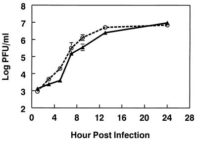
It was possible that the attenuation of s51 reflected a general defect in replication compared to the wild-type s55 virus. Therefore, the ability of the two viruses to grow in BHK-21 cells was compared. BHK cells were infected with either virus at an MOI of 5.0, and virus titers in the supernatant were evaluated over time. Both viruses grew to roughly equivalent titers with the same kinetics (Fig. 2). In fact, s51 titers were always as high as or higher than s55 titers. At 24 h postinfection, s55 plaques had an average diameter of 0.36 ± 0.1 mm, while s51 plaques had an average diameter of 0.51 ± 0.17 mm (P < 0.001; Student's t test, two tailed). These results suggest that s51 grows at least as well as s55 in vitro and may actually have a slight growth advantage.
FIG. 2.
s51 and s55 grow at equivalent rates in vitro. BHK-21 cells were infected in triplicate with the viruses s51 (○) and s55 (▴) at an MOI of 5.0. After 1 h of infection at 37°C, cells were washed three times with room temperature PBS (1% DCS), and 1 ml of growth medium was placed on the cells. One hundred microliters of supernatant was removed for titration by plaque assay at the indicated times. At the time of harvest, the sample volume removed was replaced with fresh media. Samples were titrated by plaque assay on BHK-21 cells. The data shown are from one of three representative experiments. Each data point represents the mean titer ± standard error.
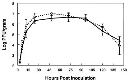
Since s51 grew as well as or better than s55 in vitro, the ability of the two viruses to replicate within the brains of infected animals was evaluated. Six-week-old female CD-1 mice were inoculated i.c. with 103 PFU of s55 or s51, and the levels of infectious virus within the brain were assessed by plaque assay at intervals ranging from 3 to 144 h postinfection. Similar to the in vitro growth curves, s51 grew to levels which were equivalent to those of s55 in the brain (Fig. 3). These results demonstrate that attenuation of s51 cannot be explained by a simple defect in viral replication, either in vitro or within the infected brain.
FIG. 3.
s51 and s55 grow with equivalent kinetics in the brains of infected mice. Six-week-old female CD-1 mice were infected i.c. with 103 PFU of s51 or s55. Mice were sacrificed by exsanguination at 12, 24, 48, 72, 96, 120, and 144 h postinfection and perfused with PBS (pH 7.4). The left hemisphere of each brain was removed and placed in three volumes of PBS (1% DCS, Ca2+-Mg2+). Samples were frozen and thawed before homogenization and titration by plaque assay on BHK-21 cells. Each data point represents the mean titer ± the standard error. ○, s51; ▴, s55. n = 6 to 12 animals for each time point from three pooled experiments.
Localization of s51 and s55 infection within the brain at early and late times postinfection.
Since s51 was attenuated for neurovirulence, but grew in brain tissue to titers equivalent to that of wild-type s55, the neuropathology of these viruses was evaluated at various times postinfection. Six-week-old CD-1 mice were inoculated i.c. with 103 PFU of s55 or s51. Mice were sacrificed at times ranging from 12 to 144 h postinfection, at which point, they were perfused with 4% paraformaldehyde, and brain sections were evaluated for the presence of viral RNA by in situ hybridization with S.A.AR86-specific riboprobes. Both viruses exhibited similar patterns of replication at early times (12 to 72 h) postinfection. Focal areas of viral infection were observed within the hippocampus at 12 to 24 h postinfection for both s51 and s55 (Fig. 4A and B). However, at late times postinfection, the two viruses exhibited dramatically different patterns of growth in the brain. By 144 h postinfection, s55 had spread throughout the brain, with large areas of in situ signal particularly prevalent in the cortex (Fig. 4E). s51 infected the same regions of the brain as s55; however, areas of s51 infection remained more focal than s55-infected areas (Fig. 4F). These results suggest that s51 and s55 established infection in a similar manner. However, s51 was prevented from spreading beyond focal areas of infection, while the wild-type s55 virus spread throughout the brain at late times postinfection. Alternatively, both viruses may spread throughout the brain equivalently, but s51 may have been cleared from the brain, while s55 infection progressed until the death of the host.
FIG. 4.
In situ hybridization to localize sites of infection by s55 or s51. Six-week-old female CD-1 mice were infected i.c. with 103 PFU of s51 (B, D, and F) or s55 (A, C, and E). At 12, 24, 48, 72, 96, 120, and 144 h postinfection, mice were exsanguinated and perfused with 4% paraformaldehyde in PBS (pH 7.4). Brains were divided sagitally down the midline, embedded in paraffin, and sectioned at 5 μm/section. Sections were subjected to in situ hybridization with riboprobes specific for S.A.AR86. Nonspecific binding controls consisted of sections from mock-infected mice probed with the S.A.AR86-specific riboprobe or sections from infected mice probed with a riboprobe specific for influenza virus strain PR/8 hemagglutinin. No specific signal was observed with either of these control groups. Shown are representative sections at 24 (A and B), 72 (C and D), and 144 (E and F) h. There were three mice per group per time point. The data shown are from one of two comparable experiments.
Introduction of a threonine at nsP1 538 of a nonneurovirulent virus.
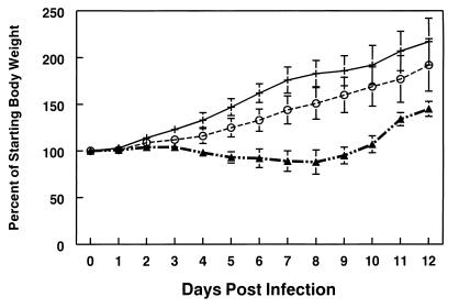
The comparison of s55 and s51 strongly suggested that nsP1 Thr 538 was associated with neurovirulence in adult mice. This hypothesis was tested further by introducing nsP1 Thr 538 into Sindbis virus clone TR339 (14, 26). The resulting virus, 39ns1, was viable as determined by growth in tissue culture, with 39ns1 growing at a similar rate to the wild-type TR339 virus (Fig. 5). At 24 h postinfection TR339 plaques had an average diameter of 0.61 ± 0.15 mm, while 39ns1 plaques had an average diameter of 0.37 ± 0.07 mm (P < 0.001, Student's t test, two tailed). Following i.c. inoculation with 103 PFU of TR339, 18- to 21-day-old mice exhibited only mild disease signs, as shown by a slight lag in weight gain compared to mock-infected animals (Fig. 6). Three out of 18 TR339-infected mice did show signs of mild hind limb paresis involving weakness in one hind limb, while the remaining 15 mice exhibited no signs of paresis. In contrast, 39ns1 infection led to a more severe disease, with mice exhibiting a transient loss in body weight (Fig. 6), as well as paresis in one or both hind limbs in 21 of 22 infected animals. Furthermore, 3 of 22 39ns1-infected mice succumbed to infection with an AST of 8.3 days. The ability of threonine at nsP1 538 to cause increased neurovirulence in a normally nonneurovirulent genetic background strongly suggests that nsP1 Thr 538 is a major determinant of S.A.AR86 neurovirulence in adult mice. However, since 39ns1 did not cause 100% mortality in infected mice, it is likely that other determinants in the S.A.AR86 genome also contribute to the adult neurovirulence phenotype.
FIG. 5.
In vitro growth kinetics of the TR339 and 39ns1 viruses. BHK-21 cells were infected in triplicate at an MOI of 5.0 with either TR339 or 39ns1. Infection was performed as in Fig. 2. Supernatant was sampled at the indicated times postinfection, and virus levels were titrated on BHK-21 cells by plaque assay. ○, TR339; ▴, 39ns1. The data shown are from one of three comparable experiments.
FIG. 6.
39ns1 but not TR339 infection causes morbidity as defined by weight loss in 18-day-old mice. Eighteen-day-old mixed groups of male and female CD-1 mice were infected i.c. with 103 PFU of either TR339 or 39ns1. Mice were weighed daily as an indicator of virus-induced morbidity. Mouse weight is plotted as a percentage of starting body weight over time. The data represent the mean percentage from five (TR339 and 39ns1) or four (mock) animals per group. ○, TR339-infected mice; ▴, 39ns1-infected mice; +, mock-infected mice. Error bars represent the standard error. The data shown represent one of four comparable experiments with 18- to 21-day-old mice.
DISCUSSION
The results presented here are relevant not only to the neurovirulence of Sindbis-group alphaviruses in adult mice, but may have general application to the pathogenesis of arbovirus-mediated encephalitis. Reciprocal genetic changes were used to demonstrate that the amino acid residue at nsP1 538 is an important determinant of neurovirulence in the adult mouse model. In one case, the nsP1 538 residue characteristic of nonneurovirulent Sindbis viruses was substituted for the wild-type residue in a neurovirulent virus, S.A.AR86. The result was a dramatic decrease in the neurovirulence of S.A.AR86. In the reciprocal experiment, the S.A.AR86 residue was introduced into the Sindbis background by using the pTR339 clone, resulting in a significant increase in the neurovirulence of this virus for older animals. These results demonstrate that nsP1 538 plays an important role in the adult neurovirulence phenotype of S.A.AR86 and provide direct evidence that the viral nonstructural genes can contribute to alphavirus neurovirulence.
The neurovirulent NSV strain replicates to higher titers within the brain than nonneurovirulent viruses (11, 40), raising the possibility that s51 was attenuated due to a general decrease in replication within the central nervous system or an inability to replicate effectively within certain cell types or regions of the brain. However, s51 and s55 grew to similar levels within the brain (Fig. 3). Furthermore, while in situ hybridization for viral transcripts did not allow detailed identification of individual cell types, s51 and s55 appeared to replicate within similar regions and cell types within the brain (Fig. 4). At early times postinfection, cells within the hippocampus were infected, while both viruses spread to other regions of the brain in a similar manner during early and intermediate times postinfection (Fig. 4). It was only at late stages of the disease that s51 differed greatly from s55. By day 5 postinfection, s51 and s55 replication was observed in the same regions of the brain. However, s55 infected large areas, while s51 was restricted to focal areas of replication within these regions (Fig. 4). It is interesting to note that while significant differences between s51 and s55 replication were observed within the brain at late times postinfection by in situ hybridization (Fig. 4), the plaque assay showed similar levels of s51 and s55 within the brain at all time points assayed (Fig. 3). However, while viral titers measured by plaque assay were falling for both viruses by day 5 postinfection, in situ hybridization clearly showed increased levels of s55 signal within the brain at day 5. This may reflect the production of anti-S.A.AR86 antibody by the infected animals at late times postinfection. The presence of antibody might decrease the amount of virus within the brain that was detectable by plaque assay, while not affecting intracellular viral replication.
The S.A.AR86 nonstructural proteins are produced as a single polyprotein, which is cleaved to produce the individual nonstructural proteins, as well as cleavage intermediates, that comprise the viral RNA replicase. nsP1 position 538 lies within the cleavage site between nsP1 and nsP2, and changes within this region may affect processing of the nonstructural proteins (reviewed in reference 35). With this in mind, our laboratory is currently investigating the effects of Thr or Ile at nsP1 538 on the cleavage kinetics of the viral nonstructural protein precursor P1234. Alteration in nonstructural protein cleavage could have several downstream effects that alter neurovirulence. Given the role that different ratios of the nonstructural protein precursors, cleavage intermediates, and mature nonstructural proteins have in regulating viral RNA synthesis (18–20), it is possible that changing the threonine to isoleucine at this position might have a positive or negative effect on viral RNA replication and viral growth. In vitro and in vivo growth curves demonstrated that s51 grew at a similar rate to virus derived from the wild-type s55 clone (Fig. 2 and 3), while 39ns1 and TR339 also grew at similar rates. However, it is still possible that the presence of Ile or Thr at nsP1 538 altered viral RNA synthesis without overtly affecting viral yield. In support of this, s51(nsP1 538 Ile) plaques were larger than those of s55 (nsP1 538 Thr), while 39ns1 (nsP1 538 Thr) produced smaller plaques than TR339 (nsP1 538 Ile). Experiments are currently under way to evaluate the effects of Thr or Ile at nsP1 538 on synthesis of the viral negative strand, positive strand, and subgenomic RNAs. Alternatively, the presence of Thr versus Ile at nsP1 538 might alter the interaction of nsP1 or one of its precursors with host cell factors, which could have downstream effects on survival of the infected cell or induction of the antiviral immune response. Precedence for this with Sindbis-group viruses is provided by the ability of histidine at position 55 of the E2 glycoprotein to overcome bcl-2-mediated protection of cells from virus-induced apoptosis in vitro (41). Furthermore, changes in the 3A protein of foot-and-mouth disease virus affect the ability of the virus both to replicate in bovine cells and to cause disease in cattle (1). Therefore, further analysis of whether s51 and s55 differ in their ability to kill neurons or other cell types, as well as the in-depth characterization of the antiviral immune response following infection with either s51 or s55, may provide insight into the mechanism(s) by which nsP1 538 contributes to neurovirulence.
There is precedent in nature for alphavirus genes other than those coding for the glycoproteins contributing to neurovirulence. Western equine encephalitis virus (WEE) is thought to have originated through a recombination event between Eastern equine encephalitis virus (EEE) and a Sindbis-like virus (8). This resulted in the glycoproteins of WEE being derived from the Sindbis-like virus and the nonstructural and capsid genes from EEE. Since Sindbis-group viruses are not associated with encephalitis in humans, this suggests that the molecular determinants of WEE neurovirulence might map to the EEE-derived sequences in the nonstructural and capsid genes. Further evaluation of this possibility may shed additional light on viral determinants of alphavirus neurovirulence.
ACKNOWLEDGMENTS
M.T.H. and D.A.S. contributed equally to this work.
This research was funded by NIH research grant RO1 AI22186. M.T.H. was supported by an institutional NIH postdoctoral training grant (T32 AI07151) and an NIH postdoctoral fellowship (F32 AI10146).
We thank William Klimstra and Kate Ryman for assistance in engineering the virus clone p39ns1. Kristen Bernard provided assistance in evaluating clinical signs in infected mice. We thank the entire Johnston laboratory for helpful discussion of this project. Cherice Connor, Michael Hawley, Jacqueline Bailey, and Dwayne Muhammed provided excellent technical support with cell culture.
REFERENCES
- 1.Beard C W, Mason P W. Genetic determinants of altered virulence of Taiwanese foot-and-mouth disease virus. J Virol. 2000;74:987–991. doi: 10.1128/jvi.74.2.987-991.2000. [DOI] [PMC free article] [PubMed] [Google Scholar]
- 2.Charles P C, Walters E, Margolis F, Johnston R E. Mechanism of neuroinvasion of Venezuelan equine encephalitis virus in the mouse. Virology. 1995;208:662–671. doi: 10.1006/viro.1995.1197. [DOI] [PubMed] [Google Scholar]
- 3.Davis N L, Brown K W, Johnston R E. A viral vaccine vector that expresses foreign genes in lymph nodes and protects against mucosal challenge. J Virol. 1996;70:3781–3787. doi: 10.1128/jvi.70.6.3781-3787.1996. [DOI] [PMC free article] [PubMed] [Google Scholar]
- 4.Ding M X, Schlesinger M J. Evidence that Sindbis virus NSP2 is an autoprotease which processes the virus nonstructural polyprotein. Virology. 1989;171:280–284. doi: 10.1016/0042-6822(89)90539-4. [DOI] [PubMed] [Google Scholar]
- 5.Dubuisson J, Lustig S, Ruggli N, Akov Y, Rice C M. Genetic determinants of Sindbis virus neuroinvasiveness. J Virol. 1997;71:2636–2646. doi: 10.1128/jvi.71.4.2636-2646.1997. [DOI] [PMC free article] [PubMed] [Google Scholar]
- 6.Gomez-de Cedron M, Ehsani N, Mikkola M L, Garcia J A, Kaariainen L. RNA helicase activity of Semliki Forest virus replicase protein NSP2. FEBS Lett. 1999;448:19–22. doi: 10.1016/s0014-5793(99)00321-x. [DOI] [PubMed] [Google Scholar]
- 7.Griffin D E, Johnson R T. Role of the immune response in recovery from Sindbis virus encephalitis in mice. J Immunol. 1977;118:1070–1075. [PubMed] [Google Scholar]
- 8.Hahn C S, Lustig S, Strauss E G, Strauss J H. Western equine encephalitis virus is a recombinant virus. Proc Natl Acad Sci USA. 1988;85:5997–6001. doi: 10.1073/pnas.85.16.5997. [DOI] [PMC free article] [PubMed] [Google Scholar]
- 9.Hahn Y S, Grakoui A, Rice C M, Strauss E G, Strauss J H. Mapping of RNA− temperature-sensitive mutants of Sindbis virus: complementation group F mutants have lesions in nsP4. J Virol. 1989;63:1194–1202. doi: 10.1128/jvi.63.3.1194-1202.1989. [DOI] [PMC free article] [PubMed] [Google Scholar]
- 10.Hardy W R, Strauss J H. Processing the nonstructural polyproteins of Sindbis virus: nonstructural proteinase is in the C-terminal half of nsP2 and functions both in cis and in trans. J Virol. 1989;63:4653–4664. doi: 10.1128/jvi.63.11.4653-4664.1989. [DOI] [PMC free article] [PubMed] [Google Scholar]
- 11.Jackson A C, Moench T R, Trapp B D, Griffin D E. Basis of neurovirulence in Sindbis virus encephalomyelitis of mice. Lab Investig. 1988;58:503–509. [PubMed] [Google Scholar]
- 12.Johnston R E, Peters C J. Alphaviruses. In: Fields B N, Knipe D M, Howley P M, editors. Fields virology. Philadelphia, Pa: Lippincott-Raven; 1996. pp. 843–898. [Google Scholar]
- 13.Kamer G, Argos P. Primary structural comparison of RNA-dependent polymerases from plant, animal and bacterial viruses. Nucleic Acids Res. 1984;12:7269–7282. doi: 10.1093/nar/12.18.7269. [DOI] [PMC free article] [PubMed] [Google Scholar]
- 14.Klimstra W B, Ryman K D, Johnston R E. Adaptation of Sindbis virus to BHK cells selects for use of heparan sulfate as an attachment receptor. J Virol. 1998;72:7357–7366. doi: 10.1128/jvi.72.9.7357-7366.1998. [DOI] [PMC free article] [PubMed] [Google Scholar]
- 15.Klimstra W B, Ryman K D, Bernard K A, Nguyen K B, Biron C A, Johnston R E. Infection of neonatal mice with Sindbis virus results in a systemic inflammatory response syndrome. J Virol. 1999;73:10387–10398. doi: 10.1128/jvi.73.12.10387-10398.1999. [DOI] [PMC free article] [PubMed] [Google Scholar]
- 16.Kobiler D, Rice C M, Brodie C, Shahar A, Dubuisson J, Halevy M, Lustig S. A single nucleotide change in the 5′ noncoding region of Sindbis virus confers neurovirulence in rats. J Virol. 1999;73:10440–10446. doi: 10.1128/jvi.73.12.10440-10446.1999. [DOI] [PMC free article] [PubMed] [Google Scholar]
- 17.Kuhn R J, Griffin D E, Zhang H, Niesters H G, Strauss J H. Attenuation of Sindbis virus neurovirulence by using defined mutations in nontranslated regions of the genome RNA. J Virol. 1992;66:7121–7127. doi: 10.1128/jvi.66.12.7121-7127.1992. [DOI] [PMC free article] [PubMed] [Google Scholar]
- 18.Lemm J A, Rice C M. Assembly of functional Sindbis virus RNA replication complexes: requirement for coexpression of P123 and P34. J Virol. 1993;67:1905–1915. doi: 10.1128/jvi.67.4.1905-1915.1993. [DOI] [PMC free article] [PubMed] [Google Scholar]
- 19.Lemm J A, Rice C M. Roles of nonstructural polyproteins and cleavage products in regulating Sindbis virus RNA replication and transcription. J Virol. 1993;67:1916–1926. doi: 10.1128/jvi.67.4.1916-1926.1993. [DOI] [PMC free article] [PubMed] [Google Scholar]
- 20.Lemm J A, Rümenapf T, Strauss E G, Strauss J H, Rice C M. Polypeptide requirements for assembly of functional Sindbis virus replication complexes: a model for the temporal regulation of minus- and plus-strand RNA synthesis. EMBO J. 1994;13:2925–2934. doi: 10.1002/j.1460-2075.1994.tb06587.x. [DOI] [PMC free article] [PubMed] [Google Scholar]
- 21.Levine B, Goldman J E, Jiang H H, Griffin D E, Hardwick J M. Bc1-2 protects mice against fatal alphavirus encephalitis. Proc Natl Acad Sci USA. 1996;93:4810–4815. doi: 10.1073/pnas.93.10.4810. [DOI] [PMC free article] [PubMed] [Google Scholar]
- 22.Levine B, Huang Q, Isaacs J T, Reed J C, Griffin D E, Hardwick J M. Conversion of lytic to persistent alphavirus infection by the bcl-2 cellular oncogene. Nature. 1993;361:739–742. doi: 10.1038/361739a0. [DOI] [PubMed] [Google Scholar]
- 23.Li G P, La Starza M W, Hardy W R, Strauss J H, Rice C M. Phosphorylation of Sindbis virus nsP3 in vivo and in vitro. Virology. 1990;179:416–427. doi: 10.1016/0042-6822(90)90310-n. [DOI] [PubMed] [Google Scholar]
- 24.Lustig S, Jackson A C, Hahn C S, Griffin D E, Strauss E G, Strauss J H. Molecular basis of Sindbis virus neurovirulence in mice. J Virol. 1988;62:2329–2336. doi: 10.1128/jvi.62.7.2329-2336.1988. [DOI] [PMC free article] [PubMed] [Google Scholar]
- 25.Malherbe H, Strickland-Cholmley M, Jackson A L. Sindbis virus infection in man. S Afr Med J. 1963;37:547–552. [PubMed] [Google Scholar]
- 26.McKnight K L, Simpson D A, Lin S-C, Knott T A, Polo J M, Pence D F, Johannsen D B, Heidner H W, Davis N L, Johnston R E. Deduced consensus sequence of Sindbis virus strain AR339: mutations contained in laboratory strains which affect cell culture and in vivo phenotypes. J Virol. 1996;70:1981–1989. doi: 10.1128/jvi.70.3.1981-1989.1996. [DOI] [PMC free article] [PubMed] [Google Scholar]
- 27.Mi S, Durbin R, Huang H V, Rice C M, Stollar V. Association of the Sindbis virus RNA methyltransferase activity with the nonstructural protein nsP1. Virology. 1989;170:385–391. doi: 10.1016/0042-6822(89)90429-7. [DOI] [PubMed] [Google Scholar]
- 28.Norder H, Lundstrom J O, Kozuch O, Magnius L O. Genetic relatedness of Sindbis virus strains from Europe, Middle East, and Africa. Virology. 1996;222:440–445. doi: 10.1006/viro.1996.0441. [DOI] [PubMed] [Google Scholar]
- 29.Rice C M, Strauss J H. Nucleotide sequence of the 26S mRNA of Sindbis virus and deduced sequence of the encoded virus structural proteins. Proc Natl Acad Sci USA. 1981;78:2062–2066. doi: 10.1073/pnas.78.4.2062. [DOI] [PMC free article] [PubMed] [Google Scholar]
- 30.Sarkar G, Sommer S S. The “megaprimer” method of site-directed mutagenesis. BioTechniques. 1990;8:404–407. [PubMed] [Google Scholar]
- 31.Scheidel L M, Stollar V. Mutations that confer resistance to mycophenolic acid and ribavirin on Sindbis virus map to the nonstructural protein nsP1. Virology. 1991;181:490–499. doi: 10.1016/0042-6822(91)90881-b. [DOI] [PubMed] [Google Scholar]
- 32.Shirako Y, Niklasson B, Dalrymple J M, Strauss E G, Strauss J H. Structure of the Ockelbo virus genome and its relationship to other Sindbis viruses. Virology. 1991;182:753–764. doi: 10.1016/0042-6822(91)90616-j. [DOI] [PubMed] [Google Scholar]
- 33.Simpson D A, Davis N L, Seh-Ching L, Russell D, Johnston R E. Complete nucleotide sequence and full-length cDNA clone of S.A.AR86, a South African alphavirus related to Sindbis. Virology. 1996;222:464–469. doi: 10.1006/viro.1996.0445. [DOI] [PubMed] [Google Scholar]
- 34.Skogh M, Espmark Å. Ockelbo disease: epidemic arthritis-exanthema syndrome in Sweden caused by Sindbis-virus like agent. Lancet. 1982;i:795–796. doi: 10.1016/s0140-6736(82)91834-7. [DOI] [PubMed] [Google Scholar]
- 35.Strauss J H, Strauss E G. The alphaviruses: gene expression, replication, and evolution. Microbiol Rev. 1994;58:491–562. doi: 10.1128/mr.58.3.491-562.1994. [DOI] [PMC free article] [PubMed] [Google Scholar]
- 36.Taylor R M, Hurlbut H S, Work T H, Kingston J R, Frothingham T E. Sindbis virus: a newly recognized arthropod-transmitted virus. Am J Trop Med Hyg. 1955;4:844–862. doi: 10.4269/ajtmh.1955.4.844. [DOI] [PubMed] [Google Scholar]
- 37.Trgovcich J, Aronson J F, Eldridge J C, Johnston R E. TNF-alpha, interferon and stress response induction as a function of age-related susceptibility to fatal Sindbis virus infection of mice. Virology. 1999;263:339–348. doi: 10.1006/viro.1999.9913. [DOI] [PubMed] [Google Scholar]
- 38.Trgovcich J, Aronson J F, Johnston R E. Fatal Sindbis virus infection of neonatal mice in the absence of encephalitis. Virology. 1996;224:73–83. doi: 10.1006/viro.1996.0508. [DOI] [PubMed] [Google Scholar]
- 39.Tucker P C, Lee S H, Bui N, Martinie D, Griffin D E. Amino acid changes in the Sindbis virus E2 glycoprotein that increase neurovirulence improve entry into neuroblastoma cells. J Virol. 1997;71:6106–6112. doi: 10.1128/jvi.71.8.6106-6112.1997. [DOI] [PMC free article] [PubMed] [Google Scholar]
- 40.Tucker P C, Strauss E G, Kuhn R J, Strauss J H, Griffin D E. Viral determinants of age-dependent virulence of Sindbis virus for mice. J Virol. 1993;67:4605–4610. doi: 10.1128/jvi.67.8.4605-4610.1993. [DOI] [PMC free article] [PubMed] [Google Scholar]
- 41.Ubol S, Tucker P C, Griffin D E, Hardwick J M. Neurovirulent strains of Alphavirus induce apoptosis in bcl-2-expressing cells: role of a single amino acid change in the E2 glycoprotein. Proc Natl Acad Sci USA. 1994;91:5202–5206. doi: 10.1073/pnas.91.11.5202. [DOI] [PMC free article] [PubMed] [Google Scholar]
- 42.Weinbren M P, Kokernot R H, Smithburn K C. Strains of Sindbis-like virus isolated from Culicine mosquitoes in the Union of South Africa. S Afr Med J. 1956;30:631–636. [PubMed] [Google Scholar]


