Abstract
Recombinant modified vaccinia virus Ankara (MVA), encoding the measles virus (MV) fusion (F) and hemagglutinin (H) (MVA-FH) glycoproteins, was evaluated in an MV vaccination-challenge model with macaques. Animals were vaccinated twice in the absence or presence of passively transferred MV-neutralizing macaque antibodies and challenged 1 year later intratracheally with wild-type MV. After the second vaccination with MVA-FH, all the animals developed MV-neutralizing antibodies and MV-specific T-cell responses. Although MVA-FH was slightly less effective in inducing MV-neutralizing antibodies in the absence of passively transferred antibodies than the currently used live attenuated vaccine, it proved to be more effective in the presence of such antibodies. All vaccinated animals were effectively protected from the challenge infection. These data suggest that MVA-FH should be further tested as an alternative to the current vaccine for infants with maternally acquired MV-neutralizing antibodies and for adults with waning vaccine-induced immunity.
Measles is a highly contagious infectious disease that continues to be a major cause of morbidity and mortality for infants, with an estimated number of 1 million deaths annually (8). Inactivated whole-virus vaccine preparations used in the 1960s did not induce long-lasting protection and were shown to predispose for severe immunopathological complications collectively referred to as the atypical measles syndrome (7, 14). In the 1970s, live attenuated measles virus (MV) vaccines which proved to be safe and effective were introduced. Application of these vaccines, which are still being used, resulted in a significant reduction of the global numbers of measles cases and largely abrogated the circulation of wild-type MV in the industrialized world. However, measles vaccination proved less effective in a number of developing countries, where measles continues to be endemic. Several factors are responsible for this reduced effectiveness, most of which are related to logistic problems like vaccination coverage and cold chain maintenance (3). However, an important additional factor is that measles frequently occurs at an early age (<9 months) in developing countries. At this age, preexisting MV-specific maternal antibody may interfere with the replication of live attenuated vaccine virus, resulting in suboptimal protection upon vaccination (16). The World Health Organization has proposed a global measles eradication strategy based on the current live attenuated MV vaccine (28). However, it is uncertain if this vaccine will be able to achieve a sufficient level of herd immunity to completely abrogate circulation of MV. Several outbreaks of clinical and subclinical measles have been described among vaccinated populations (10), and in the final stages of an eradication campaign, vaccines which are able to boost low levels of immunity may be needed.
In recent years, a number of new generation candidate MV vaccines have been developed, including immune stimulating complexes (iscoms), DNA vaccines, and recombinant poxviruses (17). An iscom-based vaccine proved to be effective in inducing protective immunity in macaques even in the presence of passively acquired MV-neutralizing antibodies (26). In contrast, recombinant vaccinia viruses (rVVs) encoding the MV fusion (F) and hemagglutinin (H) proteins, although able to induce strong MV-specific virus-neutralizing (VN) antibody and T-cell responses, were only partially effective when used in the presence of passively acquired MV-neutralizing antibodies (26). In addition, concerns about the safety of vaccinia virus made this vaccine candidate less attractive. Recently, recombinant poxviruses were developed based on the replication-deficient modified vaccinia virus Ankara (MVA) (15). This strain was proven safe for use in humans during its application in the late stage of the smallpox eradication campaign (23). Compared to fully replication-competent strains of vaccinia virus, MVA induced similar expression levels of the recombinant genes (24) and induced equal or better B- and T-cell responses in animals (11, 21, 25).
Here we describe the evaluation of a recombinant MVA-based candidate vaccine containing the MV F and H (MVA-FH) genes in an MV vaccination-challenge model in macaques. MVA-FH successfully induced MV-specific antibody and T-cell responses, including CD8+ T cells, both in the absence and presence of passively transferred MV-specific antibodies. All vaccinated macaques were still effectively protected from intratracheal challenge with wild-type MV 1 year after vaccination. The use of a nonreplicating candidate measles vaccine is not likely to predispose for atypical measles-like immunopathology. Collectively, these properties would favor MVA-FH as a candidate measles vaccine that either alone or as part of a prime-boost strategy could be used in a measles eradication program.
(This paper was presented at the XIth International Congress of Virology, Sydney, Australia, 10 August 1999.)
MATERIALS AND METHODS
Macaques.
The studies were carried out with eight captive-bred subadult healthy female cynomolgus macaques (Macaca fascicularis) which were all confirmed MV seronegative. The animals were housed together except during the vaccination and challenge periods, when they were kept as pairs in separate cages.
Viruses.
A recombinant MVA that expresses the MV (Edmonston strain) F and H glycoproteins was made by using previously described procedures (5). The F and H genes, contained within plasmids pTM-F/MV and pTM-H/MV, respectively (22), were excised by digestion with NcoI and StuI, and the overhanging ends were filled in with Klenow enzyme. The H gene was inserted into the SmaI site of pLW-17 (29), a plasmid transfer vector which contains the modified H5 promoter (20) to express the recombinant foreign gene and inserts within deletion II of MVA. The F gene was inserted into the SmaI site of the MVA vector pLW-24, which contained the 7.5 promoter of vaccinia virus (13) and MVA flanks for insertion into deletion III of MVA (24). Initial attempts to make recombinant MVA stably expressing the F protein under stronger vaccinia virus promoters, modified H5 or the strong synthetic promoter, were unsuccessful.
Recombinant MVA viruses expressing the measles virus F or H proteins were made by transfecting pLW-17 or pLW-24, containing the measles virus H or F gene, respectively, into chick embryo fibroblasts (CEF) that were infected with MVA as previously described (22). Recombinant virus was obtained by immunostaining recombinant MVA foci utilizing polyclonal anti-MV virus rabbit serum (Accurate Scientific, Westbury, N.Y.). A double recombinant MVA expressing both the F and H measles glycoproteins was made by transfecting the MV H-containing plasmid into the single recombinant MVA expressing the F gene. Double recombinant MVA foci were obtained by staining with polyclonal anti-MV antiserum that had been adsorbed with CEF cells infected with the single recombinant MVA-F. This double MVA recombinant was designated MVA-FH. Stocks of MVA and recombinant MVA-FH were prepared in secondary CEF as previously described (22).
The recombinant MVA was aliquoted and stored at −80°C. The samples were thawed and sonicated in ice for 30 s with a cup sonicator (Sonicor Instruments Corporation, Copiaque, N.Y.) shortly before inoculation.
Immunization and sampling.
Macaques were inoculated intramuscularly and intranasally (108 PFU at each site) at weeks 0 and 4. Six macaques were vaccinated with MVA-FH, of which three were naive (animal numbers H1, H5, and H7) and three had been passively immunized with anti-MV serum (BMS94) 48 h before vaccination (animal numbers UH3, UH4, and K128) (26). This serum, which has a specific virus-neutralizing (VN) antibody level of 40 IU/ml (6), was administered intravenously at a dosage of 0.7 ml per kg of body weight. This resulted in VN antibody levels at the time of the first vaccination of about 0.3 IU/ml, as was also observed in a previous study (26). The two other macaques (animal numbers H4 and H6) were vaccinated with wild-type MVA (MVA-wt) at identical dosages and vaccination routes. During the 1-year period between vaccination and challenge, heparinized blood samples were collected at regular intervals, with initial intervals of a week but later less frequently. To be able to compare the data with those from previous vaccination experiments, plasma samples obtained from macaques vaccinated with MV-Schwarz or rVV-FH were reassayed (26).
Challenge and sampling.
One year after vaccination, the macaques were challenged with wild-type MV as previously described (26, 27). Briefly, 1,000 50% tissue culture infective doses (TCID50s) of the wild-type MV strain BIL was diluted in 5 ml of phosphate-buffered saline (PBS) and administered via the intratracheal route at day 0. Pharyngeal epithelial cells (PEC), lung lavage cells (LLC), and peripheral blood mononuclear cells (PBMC), as well as plasma, were collected at days 3, 6, 9, 13, and 17, and additional plasma and PBMC samples were collected at days 0, 24, and 41. PEC were collected by using a Cytobrush Plus (Medscand Medical AB, Malmö, Sweden) that was applied into the throat by using a laryngoscope. After sampling, the brush was transferred into a tube that contained 2 ml of RPMI 1640 supplemented with penicillin (100 U/ml), streptomycin (100 μg/ml), l-glutamine (2 mM), 2-mercaptoethanol (10−5 M), and 10% fetal bovine serum (FBS) (referred to as culture medium [CM]). The tube was vortexed, and following removal of the brush, cells were pelleted by centrifugation (5 min at 400 × g). The cell pellet was then resuspended in 1 ml of CM. Lungs were lavaged with 10 ml of PBS by a small catheter that was applied into the lungs by using a laryngoscope. About 5 ml of lavage fluid containing the LLC was recovered. This LLC suspension was transferred to a tube, pelleted by centrifugation (5 min at 400 × g), and resuspended in CM. PBMC were isolated from heparinized blood by density gradient centrifugation (20 min at 600 × g) by using a gradient consisting of Optiprep (density, 1.32 g/ml; Nycomed), 6% Dextran (Sigma) in H2O, and 10 times concentrated PBS (16.7%–75%–8.3% [vol/vol/vol]).
MV reisolation.
Following MV challenge, virus replication was monitored by cocultivating the LLC, PBMC, and PEC (collected at days 3, 6, 9, 13, and 17) with a human Epstein-Barr virus-transformed B-lymphoblastic cell line (hu.B-LCL). Four replicates of 100 μl of PEC suspension were cocultivated with 2 × 10⁵ cells of hu.B-LCL in 1 ml of CM in a 24-well plate (Greiner Labor Technik, Nürtingen, Germany). The MV cytopathic effect (CPE) in one or more of the PEC-hu.B-LCL cocultivations was interpreted as positive reisolation. Virus isolations from LLC and PBMC were carried out by cocultivation of a 2-log dilution range of the macaque cells with a standard amount of the hu.B-LCL. Briefly, 3.2 × 10⁵ LLC or PBMC were divided over eight wells of a 96-well round-bottom plate (Greiner) in CM (200 μl/well). In the case of the PBMC, 1 μg of phytohemagglutinin-L (Boehringer Mannheim, Almere, The Netherlands) was added to this medium and the cells were incubated at 37°C for 2 h. Subsequently, a 2-log dilution range of the LLC or PBMC was prepared in the plate (ranging from 2−1 to 2−11), and hu.B-LCL cells were added at an amount of 1 × 10⁴ cells/well. After screening for the MV CPE during the following week, the numbers of MV-infected cells were calculated by using the formula of Reed and Muench (19).
Vaccinia virus-specific antibody response.
Rabbit kidney (RK-13) cells were infected with wild-type vaccinia virus at a multiplicity of infection of 10. Seven hours after infection, the cells were trypsinized and used as target cells in a fluorescence-activated cell sorter (FACS)-measured immunofluorescence assay. The cells (1 × 10⁵ to 3 × 10⁵ cells/100 μl) were incubated with plasma samples diluted 1:100 in PBS supplemented with fetal bovine serum (FBS). After a 1-h incubation on ice, cells were washed and subsequently incubated with fluorescein isothiocyanate (FITC)-labeled rabbit anti-human immunoglobulin G (IgG) [F(ab′)2 fragments; DAKO, Glostrup, Denmark]. After another hour on ice, cells were washed twice with PBS and fixed with 1% paraformaldehyde for 20 min on ice. Fluorescence signals were measured by using a FACScan (Becton Dickinson). Fluorescence intensity was quantified by determining the geometric mean of the fluorescence histograms.
MV-F- and MV-H-specific immunofluorescence assay.
Antibodies directed against the F and H glycoproteins were detected by a FACS-measured immunofluorescence assay using transfected human melanoma cell lines as targets, as previously described (4). Briefly, Mel-JuSo cells (wild type, MV-F, and MV-H) were incubated with plasma samples diluted 1:100 in PBS supplemented with 2% FBS (1 × 10⁵ to 3 × 10⁵ cells/100 μl). After a 1-h incubation on ice, cells were washed and subsequently stained with FITC-labeled rabbit anti-human IgG or IgM [F(ab′)2 fragments; DAKO]. Fluorescence signals were quantified by a FACScan.
MV-N-specific IgM capture ELISA.
IgM antibodies directed against the MV nucleoprotein (N) were measured in a capture enzyme-linked immunosorbent assay (ELISA) using purified recombinant baculovirus-expressed N protein (kind gift of T. F. Wild, Lyon, France) directly conjugated with horseradish peroxidase (N-HRP). ELISA plates (Greiner) coated with rabbit anti-human IgM antibodies (Meddens Diagnostics, Vorden, The Netherlands) were washed with demineralized H2O containing 0.05% Tween 80, followed by incubation with plasma samples diluted 1:100 in ELISA buffer (Meddens Diagnostics). After 1 h at 37°C, the plates were washed and incubated with N-HRP. Following another hour at 37°C, the plates were washed again and incubated with substrate solution (tetramethylbenzidin; Meddens Diagnostics). Results were expressed as the absorbance at 450 nm.
MV VN assay.
Serial 2-log dilutions (starting at 2−2) of heat-inactivated (30 min at 56°C) plasma samples were incubated in duplicate with 60 TCID50s of MV Edmonston for 1 h at 37°C in 96-well flat-bottom plates (Greiner) in Dulbecco minimal essential medium supplemented with 2% FBS. Subsequently, trypsinized Vero cells were added at a concentration of 1 × 10⁴ cells/well. Plates were incubated for 5 days at 37°C and visually monitored for the MV CPE. VN antibody titers were calculated as the means of the highest plasma dilutions still yielding a 100% reduction of cytopathic changes. The titers were transformed to international units per milliliter by using the National Institute for Biological Standards and Control reference serum (5 IU/ml; human anti-MV serum, 2nd International Standard 1990), which in our assay had a VN antibody titer of 28.5 (6). The VN antibody level in a plasma sample expressed as international units per milliliter was calculated by using the following formula: VN level = (2x/22.85) × 5 IU/ml, in which 2x is the VN titer measured in that plasma sample.
MV-specific T-cell responses.
PBMC were isolated as described above. Cells were cultured in 96-well round-bottom plates (Greiner) in CM supplemented with 1% pooled macaque serum containing MV-specific antibodies at a concentration of 1 × 10⁵ cells/well. These cells were cocultivated with UV-irradiated autologous herpesvirus papio-transformed B cells (mac.B-LCL) which had been infected with MV Edmonston 48 h before at an effector-to-target ratio of 0.1. After 3 days, recombinant human interleukin-2 (rhIL-2) or a mixture of rhIL-2 and rhIL-4 was added, and cultures were maintained for 12 to 14 days. Cells were harvested and treated with chymotrypsin (type II; Sigma) in order to remove CD69 from the membrane (12). Briefly, cells were washed with PBS, incubated with 0.1% (wt/vol) chymotrypsin in PBS for 10 min at 37°C, and subsequently washed with CM. Subsequently, cells were cultivated for 6 h with UV-irradiated autologous MV-infected mac.B-LCL cells, uninfected mac.B-LCL cells, or without mac.B-LCL cells. After this restimulation, cells were stained with anti-CD3-FITC (BPRC, Rijswijk, The Netherlands), anti-CD69-PE (Becton Dickinson), and anti-CD8-RPE/Cy5 (DAKO) and fluorescence was measured by using a FACScan. CD69 expression on CD8− and CD8+ cells was determined in the CD3+ fraction of the lymphocytes as gated in a forward light scatter versus side light scatter diagram. During previous experiments we had observed that varying percentages (ranging from 5 to 25%) of cells continued to express low levels of CD69 after 2 to 3 weeks of bulk culture. Therefore, we removed any residual CD69 by using chymotrypsin, ensuring that CD69 levels measured in the assay were indeed induced during the 6-h restimulation. Background levels of CD69-expressing cells in the medium control of our assay represent cells that either produce CD69 endogenously or express CD69 from recycled membranes.
The animal study was approved by the Local Animal Ethics Committee of the National Institute of Public Health and the Environment, Bilthoven, The Netherlands and carried out according to animal experimentation guidelines.
RESULTS
Expression of F and H glycoproteins by recombinant MVA.
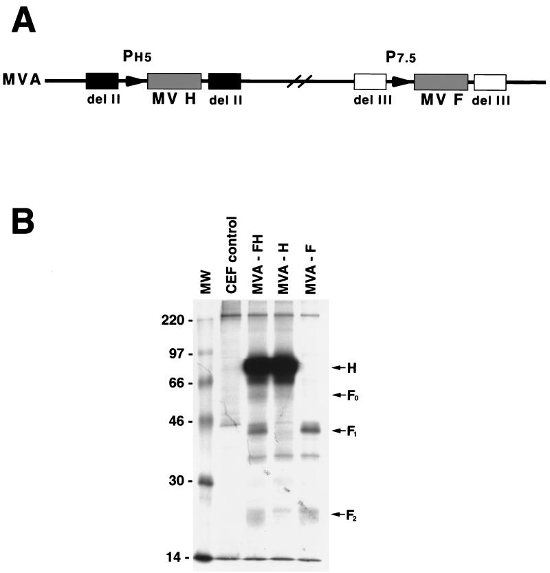
Recombinant MVA that expressed either the MV F or H proteins or both F and H proteins were constructed. The genome of the double recombinant virus is represented in Fig. 1A. Expression of the MV proteins was demonstrated by labeling MVA-infected CEF with [35S]methionine, incubating the lysates with MV polyclonal or monoclonal (not shown) antibodies, and analyzing the bound proteins by sodium dodecyl sulfate-polyacrylamide gel electrophoresis (Fig. 1B). The H glycoprotein, from cells infected with MVA-H or MVA-FH, migrated as a single major band of the expected size. The F glycoprotein was resolved as F1 and F2 subunits. When monkey BSC-1 cells were infected with the double recombinant virus but not with either of the single recombinants, syncytia formed, indicating that F and H proteins were expressed on the cell surface and were functional (data not shown). The double recombinant MVA-FH was used for all vaccination studies.
FIG. 1.
Expression of MV F and H glycoproteins by MVA-FH. (A) Diagram of the genome of MVA-FH. del II, deletion II; del III, deletion III; PH5, modified H5 promoter; P7.5, 7.5 promoter. (B) Sodium dodecyl sulfate-polyacrylamide gel electrophoresis of [35S]methione-labeled proteins from CEF infected with MVA-FH and immunoprecipitated with measles polyclonal antibody. MW, molecular weights of marker proteins in thousands. The positions of MV H, F0, F1, and F2 proteins are indicated.
MVA-FH vaccination induced antibody responses.
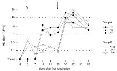
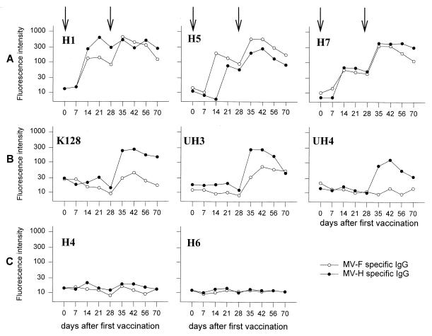
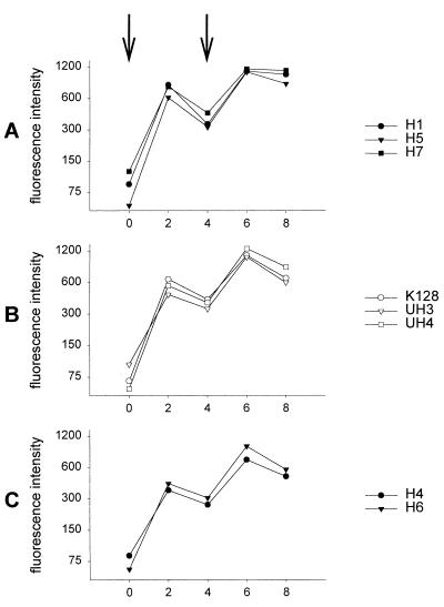
As shown in Fig. 2 and 3A, vaccination of three MV-seronegative macaques with MVA-FH induced MV-neutralizing antibodies (ranging from 2.0 to 4.0 IU/ml) and F-specific and H-specific antibodies (fluorescence signals ranging from 50 to 87 and from 56 to 294, respectively). Antibody responses proved to be boosted in all three animals by a second MVA-FH vaccination 4 weeks later (ranging from 10 to 40 IU of VN antibody/ml, and fluorescence signals ranging from 340 to 649 for F-specific antibodies and 198 to 523 for H-specific antibodies). All animals remained MV seropositive until challenge a year later. Initial vaccination of macaques passively immunized with MV-specific VN antibodies induced lower MV-specific antibody responses than in vaccinated naive animals, but after the booster vaccination, high levels of VN antibodies were induced in these animals as well (Fig. 2). At the moment of the second vaccination, all three of these macaques had VN titers of about 0.1 IU/ml. VN serum antibody titers measured 1 year after the first vaccination ranged from 0.7 to 3.6 IU/ml in these six MVA-FH-vaccinated macaques. The H antibody titers of the passively immunized monkeys were also boosted to levels comparable to those of the naïve animals, whereas the F-specific antibodies remained lower (Fig. 3). No MV-specific antibody responses were detected in the control animals vaccinated with MVA-wt (Fig. 3C). The kinetics of vaccinia virus-specific antibody responses were similar in all eight macaques upon primary and secondary vaccinations (Fig. 4), indicating that the passively acquired MV-specific antibodies did not prevent infection of cells by MVA.
FIG. 2.
Development of VN antibody responses in plasma of macaques vaccinated at weeks 0 and 4 (indicated with arrows) with MVA-FH in the absence (group A) or in the presence (group B) of passively transferred MV-specific VN antibodies.
FIG. 3.
Development of MV glycoprotein-specific plasma IgG responses in macaques vaccinated at weeks 0 and 4 (indicated with arrows) with MVA-FH in the absence (A) or in the presence (B) of passively transferred MV-specific VN antibodies. The control macaques (C) were vaccinated with MVA-wt.
FIG. 4.
Development of MVA-specific plasma IgG responses. The first and second vaccinations are indicated with arrows.
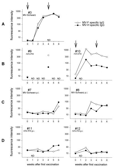
For comparison of the immunogenicity of MVA-FH with those of MV-Schwarz and rVV-FH, we reassayed plasma samples from a previous experiment (26) in which the same vaccination regimen had been used. The levels of MV-neutralizing, as well as F- and H-specific, antibodies induced by MVA-FH in seronegative macaques were similar to those induced by rVV-FH, but slightly lower than those induced by MV Schwarz (Fig. 5A and B) (26). However, when comparing the animals vaccinated in the presence of passively transferred MV-specific antibodies, the responses measured in the MVA-FH-vaccinated animals were substantially higher than those of macaques vaccinated with either MV-Schwarz or rVV-FH (Fig. 5C and D) (26).
FIG. 5.
Development of MV glycoprotein-specific plasma IgG responses in macaques vaccinated at weeks 0 and 4 with MV-Schwarz or rVV-FH in the absence (A and B, respectively) and presence (C and D, respectively) of MV-specific VN antibodies. p.i., passively immunized; ND, not done (due to unavailability of historical plasma).
MVA-FH vaccination-induced MV-specific T-cell responses.
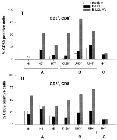
Eight weeks after the second vaccination, the phenotype of the vaccination-induced MV-specific T cells was determined. Following MV-specific bulk stimulation of PBMC with MV-infected autologous mac.B-LCL cells, the presence of MV-specific CD8− and/or CD8+ T cells could be demonstrated in the MVA-FH-vaccinated macaques. In animal H1, no CD3+CD8− cells could be expanded, while in animal H5 no specific CD3+CD8+ cells could be demonstrated (Fig. 6).
FIG. 6.
MV-specific T-cell responses in PBMC bulk cultures of macaques 8 weeks after vaccination with MVA-FH in the absence (A) or presence (B) of passively transferred MV-specific VN antibodies or with MVA-wt (C). PBMC were stimulated once in vitro with autologous MV-infected mac.B-LCL cells and expanded in the presence of rhIL-2 alone or in the presence of both rhIL-2 and rhIL-4 (indicated with an asterisk). After 12 to 14 days, cells were harvested and treated with chymotrypsin to strip preexisting CD69 molecules from the membrane surface. Subsequently, cells were restimulated for 6 h with UV-inactivated autologous MV-infected mac.B-LCL cells, uninfected mac.B-LCL cells, or without mac.B-LCL cells (medium), and the membrane expression of CD3, CD8, and CD69 was determined. The percentages of CD69-positive cells in the CD3+ lymphocytes, as gated on the basis of an FSC/SSC plot, are shown for CD8+ (I) and CD8− (II) cells. ND, not done (because no cells could be expanded).
Protection from MV challenge infection.
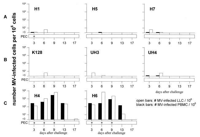
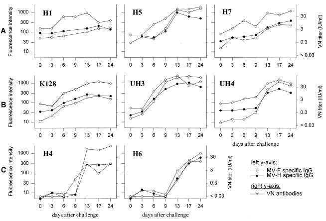
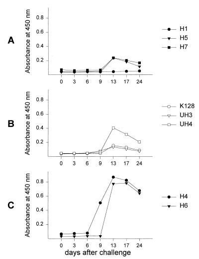
All six MVA-FH vaccinated macaques proved to be effectively protected from challenge with wild-type MV. Low MV loads (<101/106 cells) could be demonstrated in the PBMC and/or LLC of the MVA-FH-vaccinated macaques, while significant levels of MV were isolated from PEC, LLC, and PBMC of the MVA-wt-vaccinated animals (Fig. 7). The three animals from which no virus could be reisolated all showed a rise in MV-specific antibody levels, suggestive of a low-level challenge virus replication (Fig. 8). Only one of the six vaccinated animals (H1) did not show a serological booster response following challenge. As an additional parameter of infection, IgM antibodies specific for MV-N (not included in the vaccine construct) were measured following challenge. The observed levels proved to correlate well with the serological booster responses of the F- and H-specific and VN antibodies: no N-specific IgM could be measured in macaque H1, low levels of N-specific IgM were measured in the other five MVA-FH-vaccinated animals, and high levels of N-specific IgM were measured in the wild-type MVA-vaccinated animals (Fig. 9).
FIG. 7.
Number of MV-infected cells/106 LLC (open bars) or PBMC (black bars) at different times after intratracheal challenge with 103 TCID50s of MV-BIL. Macaques had been vaccinated with MVA-FH in the absence (A) or in the presence (B) of passively transferred MV-specific VN antibodies or with MVA-wt (group C). +, time point at which MV could be reisolated from PEC.
FIG. 8.
Development of MV glycoprotein-specific plasma IgG and VN antibody responses in macaques at different times after intratracheal challenge with 103 TCID50s of MV-BIL. Macaques had been vaccinated with MVA-FH in the absence (A) or in the presence (B) of passively transferred MV-specific VN antibodies or with MVA-wt (C).
FIG. 9.
Development of MV-N-specific plasma IgM responses in macaques at different times after intratracheal challenge with 103 TCID50s of MV-BIL. Macaques had been vaccinated with MVA-FH in the absence (A) or in the presence (B) of passively transferred MV-specific VN antibodies or with MVA-wt (C).
DISCUSSION
In the present study, we have shown that macaques are effectively protected from intratracheal challenge with wild-type MV 1 year after vaccination with MVA-FH irrespective of the presence of passively transferred homologous MV-specific antibody. These experiments were carried out with a macaque model for MV infection, with which the same parameters had been previously studied upon vaccination with MV-Schwarz, rVV-FH, and MV-iscom by using essentially the same regimen (26). The level of passively transferred homologous MV-specific antibodies used was also identical and corresponded to levels of serum VN antibodies that in epidemiological studies have been shown to interfere with the outcome of measles vaccination of infants (2). These levels relate to serum antibody levels that may be expected in infants of 6 to 9 months of age. In a cohort of 160 Sudanese infants aged about 6 months, we found levels below 0.1 IU/ml in 22% (n = 35), levels between 0.1 and 0.2 IU/ml in 45% (n = 72), and levels above 0.2 IU/ml in 33% (n = 53) of the cohort (unpublished data). This level had been shown to completely abolish the induction of MV-specific antibodies by MV-Schwarz vaccination and almost completely abolish the induction of this response by rVV-FH vaccination (26). In contrast, a candidate MV-iscom vaccine was shown to induce high titers of MV-specific serum antibody both in the presence and absence of passively transferred homologous MV-specific antibody (26). In the present study, MVA-FH was shown to induce higher levels of MV-specific antibodies than rVV-FH when administered in the presence of passively transferred neutralizing antibodies. We hypothesize that this is related to the relatively high MVA-FH doses used for vaccination (1 × 106.2 PFU per animal for rVV-FH versus 1 × 10⁸ PFU per animal for MVA-FH). For safety reasons (15), lower doses of rVV-FH had been administered in the previous experiment (26). The level of VN antibodies present at the time of the second vaccination (about 0.1 IU/ml; Fig. 2), which may at least in part be attributed to the passive immunization, has been shown to interfere with the replication of MV (26). The serological data also showed that the vaccinia virus-specific immune response induced by the first MVA-FH vaccination did not have a major impact on the immunogenicity of the second vaccination: it did not prevent a clear booster effect in the serological responses against either MV or MVA (Fig. 2, 3, and 4). Eight weeks after the second vaccination, all the vaccinated macaques showed a pronounced MV-specific T-cell response, as evidenced by MV-specific induction of CD69 expression by CD3+CD8− and of CD3+CD8+ bulk cultured cells (Fig. 5). This observation is of particular interest since in previous experiments we have shown that also in the absence of MV-neutralizing antibodies, vaccinated macaques may still be largely protected from challenge MV infection, indicating a protective effect of thus induced specific T-cell responses (26).
One year after vaccination, all the macaques were intratracheally challenged with MV-BIL (1, 26, 27). All the vaccinated macaques proved to be effectively protected from MV infection. Only low cell-associated virus loads could be demonstrated in lung lavages and peripheral blood, whereas, as expected, full-blown infection was demonstrated in the MVA-wt sham-vaccinated macaques. The increase in MV-neutralizing as well as F- and H-protein-specific antibody levels after challenge observed in all the macaques, and the induction of N-protein specific IgM antibodies in five of the six vaccinated macaques, confirmed that in all the vaccinated macaques, low-level virus replication had still occurred upon challenge.
Collectively, our data show that vaccination with MVA-FH in a two-dose intramuscular-intranasal regimen in the presence of passively acquired MV-neutralizing antibodies induces long-lasting protective immunity against challenge with wild-type MV. In previous experiments this was also achieved with MV-iscom, but not with live attenuated measles vaccine (MV-Schwarz), or with low doses of rVV-FH (Table 1). A clear advantage of MVA-FH over rVV-FH is its documented safety profile, since we have recently shown that even in severely immunosuppressed macaques neither virus replication nor any adverse effects occurred upon MVA infection (Stittelaar et al., submitted for publication). Furthermore, our data suggest that MVA-FH may be used to boost low levels of vaccine-induced immunity more efficiently than live attenuated MV vaccine. This could become of major importance during the final stages of the MV eradication campaign.
TABLE 1.
Average VN antibody titers in vaccinated macaques
| Vaccine | Passively immunizeda | VN antibody titers (IU/ml) at the time of:
|
|||
|---|---|---|---|---|---|
| Prime | Boosterb | Challengec | 4 weeks post-challenge | ||
| MVA-FH | Yes | 0.2 | 0.05 | 0.2 | 3 |
| No | <0.03 | 0.5 | 0.5 | 10 | |
| rVV-FHd | Yes | 0.16 | <0.08 | 0.1 | 100 |
| No | <0.08 | 1.7 | 1 | 80 | |
| MV-iscomd | Yes | 0.16 | 0.2 | 3 | 100 |
| No | <0.08 | 0.5 | 2 | 300 | |
| MV-Schwarzd | Yes | 0.16 | 0.1 | 0.2 | 90 |
| No | <0.08 | 8 | 3 | 2 | |
Passively transferred MV-specific antibody (0.2 IU/ml) 48 h before prime.
Second vaccination 4 weeks after the prime.
Intratracheal challenge with wild-type MV (103 TCID50s of BIL strain).
Data were taken from the work of Van Binnendijk et al. (26).
Finally, it is important to note that a live nonreplicating vaccine candidate such as MVA-FH may not be expected to be associated with immunopathological phenomena like the atypical measles syndrome associated with the use of inactivated MV vaccines (7, 9, 18). We conclude that our data favor the further exploration of the value of MVA-FH as a candidate replication-deficient vaccine as an alternative to the present vaccine for infants with maternally acquired MV-neutralizing antibody and for adults with waning vaccine-induced immunity.
ACKNOWLEDGMENTS
This work was supported by grants from the NIH (AI-93-06, AI-35144) and the WHO (V21/181/117) and by NIAID intramural funds.
We thank N. Schmidt for zootechnical assistance.
REFERENCES
- 1.Auwaerter P G, Rota P A, Elkins W R, Adams R J, DeLozier T, Shi Y, Bellini W J, Murphy B R, Griffin D E. Measles virus infection in rhesus macaques: altered immune responses and comparison of the virulence of six different virus strains. J Infect Dis. 1999;180:950–958. doi: 10.1086/314993. [DOI] [PubMed] [Google Scholar]
- 2.Clements C J, Cutts F T. The epidemiology of measles: thirty years of vaccination. Curr Top Microbiol Immunol. 1995;191:13–33. doi: 10.1007/978-3-642-78621-1_2. [DOI] [PubMed] [Google Scholar]
- 3.Cutts F T, Markowitz L E. Successes and failures in measles control. J Infect Dis. 1994;170(Suppl. 1):S32–S41. doi: 10.1093/infdis/170.supplement_1.s32. [DOI] [PubMed] [Google Scholar]
- 4.De Swart R L, Vos H W, UytdeHaag F G C M, Osterhaus A D M E, Van Binnendijk R S. Measles virus fusion protein- and hemagglutinin-transfected cell lines are a sensitive tool for the detection of specific antibodies in a FACS-measured immunofluorescence assay. J Virol Methods. 1998;71:35–44. doi: 10.1016/s0166-0934(97)00188-2. [DOI] [PubMed] [Google Scholar]
- 5.Earl P L, Moss B, Wyatt L S, Carroll M W. Generation of recombinant vaccinia viruses. In: Ausubel F M, Brent R, Kingston R E, Moore D D, Seidman J G, Smith J A, Struhl K, editors. Current protocols in molecular biology. New York, N.Y: Greene Publishing Associates & Wiley Interscience; 1998. pp. 16.17.1–16.17.19. [Google Scholar]
- 6.Forsey T, Heath A B, Minor P D. The 1st International Standard for anti-measles serum. Biologicals. 1991;19:237–241. doi: 10.1016/1045-1056(91)90042-i. [DOI] [PubMed] [Google Scholar]
- 7.Fulginiti V A, Eller J J, Downte A W, Kempe C H. Altered reactivity to measles virus. Atypical measles in children previously immunized with inactivated virus vaccines. JAMA. 1967;202:101–106. doi: 10.1001/jama.202.12.1075. [DOI] [PubMed] [Google Scholar]
- 8.Griffin D E, Bellini W J. Measles virus. In: Fields B N, Knipe D M, Howley P M, editors. Fields virology. 3rd ed. Philadelphia, Pa: Lippincott-Raven Publishers; 1996. pp. 1267–1312. [Google Scholar]
- 9.Harris R W, Isacson P, Karzon D T. Vaccine-induced hypersensitivity: reactions to live measles and mumps vaccine in prior recipients of inactivated measles vaccine. J Pediatr. 1969;74:552–563. doi: 10.1016/s0022-3476(69)80038-7. [DOI] [PubMed] [Google Scholar]
- 10.Helfand R F, Kim D K, Gary H E J, Edwards G L, Bisson G P, Papania M J, Heath J L, Schaff D L, Bellini W J, Redd S C, Anderson L J. Nonclassic measles infections in an immune population exposed to measles during a college bus trip. J Med Virol. 1998;56:337–341. doi: 10.1002/(sici)1096-9071(199812)56:4<337::aid-jmv9>3.0.co;2-3. [DOI] [PubMed] [Google Scholar]
- 11.Hirsch V M, Fuerst T R, Sutter G, Carroll M W, Yang L C, Goldstein S, Piatak M, Elkins W R, Alvord W G, Montefiori D C, Moss B, Lifson J D. Patterns of viral replication correlate with outcome in simian immunodeficiency virus (SIV)-infected macaques: effect of prior immunization with a trivalent SIV vaccine in modified vaccinia virus Ankara. J Virol. 1996;70:3741–3752. doi: 10.1128/jvi.70.6.3741-3752.1996. [DOI] [PMC free article] [PubMed] [Google Scholar]
- 12.Luttmann W, Knoechel B, Foerster M, Matthys H, Virchow J C, Jr, Kroegel C. Activation of human eosinophils by IL-13. Induction of CD69 surface antigen, its relationship to messenger RNA expression, and promotion of cellular viability. J Immunol. 1996;157:1678–1683. [PubMed] [Google Scholar]
- 13.Mackett M, Smith G L, Moss B. General method for production and selection of infectious vaccinia virus recombinants expressing foreign genes. J Virol. 1984;49:857–864. doi: 10.1128/jvi.49.3.857-864.1984. [DOI] [PMC free article] [PubMed] [Google Scholar]
- 14.McNair T F, Bonanno D E. Reactions to live-measles-virus vaccine in children previously inoculated with killed-virus vaccine. N Engl J Med. 1967;5:248–251. doi: 10.1056/NEJM196708032770506. [DOI] [PubMed] [Google Scholar]
- 15.Moss B. Genetically engineered poxviruses for recombinant gene expression, vaccination, and safety. Proc Natl Acad Sci USA. 1996;93:11341–11348. doi: 10.1073/pnas.93.21.11341. [DOI] [PMC free article] [PubMed] [Google Scholar]
- 16.Osterhaus A, Van Amerongen G, van Binnendijk R. Vaccine strategies to overcome maternal antibody mediated inhibition of measles vaccine. Vaccine. 1998;16:1479–1481. doi: 10.1016/s0264-410x(98)00112-1. [DOI] [PubMed] [Google Scholar]
- 17.Osterhaus A D M E, De Vries P, Van Binnendijk R S. Measles vaccines: novel generations and new strategies. J Infect Dis. 1994;170(Suppl. 1):S42–S55. doi: 10.1093/infdis/170.supplement_1.s42. [DOI] [PubMed] [Google Scholar]
- 18.Polack F P, Auwaerter P G, Lee S H, Nousari H C, Valsamakis A, Leiferman K M, Diwan A, Adams R J, Griffin D E. Production of atypical measles in rhesus macaques: evidence for disease mediated by immune complex formation and eosinophils in the presence of fusion-inhibiting antibody. Nat Med. 1999;5:629–634. doi: 10.1038/9473. [DOI] [PubMed] [Google Scholar]
- 19.Reed L J, Muench H. A simple method of estimating fifty percent endpoints. Am J Hyg. 1938;27:493–497. [Google Scholar]
- 20.Rosel J L, Earl P L, Weir J P, Moss B. Conserved TAAATG sequence at the transcriptional and translational initiation sites of vaccinia virus late genes deduced by structural and functional analysis of the HindIII H genome fragment. J Virol. 1986;60:436–449. doi: 10.1128/jvi.60.2.436-449.1986. [DOI] [PMC free article] [PubMed] [Google Scholar]
- 21.Schneider J, Gilbert S C, Blanchard T J, Hanke T, Robson K J, Hannan C M, Becker M, Sinden R, Smith G L, Hill A V. Enhanced immunogenicity for CD8+ T cell induction and complete protective efficacy of malaria DNA vaccination by boosting with modified vaccinia virus Ankara. Nat Med. 1998;4:397–402. doi: 10.1038/nm0498-397. [DOI] [PubMed] [Google Scholar]
- 22.Stern L B-L, Greenberg M, Gershoni J M, Rozenblatt S. The hemagglutinin envelope protein of canine distemper virus (CDV) confers cell tropism as illustrated by CDV and measles virus complementation analysis. J Virol. 1995;69:1661–1668. doi: 10.1128/jvi.69.3.1661-1668.1995. [DOI] [PMC free article] [PubMed] [Google Scholar]
- 23.Stickl H, Hochstein-Mintzel V, Mayr A, Huber H C, Schäfer H, Holzner A. MVA-Stufenimpfung gegen Pocken. Klinische Erprobung des attenuierten Pocken-Lebenimpfstoffes, Stamm MVA. Dtsch Med Wochenschr. 1974;99:2386–2392. doi: 10.1055/s-0028-1108143. [DOI] [PubMed] [Google Scholar]
- 24.Sutter G, Moss B. Nonreplicating vaccinia vector efficiently expresses recombinant genes. Proc Natl Acad Sci USA. 1992;89:10847–10851. doi: 10.1073/pnas.89.22.10847. [DOI] [PMC free article] [PubMed] [Google Scholar]
- 25.Sutter G, Wyatt L S, Foley P L, Bennink J R, Moss B. A recombinant vector derived from the host range-restricted and highly attenuated MVA strain of vaccinia virus stimulates protective immunity in mice to influenza virus. Vaccine. 1994;12:1032–1040. doi: 10.1016/0264-410x(94)90341-7. [DOI] [PubMed] [Google Scholar]
- 26.Van Binnendijk R S, Poelen M C M, Van Amerongen G, De Vries P, Osterhaus A D M E. Protective immunity in macaques vaccinated with live attenuated, recombinant and subunit measles vaccines in the presence of passively acquired antibodies. J Infect Dis. 1997;175:524–534. doi: 10.1093/infdis/175.3.524. [DOI] [PubMed] [Google Scholar]
- 27.Van Binnendijk R S, van der Heijden R W J, Van Amerongen G, UytdeHaag F G C M, Osterhaus A D M E. Viral replication and development of specific immunity in macaques after infection with different measles virus strains. J Infect Dis. 1994;170:443–448. doi: 10.1093/infdis/170.2.443. [DOI] [PubMed] [Google Scholar]
- 28.Wild T F. Measles vaccines, new developments and immunization strategies. Vaccine. 1999;17:1726–1729. doi: 10.1016/s0264-410x(98)00428-9. [DOI] [PubMed] [Google Scholar]
- 29.Wyatt L S, Shors S T, Murphy B R, Moss B. Development of a replication-deficient recombinant vaccinia virus vaccine effective against parainfluenza virus 3 infection in an animal model. Vaccine. 1996;14:1451–1458. doi: 10.1016/s0264-410x(96)00072-2. [DOI] [PubMed] [Google Scholar]


