Fig. 4.
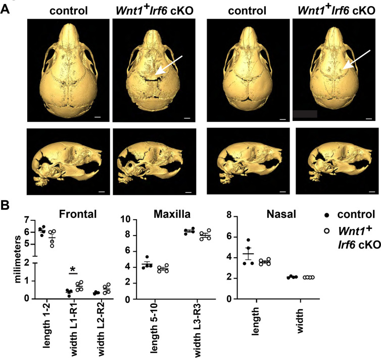
Cranial bone development is impaired in Wnt1-Cre Irf6 cKO mice. A. Representative microCT reconstructions of P10 Wnt1-Cre+;Irf6fl/fl cKO mice and littermate sex-matched controls. Wnt1-Cre+;Irf6fl/fl cKO mice have decreased formation or mineralization of the cranial bones at the midline with variable penetrance (arrows). Scale: 1 mm. B. MicroCT reconstructions were utilized for cranial bone measurements. The space between the left and right frontal bones of Wnt1-Cre+;Irf6fl/fl cKO mice was significantly wider than controls (L1-R1, *p<0.05) and the frontal bones tended to have decreased total length (length 1–2). Maxilla of Wnt1-Cre+;Irf6fl/fl cKO mice tended to be smaller (lower length and width measurements) and the frontal bone of Wnt1-Cre+;Irf6fl/fl cKO mice tended to be shorter, however, these differences were not significantly different. N=4.

