Abstract
Aims
Advanced echocardiographic imaging (AEI) techniques, such as three-dimensional (3D) and multi-chamber speckle-tracking deformation imaging (strain) analysis, have been shown to be more accurate in assessing heart chamber geometry and function when compared with conventional echocardiography providing additional prognostic value. However, incorporating AEI alongside standard examinations may be heterogeneous between echo laboratories (echo labs). Thus, our goal was to gain a better understanding of the many AEI modalities that are available and employed in Italy.
Methods and results
The Italian Society of Echocardiography and Cardiovascular Imaging (SIECVI) conducted a national survey over a month (November 2022) to describe the use of AEI in Italy. Data were retrieved via an electronic survey based on a structured questionnaire uploaded on the SIECVI website. Data obtained from 173 echo labs were divided into 3 groups, according to the numbers of echocardiograms performed: <250 exams (low-volume activity, 53 centres), between 251 and 550 exams (moderate-volume activity, 62 centres), and ≥550 exams (high-volume activity, 58 centres). Transthoracic echocardiography (TTE) 3D was in use in 75% of centres with a consistent difference between low (55%), medium (71%), and high activity volume (85%) (P = 0.002), while 3D transoesophageal echocardiography (TEE) was in use in 84% of centres, reaching the 95% in high activity volume echo labs (P = 0.006). In centres with available 3D TTE, it was used for the left ventricle (LV) analysis in 67%, for the right ventricle (RV) in 45%, and for the left atrium (LA) in 40%, showing greater use in high-volume centres compared with low- and medium-volume centres (all P < 0.04). Strain analysis was utilized in most echo labs (80%), with a trend towards greater use in high-volume centres than low- and medium-volume centres (77%, 74%, and 90%, respectively; P = 0.08). In centres with available strain analysis, it was mainly employed for the LV (80%) and much less frequently for the RV and LA (49% and 48%, respectively).
Conclusion
In Italy, the AEI modalities are more frequently available in centres with high-volume activity but employed only in a few applications, being more frequent in analysing the LV compared with the RV and LA. Therefore, the echocardiography community and SIECVI should promote uniformity and effective training across the Italian centres. Meanwhile, collaborations across centres with various resources and expertise should be encouraged to use the benefits of the AEI.
Keywords: national survey, advanced echocardiography imaging, technologies
Introduction
Advanced echocardiographic imaging (AEI) techniques, such as three-dimensional (3D) and multi-chamber speckle-tracking deformation imaging (strain) analysis, have been shown to be more accurate in assessing heart chamber geometry and function, as well as providing additional prognostic value, when compared with conventional two-dimensional echocardiography.1–3 Also, new automated on-cart equipment has recently been proven to perform precise, quick, and repeatable strain and 3D evaluations of the heart chambers.4–6 Therefore, AEI is frequently highlighted by the current recommendation documents from the European Association of Cardiovascular Imaging (EACVI) and the American Society of Echocardiography (ASE) as a state-of-the-art approach for patient evaluation.7–10 However, each echocardiographic laboratory’s (echo labs) capacity to incorporate AEI alongside standard examinations may differ based on internal structure, workload, financial resources, experience, and patient population. Accordingly, this survey aims to obtain more knowledge about the AEI methods used in Italy to influence future strategies for optimizing their integration and widespread clinical deployment into regular patient evaluation.
Methods
Our recent publication described the national survey methodology in detail.11 Compared with the initial database, we analysed only the echo labs within cardiology units and departments over 1 month of activity. November 2022 was chosen as an ideal reference month (regular planning of activities in the absence of national holidays). A list of SIECVI-accredited echo labs was reviewed to contact each member by e-mail. Data from members were retrieved via an electronic survey based on a structured questionnaire uploaded on the SIECVI website (www.siec.it.). For allocation of the response, the questionnaire required general information, such as the name of the hospital, the investigator, and the interviewed person’s name: (i) general information: date, hospital’s name, department, name of the interviewed physician, city, and region of Italy; (ii) the number of exams performed, divided by type; and (iii) the number of echocardiographic machines/transducers/software according to AEI analysis divided for cardiac chambers.
Statistical analysis
Categorical data are expressed in terms of the number of subjects and percentage, while continuous data are expressed as mean ± standard deviation or median (minimum–maximum) depending on the variables’ distribution. For continuous variables, inter-group differences were tested with a one-way analysis of variance and inter-group comparison by Bonferroni or Kruskal–Wallis, followed by the Mann–Whitney test as appropriate. The χ2 test or Fisher exact test was used to compare the distribution of categorical variables among groups. Statistical analysis was performed using the JMP PRO software package, version 16 (SAS Institute Inc., Cary, NC).
Results
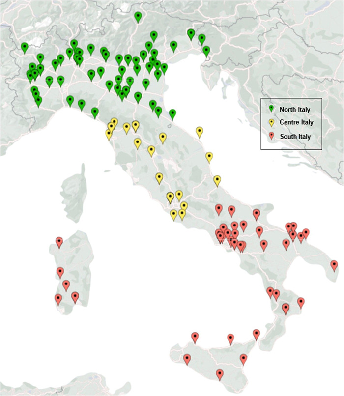
Data were obtained from 173 cardiology units and department echo labs (Table 1). The median of echocardiographic exams was 400 (IQ range 250–650). Echo labs were divided into 3 groups according to the volume of activity: <250 exams/month (low-volume, 53 centres, 31%, mean 172 ± 72), 251–550 exams/month (medium-volume, 62 centres, 36%, mean 391 ± 76), and ≥550 exams/months (high-volume, 58 centres, 33%, mean 1001 ± 537). Participant echo labs composed an adequate coverage of the national territory but with a higher distribution in the north (88 centres, 51%) compared with the centre (32, 18%) and south (53, 31%) of Italy (Figure 1). The volume of activity was also more pronounced in the north (high volume 62%) compared with the centre (17%) and south of Italy (21%), P = 0.005. The mean number of transoesophageal echocardiogram (TEE) was 30 ± 25, rising proportionally to the activity of the centres (low-volume 15 ± 15, medium-volume 25 ± 23, high-volume 48 ± 33), P < 0.0001.
Table 1.
General and technological results in the Italian echo lab overall and according to volume of activity
| Overall (n = 173) | Low- volume <250 ex/months (n = 53, 31%) | Moderate-volume ex/month 250–550 (n = 62, 36%) | High-volume ex/month >550 (n = 58, 33%) | P-value | |
|---|---|---|---|---|---|
| North Italy | 88 (51%) | 16 (30%) | 36 (58%) | 36 (62%) | 0.005 |
| Centre Italy | 32 (18%) | 12 (23%) | 10 (16%) | 10 (17%) | |
| South Italy | 53 (31%) | 25 (47%) | 16 (26%) | 12 (21%) | |
| Echocardiography machines, n | 5.0 ± 3.0 | 3.5 ± 1.8 | 4.0 ± 1.8 | 7.5 ± 4.5 | <0.001 |
| Multivendor | 114 (66%) | 33 (62%) | 35 (56%) | 46 (79%) | 0.02 |
| 3D echocardiography machines/transducer | 2.1 ± 1.5 | 1.5 ± 1.1 | 1.8 ± 1.3 | 2.9 ± 2.0 | <0.0001 |
| Use of 3D transthoracic | 122 (75%) | 29 (55%) | 44 (71%) | 49 (85%) | 0.002 |
| Use of 3D transoesophageal | 146 (84%) | 38 (72%) | 54 (87%) | 54 (95%) | 0.006 |
| Global longitudinal strain, yes | 139 (80%) | 41 (77%) | 46 (74%) | 52 (90%) | 0.08 |
| LV GLS | |||||
| No | 34 (20%) | 12 (23%) | 16 (26%) | 6 (10%) | 0.04 |
| 1–20% | 27 (16%) | 8 (15%) | 13 (21%) | 6 (10%) | |
| 21–49% | 35 (20%) | 13 (25%) | 8 (13%) | 14 (24%) | |
| 50–99% | 48 (28%) | 9 (17%) | 15 (24%) | 24 (41%) | |
| 100% | 29 (17%) | 11 (21%) | 10 (16%) | 8 (14%) | |
| RV GLS | |||||
| No | 89 (51%) | 34 (64%) | 39 (63%) | 16 (28%) | 0.001 |
| 1–20% | 47 (27%) | 10 (19%) | 12 (19%) | 25 (43%) | |
| 21–49% | 23 (13%) | 7 (13%) | 6 (10%) | 10 (17%) | |
| 50–99% | 13 (8%) | 1 (2%) | 5 (8%) | 7 (12%) | |
| 100% | 1 (1%) | 1 (2%) | 0 | 0 | |
| LA GLS | |||||
| No | 90 (52%) | 36 (68%) | 36 (58%) | 18 (31%) | 0.001 |
| 1–20% | 50 (29%) | 11 (20%) | 13 (21%) | 26 (45%) | |
| 21–49% | 22 (13%) | 5 (9%) | 10 (16%) | 7 (12%) | |
| 50–99% | 10 (6%) | 0 | 3 (5%) | 7 (12%) | |
| 100% | 1 (1%) | 1 (2%) | 0 | 0 | |
| 3D transthoracic, yes | 122 (71%) | 29 (55%) | 44 (71%) | 49 (84%) | 0.002 |
| LV 3D | |||||
| No | 57 (33%) | 25 (47%) | 23 (37%) | 9 (16%) | 0.04 |
| 1–20% | 44 (26%) | 12 (23%) | 16 (26%) | 16 (28%) | |
| 21–49% | 30 (17%) | 5 (9%) | 9 (15%) | 16 (28%) | |
| 50–99% | 30 (17%) | 8 (15%) | 10 (16%) | 12 (21%) | |
| 100% | 11 (7%) | 3 (6%) | 4 (6%) | 4 (7%) | |
| RV 3D | |||||
| No | 94 (55%) | 40 (75%) | 37 (60%) | 17 (30%) | 0.0008 |
| 1–20% | 41 (24%) | 6 (11%) | 13 (21%) | 22 (39%) | |
| 21–49% | 21 (12%) | 2 (4%) | 8 (13%) | 11 (19%) | |
| 50–99% | 13 (8%) | 4 (8%) | 3 (5%) | 6 (11%) | |
| 100% | 3 (2%) | 1 (2%) | 1 (2%) | 1 (2%) | |
| LA 3D | |||||
| No | 102 (60%) | 41 (77%) | 35 (57%) | 26 (45%) | 0.04 |
| 1–20% | 45 (26%) | 5 (9%) | 18 (30%) | 22 (39%) | |
| 21–49% | 17 (10%) | 4 (8%) | 7 (12%) | 6 (11%) | |
| 50–99% | 5 (3%) | 2 (4%) | 1 (2%) | 2 (4%) | |
| 100% | 2 (1%) | 1 (2%) | 0 | 1 (2%) | |
| TEE 3D | |||||
| No | 27 (16%) | 15 (28%) | 8 (13%) | 4 (7%) | 0.008 |
| 21–49% | 25 (14%) | 4 (8%) | 11 (18%) | 10 (17%) | |
| 50–99% | 61 (35%) | 20 (38%) | 25 (40%) | 16 (28%) | |
| 100% | 60 (35%) | 14 (26%) | 18 (29%) | 28 (48%) |
Figure 1.
Geographical distribution of the participating centres.
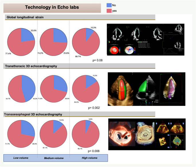
The facility to perform any 3D evaluation shows a good distribution on a national level (mean number of 3D machines and/or transducers 2.1 ± 1.5), with increasing accessibility according to the volume of activity (low 1.5 ± 1.1, medium 1.8 ± 1.3, high 2.9 ± 2.0, P < 0.0001). Specifically, transthoracic echocardiography (TTE) 3D was in use in 75% of centres with a consistent difference between low (55%), medium (71%), and high volume (85%), P = 0.002, while TEE 3D was in use in 84% of centres, reaching the 95% in high-volume echo labs (P = 0.006) (Figure 2).
Figure 2.
Advanced echocardiography imaging present in echo lab in Italy according to type (from above: strain, 3D transthoracic echocardiography, and 3D transoesophageal echocardiography). Blue, absent; red, present. P for statistical significance.
In centres with available TTE 3D, it was used for the left ventricle (LV) in 67%, for the right ventricle (RV) in 45%, and for the left atrium (LA) in 40%, showing greater use in high-volume centres compared with low- and medium-volume centres (all P < 0.04).
Strain analysis was utilized in most echo labs (80%), with a trend towards greater use in high-volume centres than low- and medium-volume centres (77%, 74%, and 90%, respectively), P = 0.08 (Figure 2). In centres with available strain analysis, it was mainly employed for the LV (80%) and much less frequently for the RV and LA (49% and 48%, respectively).
Discussion
The present survey provides unique real-world data about AEI distribution at a national level. The main findings were as follows: (i) currently, AEI is not part of the routine examination in most laboratories, especially in those with low- and medium-volume activity; (ii) nearly three-quarters of the centres have 3D TTE available for the assessment of LV, but less than half for the assessment of RV and LA; (iii) the vast majority of centres has the chance of performing a 3D TEE, which almost universal in centres with a high volume of activity; and (iv) although the strain technology is available in most echo labs, it is rarely used for the RV and LA analysis.
In the present survey, many centres (75%) answered that 3D TTE was available in their echo lab but with consistent activity volume differences. Still, most of them used 3D TTE for the LV analysis, according to the ASE and the EACVI guidelines.12 Similarly, in a recent EACVI survey on standardization of cardiac chambers quantification by TTE, >90% of centres had access to 3D TTE; however, most centres reserved these techniques for selected cases, particularly for measuring LV volumes and ejection fraction.13
Disappointingly, we found that most survey participants infrequently performed RV measurements using 3D TTE (45% of centres with the technology available). Similar data were reported in another recent EACVI survey on the multi-modality imaging assessment of the right heart.14 This observation probably reflects the lack of dedicated software (as compared with the LV) for this assessment.
Likewise, the present survey highlighted that less than half of the laboratories equipped with the modality analysed the LA with 3D TTE. Our finding parallels the result of the EACVI survey on standardization of cardiac chamber quantification by transthoracic echocardiography, in which only 10% of centres used 3D TTE to assess LA volumes.13
In the present survey, 80% of centres had access to strain analysis, suggesting the wide availability of this modality in most Italian echo labs. However, most centres appear to reserve strain only for LV analysis. Indeed, it was unexpected to report that, despite growing evidence of their additional value in the literature, only 49% of the centres used RV and 48% LA strain analysis. Our observations are consistent with a recent worldwide survey from the EACVI, which highlighted how, despite the almost universal availability, only 39% of the participants performed and reported strain results frequently (>50%), which was mainly used to assess the LV (99%) and less frequently the RV (57%) and the LA (46%) function.15
The recent innovations and advantages of AEI are unquestionable. A growing body of evidence demonstrates the effectiveness of AEI in identifying cardiac disorders at an early stage, showing its superiority over traditional methods in terms of repeatability, timeliness, affordability, and feasibility in a wide range of clinical scenarios such as valvular heart diseases,16 cardio-oncology,17 immune-mediated18 and infiltrative diseases,19 arterial hypertension and metabolic disorders,20 heart failure with preserved ejection fraction,21 hypertrophic cardiomyopathy and phenocopies,22 acute coronary syndrome,23 chronic ischaemic cardiomyopathy,24 adult with congenital heart disease,25 pulmonary arterial hypertension,26 and acute myocarditis.27 The proof that AEI has a place in everyday practice is indicated by its role in the COVID-19 pandemic.28 Despite this amount of evidence, our data highlight that numerous obstacles prevent a wider spread of AEI in clinical practice. Most likely, inadequate training and time constraints are the primary reasons for not adopting AEI more frequently. Indeed, sonographers and cardiologists must be educated in image capture and analysis techniques that allow for reliable post-processing and robust results, but integrating AEI requires many other crucial resources, such as suitable equipment, patient selection, adoption of protocols into ordinary clinical practice, modification of echo lab workflow, storage, and reporting.29 In addition, hospital administration must acknowledge and believe in the clinical usefulness of AEI and necessary billing and reimbursement adaptations, as AEI also involves a discussion around cost justification. It’s also essential to define more robust reference values and standardization of values, considering that 66% of centres in the present survey use two or more different vendors within the same laboratory.30
Certainly, additional study is needed to determine whether AEI can enhance patient care and results. Clinical trials incorporating AEI features will be critical in identifying the most relevant and robust patient care parameters in various clinical settings. Nonetheless, the widespread adoption of AEI necessitates, first and foremost, a willingness to adapt based on the recognition that AEI adds practical value to our daily practice. Accordingly, recent data demonstrated that using AEI is timesaving compared with conventional evaluation.31 Therefore, if AEI is not part of the routine practice yet, scientific societies should designate the inclusion of these procedures in standard transthoracic echocardiographic examinations among their responsibilities. To fulfil this objective, the SIECVI is now working to standardize AEI acquisition, reporting, dedicated training, certification, and quality control methods across most echo labs in Italy.32
Study limitations
We used the SIECVI’s electronic e-mail list, which includes the majority—but certainly not all—of the echocardiographic activity in Italy.11 Some extra-SIECVI centres have high volumes and high-quality standards. However, although the survey may have underestimated the diffusion of AEI activities in selected centres of excellence, it most likely accurately reflected the quality and pattern of practice.
As with any survey, there will be non-responders for various reasons, including a lack of time or a reluctance to engage in the study. Moreover, the replies may be skewed due to the respondents’ possible differing perspectives or interpretations of the questions.
Finally, no independent, external validation of the data provided by the cardiologist head of the participating echo lab was possible.11,33,34
Conclusions
In Italy, the AEI modalities are more frequently available in centres with high-volume activity but employed only in a few cardiac chamber applications, being more frequent in analysing the LV compared with the RV and LA. Therefore, the echocardiography community and SIECVI should promote uniformity and effective training across the Italian centres. Meanwhile, collaborations across centres with various resources and expertise should be encouraged to use the benefits of the AEI.
Acknowledgements
Abbate Massimiliana (Cardiology Division, Vanvitelli Monaldi Hospital, Napoli), Accadia Maria (Cardiology Division, Del Mare Ponticelli Hospital, Napoli), Alemanni Rossella (Cardiac Surgery Division, Casa Sollievo Della Sofferenza Hospital, San Giovanni Rotondo), Angelini Andrea (Cardiology Division, Cardinal Massaia Hospital, Asti), Anglano Francesco (Cardiology Division, Ravenna Medical Center, Ravenna), Anselmi Maurizio (Cardiology Division, Fracastoro Di San Bonifacio Hospital, Verona), Aquila Iolanda (Cardiology Division, University Hospital Mater Domini, Catanzaro), Aramu Simona (Cardiology Division, San Martino Hospital, Oristano), Avogadri Enrico (Rehabilitative Cardiology, S.S. Trinità Hospital, Fossano), Azzaro Giuseppe (Cardiology Division, Cardinale Massaia Hospital, Asti), Badano Luigi (Integrated Cardiovascular Diagnostic Unit, Auxologico San Luca IRCCS Hospital, Milano), Balducci Anna (Pediatric Cardiology Division, Polyclinic St Orsola-Malpighi IRCCS Hospital, Bologna), Ballocca Flavia (Cardiology Division, Maria Vittoria Hospital, Torino), Barbarossa Alessandro (Clinic of Cardiology and Arrhythmology, Marche University Hospital, Ancona), Barbati Giovanni (Cardiology Division, St Bortolo Hospital, Vicenza), Barletta Valentina (Cardiology 2 Division, Cardiac Vascular Thoracic Department. Pisa University Hospital, Pisa), Barone Daniel (Cardiology Division, St Andrea Hospital, La Spezia), Becherini Francesco (Cardiology and Cardiovascular Medicine Division, Fondazione Toscana Gabriele Monasterio, Pisa), Benfari Giovanni (Cardiology Division, University of Verona, Verona), Beraldi Monica (Cardiology Division, ASST-Mantova Carlo Poma Hospital, Mantova), Bergandi Gianluigi (Cardiology Division, Civil Hospital, Ivrea), Bilardo Giuseppe (Cardiology Division, Civil Hospital Fetre), Binno Simone Maurizio (Cardiology Division, G. Da Saliceto Hospital, Piacenza), Bolognesi Massimo (Center for Internal Medicine and Sports Cardiology, Local Health Unit of Romagna, Cesena), Bongiovi Stefano (Cardiology Division, Immacolata Concezione Civil Hospital, Piove di Sacco), Bragato Renato Maria (Echocardiography and Emergency Cardiovascular Care Division, Humanitas Clinical and Research Centre, Rozzano), Braggion Gabriele (Cardiology Division, Santa Maria Regina Degli Angeli Hospital, Adria), Brancaleoni Rossella (Cardiology Division, A. Costa Civil Hospital, Porretta Terme), Bursi Francesca (Cardiology Division, San Paolo Hospital, Milano), Cadeddu Dessalvi Christian (Cardiology Division, University Hospital of Cagliari, Cagliari), Cameli Matteo (Cardiology Division, Polyclinic Le Scotte Hospital, Siena), Canu Antonella (Cardiology Division, St Annunziata Hospital, Sassari), Capitelli Mariano (Internal Medicine Division, Pavullo Hospital, Pavullo nel Frignano), Capra Anna Clara Maria (Cardiological Diagnostics Division, Synlab San Nicolò Diagnostic Center, Lecco), Carbonara Rosa (Cardiology Division, Maugeri Hospital, Bari), Carbone Maria (Emergency Medicine Division, St Anna and St Sebastiano Hospital, Caserta), Carbonella Marco (Cardiology Division, SS Maria Addolorata Hospital, Eboli), Carrabba Nazario (Cardiology Division, Careggi Hospital, Firenze), Casavecchia Grazia, (Cardiology Division, University Hospital Ospedali Riuniti, Foggia), Casula Margherita (Cardiology Division, Nostra Signora di Bonaria Hospital, San Gavino Monreale), Chesi Elena (Neonatology Division, St Maria Nuova Hospital, Reggio Emilia), Cicco Sebastiano (Unit of Internal Medicine ‘G. Baccelli’ and Unit of Hypertension ‘A.M. Pirrelli’, Department of Precision and Regenerative Medicine and Ionian Area, University of Bari Aldo Moro Medical School, AUOC Bari Polyclinic, Bari), Citro Rodolfo (Echocardiography Division, University Hospital San Giovanni di Dio and Ruggi d'Aragona, Salerno), Civelli Maurizio (Cardiology Division, European Institute of Oncology, Milano), Cocchia Rosangela (Rehabilitative Cardiology, Cardarelli Hospital, Napoli), Colombo Barbara Maria (Clinic of Emergency Medicine, IRCCS San Martino Polyclinic Hospital, Genova), Colonna Paolo (Cardiology Division, Polyclinic Hospital, Bari), Conte Maddalena (Department of Translational Medical Sciences, University of Naples Federico II, Naples), Corrado Giovanni (Cardiology Division, Valduce Hospital, Como), Cortesi Pietro (Cardioncology Division, IRCCS Istituto Romagnolo per lo Studio dei Tumori (IRST) ‘Dino Amadori’, Meldola), Cortigiani Lauro (Cardiology Division, San Luca Hospital, Lucca), Costantino Marco Fabio (Cardiology Division, San Carlo Hospital, Potenza), Cozza Fabiana (Cardiology Division, Fondazione Poliambulanza Hospital, Brescia), Cucchini Umberto (Cardiology Division, San Bassiano Hospital, Bassano Del Grappa), D'Andrea Fabrizio (Cardiology Division, St Andrea Hospital, Roma), D'Andrea Antonello (Cardiology Division, Umberto I Hospital, Nocera Inferiore), D'Auria Francesca (Vascular—Endovascular Surgery Division, University Hospital San Giovanni di Dio e Ruggi d'Aragona, Salerno), D’Angelo Myriam (Cardiology Division, Bonino Pulejo IRCCS Hospital, Messina), Da Ros Santina (Cardiology Division, Riuniti Padova Sud Hospital, Monselice), De Caridi Giovanni (Vascular Surgery Division, University Hospital Polyclinic G. Martino, University of Messina, Messina), De Feo Stefania (Cardiology Division, P. Pederzoli Hospital, Peschiera Del Garda), De Matteis Giovanni Maria (Cardiology Division, Sandro Pertini Hospital, Roma), De Vecchi Simona (Cardiology Division, Major University Hospital of Charity, Novara), Del Giudice Carmen (Cardiology Division, AORN dei Colli, Monaldi Hospital, Napoli), Dell'Angela Luca (Cardiology Division, Gorizia & Monfalcone Hospital, Gorizia), Delli Paoli Lucrezia (Cardiological Intensive Care Unit, St Michele Clinic, Maddaloni), Dentamaro Ilaria (Cardiology Division, Miulli Hospital, Acquaviva delle Fonti), Destefanis Paola (Cardiology Division, San Luigi Gonzaga University Hospital, Orbassano), Di Fulvio Maria (Cardiology-ICCU Division, Ss. Annunziata Hospital, Chieti), Di Gaetano Renato (Cardiology Division, Bolzano Hospital, Bolzano), Di Giannuario Giovanna (Cardiology Division, Infermi Hospital, Rimini), Di Gioia Angelo (Cardiology Division, St Giuliano Hospital, Giugliano in Campania), Di Martino Luigi Flavio Massimiliano (Cardiology Division, Santa Maria Degli Angeli Hospital, Putignano), Di Muro Carmine (Sports Medicine Division, Livorno Hospital, Livorno), Di Nora Concetta (Cardiology Division, St Maria Della Misericordia Hospital, Udine), Di Salvo Giovanni (Pediatric Cardiology and Congenital Heart Disease Division, Padova University Hospital, Padova), Dodi Claudio (Cardiology Division, San Antonino Clinic, Piacenza), Dogliani Sarah (Cardiology Division, St Annunziata Hospital, Savigliano), Donati Federica (PASCIA Center, Polyclinic, Modena), Dottori Melissa (Cardiology Division, Marche University Hospital, Ancona), Epifani Giuseppe (Internal Medicine Division, Camberlingo Hospital, Francavilla Fontana), Fabiani Iacopo (Cardiology and Cardiovascular Medicine Division, Fondazione Toscana Gabriele Monasterio, Pisa), Ferrara Francesca (Internal Medicine Division, University Hospital Modena Polyclinic, Modena), Ferrara Luigi (Cardiology Division, Villa Dei Fiori Hospital, Acerra), Ferrua Stefania (Cardiology Division, Degli Infermi Hospital, Rivoli), Filice Gemma (Cardiology Division, P.O. Annunziata Hospital, Cosenza), Fiorino Maria (Cardiology Division, Civico Hospital, Palermo), Forno Davide (Cardiology Division, Maria Vittoria Hospital, Torino), Garini Alberto (Cardiology Division, Cremona Hospital, Cremona), Giarratana Gioachino Agostino (Cardiac Surgery Division, Polyclinic P. Giaccone Hospital, Palermo), Gigantino Giuseppe (Cardiology Division, University Hospital San Giovanni di Dio e Ruggi d'Aragona, Salerno), Giorgi Mauro (Cardiology Division, Molinette Hospital—Città della Salute e della Scienza, Torino), Giubertoni Elisa (Cardiology Division Civil Hospital, Guastalla), Greco Cosimo Angelo (Cardiology Division, Veris Delli Ponti Hospital, Scorrano), Grigolato Michele (Polycardiography Division, Civil Hospital, Brescia), Grosso Marra Walter (Cardiology Division, Civil Hospital, Ivrea), Holzl Anna (Internal Medicine Division, Quisisana Clinic, Ferrara), Iaiza Alessandra (Cardiac surgery Division, San Camillo-Fornalinini Hospital, Roma), Iannaccone Andrea (Internal Medicine Division, Ordine Mauriziano Hospital, Torino), Ilardi Federica (Cardiology Division, Federico II University Hospital, Napoli), Imbalzano Egidio (Internal Medicine Division, University Hospital Polyclinic G. Martino, University of Messina, Messina), Inciardi Riccardo (Cardiology Division, Civil Hospital, Brescia), Inserra Corinna Antonia (Cardiology Division, Civil Hospital, Legnano), Iori Emilio (Cardiology Division, New Civil Hospital, Sassuolo), Izzo Annibale (Cardiology Division, St Anna and St Sebastiano Hospital, Caserta), La Rosa Giuseppe (Cardiology Division, Santa Barbara Hospital, Gela), Labanti Graziana (Cardiology Division, Bellaria Hospital, Bologna), Lanzone Alberto Maria (Cardiology Division, San Rocco Clinical Institute, Ome), Lanzoni Laura (Cardiology Division, Sacro Cuore Don Calabria IRCCS Hospital, Verona), Lapetina Ornella (Cardiology Division, San Carlo Hospital, Melfi), Leiballi Elisa (Cardiological and Cardio-Oncological Rehabilitation Unit, CRO Hospital, Sacile), Librera Mariateresa (Cardiology Division, Mediterranea Clinic, Napoli), Lo Conte Carmenita (Cardiology Division, St Ottone Frangipane Hospital, Ariano Irpino), Lo Monaco Maria (Cardiology Division, Humanitas Gavazzeni Hospital, Bergamo), Lombardo Antonella (Cardiology Division, Polyclinic Foundation A. Gemelli-IRCCS, Roma), Luciani Michelangelo (Cardiology Division, Belcolle Hospital, Viterbo), Lusardi Paola (Cardiology and Cardiac Surgery Division, Maria Pia Hospital, Torino), Magnante Antonio (Cardiology Division, Madonna Delle Grazie Hospital, Matera), Malagoli Alessandro (Cardiology Division, Baggiovara Civil Hospital, Modena), Malatesta Gelsomina (Cardiology Division, IRCCS INRCA Hospital, Ancona), Mancusi Costantino (Hypertension Center, Federico II University Hospital, Napoli), Manes Maria Teresa (Cardiology Division, St Francesco Hospital, Cosenza), Manganelli Fiore (Cardiology Division, St Giuseppe Moscati Hospital, Avellino), Manuppelli Vincenzo (Cardiology Division, University Hospital Ospedali Riuniti, Foggia), Marchese Valeria (Cardiology Division, Santa Maria Della Speranza Hospital, Battipaglia), Marinacci Lina (Cardiology Division, Civil Hospital, Città di Castello), Mattioli Roberto (Cardiology Division, IRCCS Multimedica Hospital, Sesto San Giovanni), Mazza Giuseppe Antonio (Pediaric Cardiology Division, Regina Margherita Hospital—Città della Salute e della Scienza, Torino), Mazza Stefano (Cardiology Division, Maggiore St Andrea Hospital, Vercelli), Melis Marco (Cardiology Division, Arnas Brotzu Hospital, Cagliari), Meloni Giulia (Center for Prevention, Diagnosis and Therapy of Arterial Hypertension and Cardiovascular Complications, St Camillo Hospital, Sassari), Merli Elisa (Cardiology Division, Per Gli Infermi Hospital, Faenza), Milan Alberto (Internal Medicine 4 Division, Molinette Hospital—Città della Salute e della Scienza, Torino), Minardi Giovanni (Echolab Salvator Mundi International Hospital, Roma), Monaco Antonella (Cardiology Outpatient Clinic, Cardiology Outpatient Clinic, Civitanova Marche), Monte Ines (Cardiology Division, University Hospital Polyclinic ‘G. Rodolico-S. Marco’, University of Catania, Catania), Montresor Graziano (Cardiology Division, Gavardo Hospital, Brescia), Moreo Antonella (De Gasperis Cardio Center, ASST Grande Ospedale Metropolitano Niguarda, Milano), Mori Fabio (Non-invasive Cardiovascular Diagnostic Division, Careggi University Hospital, Firenze), Morini Sofia (Cardiology Division, Riuniti della Valdichiana Hospital, Montepulciano), Moro Claudio (Cardiology Division, Pio Xi Hospital, Desio), Morrone Doralisa (Cardiology Division, Cisanello Hospital, Pisa), Negri Francesco (Cardiology Division, Azienda Sanitaria Universitaria Friuli Centrale, Udine), Nipote Carmelo (Cardiology Division, Civil Hospital, S. Agata Militello), Nisi Fulvio (Anesthesia and intensive care Division, IRCCS Humanitas Research Hospital, Rozzano), Nocco Silvio (Cardiology Division, Sirai Hospital, Carbonia), Novello Luigi (Geriatric Division, Valdagno Hospital, Arzignano), Nunziata Luigi (Cardiology Division, St Maria della Pietà Hospital, Nola), Paoletti Perini Alessandro (Cardiology Division, St Maria Nuova Hospital, Firenze), Parodi Antonello (Cardiology Division, Padre Antero Micone Hospital, Genova), Pasanisi Emilio Maria (Cardiology Division, Livorno Hospital, Livorno), Pastorini Guido (Cardiology Division, Regina Montis Regalis Hospital, Mondovì), Pavasini Rita (Cardiology Division, St Anna University Hospital, Ferrara), Pavoni Daisy (Cardiology Division, Azienda Sanitaria Universitaria Friuli Centrale, Udine), Pedone Chiara (Cardiology Division, Maggiore Hospital, Bologna), Pelliccia Francesco (Cardiology Division, Umberto I Hospital, Roma), Pelliciari Giovanni (Internal Medicine Division, Gruppioni Clinic, Pianoro), Pelloni Elisa (Cardiology Division, Parini Hospital, Aosta), Pergola Valeria (Cardiology Division, Padova University Hospital, Padova), Perillo Giovanni (Cardiology Division, Policlinico Celio Hospital, Roma), Petruccelli Enrica (Cardiology Division, St Giacomo Hospital, Monopoli), Pezzullo Chiara (Cardiology Division, G.B. Grassi Hospital, Ostia), Piacentini Gerardo (Fetal and Neonatal Cardiology Unit—Fatebenefratelli Isola Tiberina Gemelli Isola Hospital, Roma), Picardi Elisa (Cardiology Division, Civic Hospital, Chivasso), Pinna Giovanni (Neonatology and Neonatal Intensive Care Division, San Camillo-Forlanini Hospital, Roma), Pizzarelli Massimiliano (Cardiology outpatient clinic, Quisisana Clinic, Ferrara), Pizzuti Alfredo (Cardiology outpatient clinic, Koelliker Hospital, Torino), Poggi Matteo Maria (Interdisciplinary Internal Medicine Division, Careggi University Hospital, Firenze), Posteraro Alfredo (Cardiology Division, San Giovanni Evangelista Hospital, Tivoli), Privitera Carmen (Pediatric Division, St Chiara Hospital, Trento), Rampazzo Debora (Cardiology Division, Madonna della Navicella Hospital, Chioggia), Ratti Carlo (Cardiology Division, Santa Maria Bianca Hospital, Mirandola), Rettegno Sara (Cardiology Division, St Croce Hospital, Moncalieri), Ricci Fabrizio (Cardiology Division, Ss. Annunziata Hospital, Chieti), Ricci Caterina (Cardiology Outpatient Clinic, CDS Regina Margherita, Castelfranco Emilia), Rolando Cristina (Cardiology Division, Civil Hospital, Ciriè), Rossi Stefania (Cardiology Division, Civil Hospital, Lavagna), Rovera Chiara (Cardiology Division, Civic Hospital, Chivasso), Ruggieri Roberta (Cardiology Division, Di Venere Hospital, Bari), Russo Maria Giovanna (Pediatric cardiology Division, AORN dei Colli, Monaldi Hospital, Napoli), Sacchi Nicola (Medical Division, St Agostino Hospital, Castiglione del Lago), Saladino Antonino (Cardiology Division, Giovanni Paolo II Hospital, Sciacca), Sani Francesca (Cardiology Division, San Giovanni di Dio Hospital, Firenze), Sartori Chiara (Cardiology Division, St Antonio and Biagio Hospital, Alessandria), Scarabeo Virginia (Cardiology Division, Camposampiero Hospital, Camposampiero), Scillone Antonio (Intensive Cardiac Rehabilitation Unit, Villa del Sole Clinic, Cosenza), Scopelliti Pasquale Antonio (Cardiology Division, Pesenti Fenaroli Hospital, Alzano Lombardo), Scorza Alfredo (Cardiology Division, Riuniti Anzio-Nettuno Hospital, Anzio), Scozzafava Angela (Cardiology Division, Pugliese Hospital, Catanzaro), Serafini Francesco (Medical Division, Dell'Angelo Hospital, Mestre), Serra Walter (Cardiology Division, University Hospital, Parma), Severino Sergio (Cardiology Division, Cotugno Hospital, Napoli), Simeone Beatrice (Cardiology Division, ICOT Marco Pasquali Clinic, Latina), Sirico Domenico (Pediatric Cardiology and Congenital Heart Disease Division, Padova University Hospital, Padova), Solari Marco (Cardiology Division, St Giuseppe Hospital, Empoli), Spadaro Gian Luca (Cardiology Division, San Bortolo Hospital, Vicenza), Stefani Laura (Sports Medicine Division, Careggi University Hospital, Firenze), Strangio Antonio (Cardiology Division, San Giovanni Di Dio Hospital, Crotone), Surace Francesca Chiara (Pediatric Cardiac Surgery and Cardiology Division, Marche University Hospital, Ancona), Tamborini Gloria (Cardiology Division, Centro Cardiologico Monzino, IRCCS, Milano), Tarquinio Nicola (Internal Medicine Division, IRCCS INRCA Hospital, Osimo), Tassone Eliezer Joseph (Echocardiography and Ergometry Laboratory, Medicare, Lamezia Terme), Tavarozzi Isabella (Cardiology Division, Ferdinando Veneziale Hospital, Isernia), Tchana Bertrand (Pediatric Cardiology Division, University Hospital, Parma), Tedesco Giuseppe (Cardiology Division, Civil Hospital, Bitonto), Tinto Monica (Cardiology Division, Mater Salutis Hospital, Legnago), Torzillo Daniela (Internal Medicine Division, L. Sacco Hospital, University of Milan), Totaro Antonio (Cardiology Division, Gemelli Molise Hospital, Campobasso), Triolo Oreste Fabio (Cardiology Division, Polyclinic P. Giaccone, Palermo), Troisi Federica (Cardiology Division, Miulli Hospital, Acquaviva delle Fonti), Tusa Maurizio (Cardiology Division, St Donato Polyclinic, San Donato Milanese), Vancheri Federico (Medical Division, St Elia Hospital, Caltanissetta), Varasano Vincenzo (Internal and Emergency Medicine Division, Civil Hospital, Policoro), Venezia Amedeo (Geriatric Division, Miulli Hospital, Acquaviva delle Fonti), Vermi Anna Chiara (Cardiology Division, Civil Hospital, Castel San Giovanni), Zampi Giordano (Cardiology Division, Belcolle Hospital, Viterbo), Zannoni Jessica (Cardiology Division, St Donato Polyclinic, San Donato Milanese), Zito Concetta (Cardiology Division, University Hospital Polyclinic G.Martino, University of Messina, Messina), Zugaro Antonello (Intensive Care Unit, St Salvatore Hospital, L'Aquila).
Contributor Information
Andrea Barbieri, Cardiology Division, Department of Biomedical, Metabolic and Neural Sciences, University of Modena and Reggio Emilia, Policlinico di Modena, Via del Pozzo, 71, Modena 41124, Italy.
Francesca Mantovani, Cardiology Division, Azienda USL—IRCCS di Reggio Emilia, Reggio Emilia, Italy.
Quirino Ciampi, Cardiology Division, Fatebenefratelli Hospital, Benevento, Italy.
Agata Barchitta, Semi-Intensive Care Unit, Padova University Hospital, Padova, Italy.
Giorgio Faganello, Cardiovascular Center, Maggiore Hospital, Trieste, Italy.
Sofia Miceli, Geriatric Division, University Hospital Mater Domini, Catanzaro, Italy.
Vito Maurizio Parato, Cardiology Division, Madonna del Soccorso Hospital, San Benedetto del Tronto (AP), Italy.
Antonio Tota, Cardiology Division, Polyclinic Hospital, Bari, Italy.
Giuseppe Trocino, Non Invasive Cardiac Imaging Department, Fondazione IRCCS San Gerardo dei Tintori, Monza, Italy.
Francesco Antonini-Canterin, Rehabilitative Cardiology, Rehabilitative Hospital High Speciality, Motta di Livenza (TV), Italy.
Scipione Carerj, Cardiology Division, University Hospital Polyclinic G. Martino, University of Messina, Messina, Italy.
Mauro Pepi, Cardiology Division, Centro Cardiologico Monzino, IRCCS, Milano, Italy.
Funding
None declared.
Data availability
The article’s data are provided by the SIECVI by permission. Data will be shared on request to the corresponding author with the permission of the SIECVI.
Lead author biography
 Andrea Barbieri currently works as senior cardiology at Azienda Ospedaliero-Universitaria di Modena-Policlinico, Italy, where he is the Head of Echocardiography Laboratory since 2001. He is author of 104 papers in peer-reviewed international journals and 99 abstracts in ISI journals and/or meeting proceedings, with an H-index 29 (Scopus), and reviewer for several international journals. His main research interests are valvular heart disease, heart failure, left ventricular hypertrophy, and stress echocardiography. Of note, he participates to Mitral Regurgitation International Database (MIDA) International Registry. He is part of the National Board of SIECVI (società italiana di ecocardiografia e cardiovascular imaging) from 2019.
References
- 1.Potter E, Marwick TH. Assessment of left ventricular function by echocardiography: the case for routinely adding global longitudinal strain to ejection fraction. JACC Cardiovasc Imaging 2018;11:260–74. [DOI] [PubMed] [Google Scholar]
- 2.Thomas L, Muraru D, Popescu BA, Sitges M, Rosca M, Pedrizzetti Get al. . Evaluation of left atrial size and function: relevance for clinical practice. J Am Soc Echocardiogr 2020;33:934–52. [DOI] [PubMed] [Google Scholar]
- 3.Gavazzoni M, Badano LP, Vizzardi E, Raddino R, Genovese D, Taramasso Met al. . Prognostic value of right ventricular free wall longitudinal strain in a large cohort of outpatients with left-side heart disease. Eur Heart J Cardiovasc Imaging 2020;21:1013–21. [DOI] [PubMed] [Google Scholar]
- 4.Narang A, Mor-Avi V, Prado A, Volpato V, Prater D, Tamborini Get al. . Machine learning based automated dynamic quantification of left heart chamber volumes. Eur Heart J Cardiovasc Imaging 2019;20:541–9. [DOI] [PMC free article] [PubMed] [Google Scholar]
- 5.Genovese D, Rashedi N, Weinert L, Narang A, Addetia K, Patel ARet al. . Machine learning-based three-dimensional echocardiographic quantification of right ventricular size and function: validation against cardiac magnetic resonance. J Am Soc Echocardiogr 2019;32:969–77. [DOI] [PubMed] [Google Scholar]
- 6.Barbieri A, Albini A, Chiusolo S, Forzati N, Laus V, Maisano Aet al. . Three-dimensional automated, machine-learning-based left heart chamber metrics: associations with prevalent vascular risk factors and cardiovascular diseases. J Clin Med 2022;11:7363. [DOI] [PMC free article] [PubMed] [Google Scholar]
- 7.Lang RM, Badano LP, Mor-Avi V, Afilalo J, Armstrong A, Ernande Let al. . Recommendations for cardiac chamber quantification by echocardiography in adults: an update from the American Society of Echocardiography and the European Association of Cardiovascular Imaging. Eur Heart J Cardiovasc Imaging 2015;16:233–70. [DOI] [PubMed] [Google Scholar]
- 8.Voigt JU, Pedrizzetti G, Lysyansky P, Marwick TH, Houle H, Baumann Ret al. . Definitions for a common standard for 2D speckle tracking echocardiography: consensus document of the EACVI/ASE/Industry Task Force to standardize deformation imaging. Eur Heart J Cardiovasc Imaging 2015;16:1–11. [DOI] [PubMed] [Google Scholar]
- 9.Mitchell C, Rahko PS, Blauwet LA, Canaday B, Finstuen JA, Foster MCet al. . Guidelines for performing a comprehensive transthoracic echocardiographic examination in adults: recommendations from the American Society of Echocardiography. J Am Soc Echocardiogr 2019;32:1–64. [DOI] [PubMed] [Google Scholar]
- 10.Badano LP, Kolias TJ, Muraru D, Abraham TP, Aurigemma G, Edvardsen Tet al. . Standardization of left atrial, right ventricular, and right atrial deformation imaging using two-dimensional speckle tracking echocardiography: a consensus document of the EACVI/ASE/Industry Task Force to standardize deformation imaging. Eur Heart J Cardiovasc Imaging 2018;19:591–600. [DOI] [PubMed] [Google Scholar]
- 11.Ciampi Q, Pepi M, Antonini-Canterin F, Barbieri A, Barchitta A, Faganello Get al. . Organization and activity of Italian echocardiographic laboratories: a survey of the Italian Society of Echocardiography and Cardiovascular Imaging. J Cardiovasc Echogr 2023;33:1–9. [DOI] [PMC free article] [PubMed] [Google Scholar]
- 12.Galderisi M, Cosyns B, Edvardsen T, Cardim N, Delgado V, Di Salvo Get al. . Standardization of adult transthoracic echocardiography reporting in agreement with recent chamber quantification, diastolic function, and heart valve disease recommendations: an expert consensus document of the European Association of Cardiovascular Imaging. Eur Heart J Cardiovasc Imaging 2017;18:1301–10. [DOI] [PubMed] [Google Scholar]
- 13.Ajmone Marsan N, Michalski B, Cameli M, Podlesnikar T, Manka R, Sitges Met al. . EACVI survey on standardization of cardiac chambers quantification by transthoracic echocardiography. Eur Heart J Cardiovasc Imaging 2020;21:119–23. [DOI] [PubMed] [Google Scholar]
- 14.Soliman-Aboumarie H, Joshi SS, Cameli M, Michalski B, Manka R, Haugaa Ket al. . EACVI survey on the multi-modality imaging assessment of the right heart. Eur Heart J Cardiovasc Imaging 2022;23:1417–22. [DOI] [PubMed] [Google Scholar]
- 15.Sade LE, Joshi SS, Cameli M, Cosyns B, Delgado V, Donal Eet al. . Current clinical use of speckle tracking strain imaging: insights from a worldwide survey from the European Association of Cardiovascular Imaging-EACVI. Eur Heart J Cardiovasc Imaging 2023;24:1583–92. [DOI] [PubMed] [Google Scholar]
- 16.Fortuni F, Bax JJ, Delgado V. Changing the paradigm in the management of valvular heart disease: in addition to left ventricular ejection fraction, focus on the myocardium. Circulation 2021;143:209–11. [DOI] [PubMed] [Google Scholar]
- 17.Addison D, Neilan TG, Barac A, Scherrer-Crosbie M, Okwuosa TM, Plana JCet al. . Cardiovascular imaging in contemporary cardio-oncology: a scientific statement from the American Heart Association. Circulation 2023;148:1271–86. [DOI] [PubMed] [Google Scholar]
- 18.Iskander J, Kelada P, Rashad L, Massoud D, Afdal P, Abdelmassih AF. Advanced echocardiography techniques: the future stethoscope of systemic diseases. Curr Probl Cardiol 2022;47:100847. [DOI] [PMC free article] [PubMed] [Google Scholar]
- 19.Kottam A, Hanneman K, Schenone A, Daubert MA, Sidhu GD, Gropler RJet al. . State-of-the-art imaging of infiltrative cardiomyopathies: a scientific statement from the American Heart Association. Circ Cardiovasc Imaging 2023;16:e000081. [DOI] [PubMed] [Google Scholar]
- 20.Zito C, Longobardo L, Citro R, Galderisi M, Oreto L, Carerj MLet al. . Ten years of 2D longitudinal strain for early myocardial dysfunction detection: a clinical overview. Biomed Res Int 2018;2018:8979407. [DOI] [PMC free article] [PubMed] [Google Scholar]
- 21.Baron T, Gerovasileiou S, Flachskampf FA. The role of imaging in the selection of patients for HFpEF therapy. Eur Heart J Cardiovasc Imaging 2023;24:1343–51. [DOI] [PMC free article] [PubMed] [Google Scholar]
- 22.Nagueh SF, Phelan D, Abraham T, Armour A, Desai MY, Dragulescu Aet al. . Recommendations for multimodality cardiovascular imaging of patients with hypertrophic cardiomyopathy: an update from the American Society of Echocardiography, in collaboration with the American Society of Nuclear Cardiology, the Society for Cardiovascular Magnetic Resonance, and the Society of Cardiovascular Computed Tomography. J Am Soc Echocardiogr 2022;35:533–69. [DOI] [PubMed] [Google Scholar]
- 23.Sjoli B, Orn S, Grenne B, Ihlen H, Edvardsen T, Brunvand H. Diagnostic capability and reproducibility of strain by Doppler and by speckle tracking in patients with acute myocardial infarction. JACC Cardiovasc Imaging 2009;2:24–33. [DOI] [PubMed] [Google Scholar]
- 24.Virani SS, Newby LK, Arnold SV, Bittner V, Brewer LC, Demeter SHet al. . 2023 AHA/ACC/ACCP/ASPC/NLA/PCNA guideline for the management of patients with chronic coronary disease: a report of the American Heart Association/American College of Cardiology Joint Committee on Clinical Practice Guidelines. Circulation 2023;148:e9–e119. [DOI] [PubMed] [Google Scholar]
- 25.Di Salvo G, Miller O, Babu Narayan S, Li W, Budts W, Valsangiacomo Buechel ERet al. . Imaging the adult with congenital heart disease: a multimodality imaging approach-position paper from the EACVI. Eur Heart J Cardiovasc Imaging 2018;19:1077–98. [DOI] [PubMed] [Google Scholar]
- 26.Stolfo D, Albani S, Biondi F, De Luca A, Barbati G, Howard Let al. . Global right heart assessment with speckle-tracking imaging improves the risk prediction of a validated scoring system in pulmonary arterial hypertension. J Am Soc Echocardiogr 2020;33:1334–44.e2. [DOI] [PubMed] [Google Scholar]
- 27.Logstrup BB, Nielsen JM, Kim WY, Poulsen SH. Myocardial oedema in acute myocarditis detected by echocardiographic 2D myocardial deformation analysis. Eur Heart J Cardiovasc Imaging 2016;17:1018–26. [DOI] [PubMed] [Google Scholar]
- 28.Bursi F, Santangelo G, Sansalone D, Valli F, Vella AM, Toriello Fet al. . Prognostic utility of quantitative offline 2D-echocardiography in hospitalized patients with COVID-19 disease. Echocardiography 2020;37:2029–39. [DOI] [PMC free article] [PubMed] [Google Scholar]
- 29.Barbieri A, Pepi M. Three-dimensional echocardiography based on automation and machine learning principles and the renaissance of cardiac morphometry. J Clin Med 2022;11:4357. [DOI] [PMC free article] [PubMed] [Google Scholar]
- 30.Eriksen-Volnes T, Grue JF, Hellum Olaisen S, Letnes JM, Nes B, Lovstakken Let al. . Normalized echocardiographic values from guideline-directed dedicated views for cardiac dimensions and left ventricular function. JACC Cardiovasc Imaging 2023;16:1501–15. [DOI] [PubMed] [Google Scholar]
- 31.Volpato V, Ciampi P, Johnson R, Hipke K, Tomaselli M, Oliverio Get al. . Feasibility and time analysis of three-dimensional and myocardial deformation versus conventional two-dimensional echocardiography to assess cardiac chambers. J Am Soc Echocardiogr 2022;35:1102–5. [DOI] [PubMed] [Google Scholar]
- 32.Monte IP, De Chiara B, Demicheli G, Aragona P, Ancona R, Antonini-Canterin Fet al. . Update on the organizational aspects of echocardiography in Italy (from operator training to the report: 2007–2019): a consensus document by the “Societa Italiana di Ecocardiografia e CardioVascular Imaging” accreditation area and board 2017–2019. J Cardiovasc Echogr 2019;29:133–8. [DOI] [PMC free article] [PubMed] [Google Scholar]
- 33.Ciampi Q, Antonini-Canterin F, Barbieri A, Barchitta A, Benedetto F, Cresti Aet al. . Remodeling of activities of Italian echocardiographic laboratories during the coronavirus disease 2019 lockdown: the SIECoVId study. J Cardiovasc Med (Hagerstown) 2021;22:600–2. [DOI] [PubMed] [Google Scholar]
- 34.Ciampi Q, Antonini-Canterin F, Barbieri A, Barchitta A, Benedetto F, Cresti Aet al. . Reshaping of Italian echocardiographic laboratories activities during the second wave of COVID-19 pandemic and expectations for the post-pandemic era. J Clin Med 2021;10:3466. [DOI] [PMC free article] [PubMed] [Google Scholar]
Associated Data
This section collects any data citations, data availability statements, or supplementary materials included in this article.
Data Availability Statement
The article’s data are provided by the SIECVI by permission. Data will be shared on request to the corresponding author with the permission of the SIECVI.


