Abstract
We report the generation of retroviral vectors based on Moloney murine leukemia virus that specifically transduce cells infected with T-cell-tropic human immunodeficiency virus type 1 (HIV-1). This vector was pseudotyped with T-cell-tropic HIV-1 receptors CD4 and CXCR4. We demonstrate that transduction is contingent upon HIV-1 gp120 and gp41 expression.
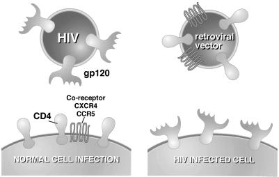
Gene therapy protocols involve the transfer of genetic material to target cells, where expression can be of therapeutic benefit. To this end a number of viral vectors have been developed (27). The cell types which are infected by these vectors are restricted by the tropism of the virus on which the vector is based. There is considerable utility in achieving targeted delivery to certain cell types—especially where the target cell types are in a heterogeneous mix (e.g., specific neurons in the brain) or dispersed (e.g., stem cells). To date, a number of strategies to alter the tropism of viral vectors have been tested. These include using adapter antibodies (10, 12), pseudotyping the viral molecules responsible for cell binding from different strains or different viruses altogether (3, 15, 29), and redesigning these recognition molecules (6). Though pseudotyping with other viral molecules has been the most successful approach in terms of generating high-titer vectors, the “rational” redesign approach has generated only low-titer targeting vectors. This is probably due to uncoupling of binding and membrane fusion in the designed envelopes, whereas in naturally evolved envelopes these functions are coupled. In the present study, we followed up on an observation by Young et al. (30), who reported that the cell surface glycoprotein CD4 could be efficiently incorporated into avian leukosis virus (ALV). CD4 is the primary receptor for the human immunodeficiency type 1 (HIV-1) envelope protein gp120 (7). However, CD4-pseudotyped virus could not infect cells expressing HIV-1 proteins gp120 and gp41, because a coreceptor is required for membrane fusion following the binding of gp120 to CD4 (5). The coreceptor is a chemokine receptor and varies according to the tropism of the HIV-1 isolate: T-cell-tropic HIV-1 strains require the chemokine receptor CXCR4 (also termed fusin) (14), while macrophage-tropic HIV-1 strains require CCR5 (1). In this study, we set out to test whether murine leukemia virus (MLV)-based vectors could incorporate CD4, as is the case for ALV. Furthermore, we tested whether a chemokine receptor could also be incorporated into the virus such that this vector could infect gp120- and gp41-expressing cells (Fig. 1).
FIG. 1.
Schematic illustration for the experimental approach described in the text. Infection by HIV is mediated by gp120 binding to CD4 and a coreceptor (CXCR4 for T-cell-tropic envelopes or CCR5 for macrophage-tropic envelopes). During the life cycle of HIV, gp120 is expressed on the cell surface, and so a Moloney MLV-based retroviral vector, displaying CD4 and a coreceptor, was generated to test if it was specific for infection of gp120-expressing cells.
MLV vectors are typically generated by providing in a cell the gag, pol, and env gene products in trans, together with a vector RNA that contains retroviral long terminal repeats (LTRs), the packaging signal, and a reporter gene. Since in this study we were altering env-mediated functions, we used a cell line derived from 293 human embryonic kidney cells that stably express only the gag and pol genes of MLV (293gp/bsr [N. V. Somia and I. M. Verma, unpublished data]). This cell line was used to generate a virus that transduces the gene for green fluorescent protein (GFP) on transfection of a retroviral vector, pCLMFG-GFP (24). Another cell line that further expresses the transcript for the retroviral vector pCLMFG-lacZ (293gp/lacZ [Somia and Verma, unpublished data]) was used to generate a virus that transduces the gene for β-galactosidase (β-Gal). CD4, CXCR4, and CCR5 expression constructs (singly or in combinations) or an expression plasmid for the amphotropic env were transfected as indicated below to substitute for the envelope.
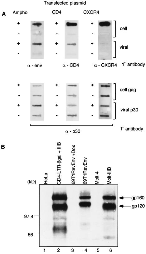
We first determined the expression of the amphotropic envelope, CD4, and CXCR4 in the 293gp/lacZ cells, and their relative levels of incorporation into viral particles, by immunoblot analysis. Figure 2A shows the efficiency of incorporation of the amphotropic envelope into viral particles. It further shows that CD4 can be incorporated into viral particles, though not as efficiently as the amphotropic envelope. This extends to MLV the observations regarding CD4 incorporation into ALV. Since envelopes of retroviruses are not necessarily interchangeable, pseudotyping of other molecules needs to be tested empirically. Finally, CXCR4 is incorporated, but at a very low level compared to the amphotropic envelope and CD4. The Gag polyprotein in the cell pellet and the processed p30 protein in the viral pellets provide loading controls. The relative level of incorporation of CXCR4 into the viral particle is much lower than that of CD4.
FIG. 2.
(A) Immunoblot analysis of amphotropic envelope, CD4, and CXCR4 proteins in transfected cells and viral particles. Twenty micrograms of expression constructs for the amphotropic envelope (SV-A-MLV-env) (Ampho) (21), CD4 (CMX-CD4, a gift from Didier Trono), or CXCR4 (pc.Fusin) (9) were transfected into 5 × 10⁵ 293gp/lacZ cells, according to the method of Chen and Okayama (4). Mock-transfected cells (−) were similarly treated, but no plasmid was transfected. After overnight incubation, the medium was changed, and the vector was harvested 48 h later. The cells were collected and lysed in 1 ml of lysis buffer (20 mM Tris-HCl [pH 8.0], 1 mM CaCl2, 150 mM NaCl, 1% Triton), and the cell extract (1 μl for the amphotropic envelope, CD4, and loading controls; 10 μl for CXCR4) was transferred to a nitrocellulose membrane for immunoblot analysis. The supernatant was filtered through a 0.45-μm-pore-size filter, the viral particles were pelleted through a 20% sucrose cushion by ultracentrifugation (90,000 × g for 1.5 h) and resuspended in 1 ml of lysis buffer, and the viral extract (1 μl for the amphotropic envelope, CD4, and loading controls; 10 μl for CXCR4) was transferred to a nitrocellulose membrane for immunoblot analysis. Primary (1°) antibodies used were goat anti-Raushers gp69/71 (National Cancer Institute/Biological Carcinogenesis Branch repository) (α-env), rabbit anti-human CD4 (8) (α-CD4), and mouse anti-human CXCR4 (monoclonal antibody 4G10, a gift from Edward Berger) (α-CXCR4). Secondary antibodies were rabbit anti-goat immunoglobulin G (IgG)-horseradish peroxidase (HRP) conjugate (Pierce), donkey anti-rabbit IgG-HRP conjugate (Amersham), and rabbit anti-mouse IgG-HRP (Pierce). The secondary antibodies were detected by using the ECL Western blotting detection system (Amersham). The loading controls were probed with a goat anti-Raushers p30 antibody (NCI/BCB repository). The Gag polyprotein (for cell pellets) and p30 (for viral pellets) are shown as loading controls in the bottom panel. (B) Immunoblot analysis of gp120 expression. The following cell extracts (100 μg) were used: uninfected HeLa cells, HeLa-CD4-LTR/β-gal cells infected with T-cell-tropic HIV-1 IIIB, 69T1RevEnv cells grown in the presence of Dox (1 μg/ml), 69T1RevEnv cells, uninfected Molt-4 cells, and the HIV-1 IIIB-infected cell line Molt-IIIB. The primary antibody was sheep anti-gp120 (from Michael Phelan), and the secondary antibody was donkey anti-sheep IgG-HRP conjugate (Sigma). The slower-migrating band is the unprocessed env gene product gp160. (C) CCR5 incorporation into a Moloney MLV vector. 293 cells were transfected with 15 μg of an expression plasmid for macrophage-tropic gp120 (pSV-JRFL-env) and 5 μg of an expression plasmid for Rev. The cells were infected 48 h later with a β-Gal-transducing retroviral vector. The plasmid combinations that were transfected into 293/lacZ cells to pseudotype and generate the β-Gal vector are shown at the top.
To test if expression of CD4 and CXCR4 in 293gp/lacZ cells produced a vector capable of infecting gp120-expressing cells, we utilized a cell line, 69T1RevEnv (31), which conditionally expresses T-cell-tropic gp120 by placing the HIV-1 env gene under the control of a tetracycline-responsive promoter (16). Expression of gp120 is repressed by the presence of tetracycline or its analogue doxycycline (Dox). Figure 2B shows an immunoblot analysis for gp120 expression in 69T1RevEnv cells in the presence or absence of Dox. The expression of gp120 was detected only in the absence of Dox (Fig. 2B, lane 4). MLV vectors were produced by the transfection of various expression constructs into 293gp/lacZ cells and were used to infect 69T1RevEnv cells with and without the addition of Dox. Table 1 illustrates that infection was contingent upon gp120 expression and was observed only when both CD4 and CXCR4 are used to generate the vectors, with an average titer of 2.6 × 10³/ml. Dox had no adverse effect on viral infection, since the amphotropic envelope (using a phosphate transporter as a receptor) (23) enabled infection with equal efficiencies in the absence and presence of Dox. Table 1 also illustrates that the viral vector can be quantitatively concentrated 100-fold by ultracentrifugation. The infection is specific for the tropism of the gp120 moiety, since the CCR5-pseudotyped vector did not mediate infection. The fact that the CCR5 molecule is functional and incorporated into the vector is illustrated in Fig. 2C, where transduction by a vector pseudotyped with CCR5 and CD4 is shown on 293 cells expressing a macrophage-tropic gp120 molecule. In this case, pseudotyping CXCR4 and CD4 did not result in transduction. It is interesting that CD4 expression alone was unable to produce a vector capable of infecting gp120- and gp41-expressing cells, even though the 69T1RevEnv cells express endogenous CXCR4. This suggests that both CD4 and CXCR4 are required on the same face for infection to be productive, and this stero-restriction is consistent with the recently defined structure of the gp120-CD4 complex (20).
TABLE 1.
Titers of amphotropic and pseudotyped vectorsa
| Construct | Titer on 69T1RevEnv cells/ml
|
|
|---|---|---|
| +Dox | −Dox | |
| CD4 | <10 | <10 |
| CXCR4 | <10 | <10 |
| CD4 + CXCR4 | <10 | 2.6 × 10³ |
| CD4 + CCR5 | <10 | <10 |
| Amphotropic envelope | 1.9 × 10⁵ | 2.1 × 10⁵ |
| CD4 + CXCR4 (×100) | <10 | 4.9 × 10⁵ |
293gp/lacZ cells were transfected (4) with 10 μg of CMX-CD4, 15 μg of pc.Fusin (CXCR4), or 15 μg of pcCCR5 or the combinations indicated. The total DNA was kept constant at 25 μg by the addition of a carrier plasmid pEGFP (Clontech). Vectors were harvested as described in the legend for Fig. 1, and 1 ml of medium was used to infect 5 × 10⁴ 69T1RevEnv cells overnight in the presence (1 μg/ml) or absence of Dox. One hundred microliters was used in the case of the amphotropic envelope. The infection was carried out in a total volume of 2 ml of medium. The medium was changed 48 h later, and the cells were stained for β-Gal expression (25) with 5-bromo-4-chloro-3-indolyl-β-d-galactopyranoside. Concentrated virus was prepared by ultracentrifugation of the supernatant (40 ml) at 50,000 × g and resuspension in 400 μl of cell culture medium. Viral suspension (100 μl) was used for infection of 69T1RevEnv cells, and the titer was determined according to the number of cells expressing β-Gal. The values are the means of the results from two independent experiments.
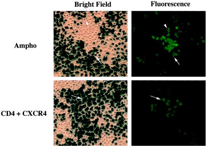
We next tested the ability of the CD4- and CXCR4-pseudotyped MLV vector to infect cells that have been productively infected with wild-type HIV-1. To this end we utilized a cell line, HeLa CD4-LTR/β-gal, that was constructed by Kimpton and Emerman (18). This is a HeLa cell line that has been engineered to ectopically express CD4. Since this cell line already expresses endogenous CXCR4, it can be infected with T-cell-tropic HIV-1 (HIV-1 IIIB). Kimpton and Emerman further manipulated these cells so that they harbor an Escherichia coli β-Gal reporter gene under the control of the HIV-1 LTR. HIV-1 infection of HeLa CD4-LTR/β-gal cells results in the production of the transactivator Tat, which in turn activates expression of the β-Gal gene. Hence, expression of β-Gal is an indicator of infection by HIV-1. HeLa CD4-LTR/β-gal cells were infected with HIV-1 IIIB. Figure 2B indicates that these infected cells express significant amounts of gp120 (Fig. 2B, lane 2), which comigrates with gp120 from T cells infected with HIV-1 IIIB (Fig. 2B, lane 6) (13). The CD4- and CXCR4-pseudotyped vector containing the GFP gene was used to infect HIV-infected HeLa CD4-LTR/β-gal cells. Figure 3 shows that infection by this vector was observed only in HIV-1-infected cells, since only cells infected with HIV-1 (blue, β-Gal-positive cells) were also found to express GFP. A vector with an amphotropic envelope transduced both HIV-1-infected and uninfected cells, since GFP expression was found in both β-Gal-positive and -negative cells. The titer was 102/ml, which is 1 log lower than that observed on 69T1RevEnv cells; however, only a proportion of the HeLa CD4-LTR/β-gal cells were infected with HIV-1, as shown by the number of blue, β-Gal-positive cells in Fig. 3.
FIG. 3.
CD4- and CXCR4-pseudotyped MLV vector selectively infects HIV-1-infected cells. HeLa CD4-LTR/β-gal cells (18) were incubated with a supernatant from HIV-1-producing Molt-IIIB cells for 2 days. The cells were then infected with a CD4- and CXCR4-pseudotyped or amphotropic (Ampho) MLV vector containing the GFP gene. At 2 days postinfection, the cells were analyzed for GFP expression by fluorescence microscopy and photographed. The cells were then stained for β-Gal expression (25) with 5-bromo-4-chloro-3-indolyl-β-d-galactopyranoside. The same field was photographed under bright-field microscopy; β-Gal expression (blue) indicates HIV-1 infection. Note that the amphotropic vector transduces both HIV-1-infected and uninfected cells (arrow and arrowhead, respectively). The CD4- and CXCR4-pseudotyped vector transduces only HIV-1-infected cells (all cells that are green are also blue).
We have demonstrated above that MLV-based vectors can be pseudotyped with CD4 and CXCR4, directing infection to cells expressing T-cell-tropic HIV-1 env. Other groups have reported targeting to gp120-expressing cells. Endres et al. (11) have shown incorporation of CD4 and CCR5 or CXCR4 into an HIV-1-based vector with titers of 104/ml. This vector can be used to transduce HIV-1-infected cells, though the vector will be remobilized with a gp120 envelope-based tropism. This can lead to an overestimation of the titer and may also limit the types of molecules that could be transduced to HIV-1-infected cells in vivo, since a remobilized vector may infect a CD4 cell that has not been infected with HIV-1. This may limit the transgene in the vector to nontoxic molecules. Furthermore, this may seem an attractive approach in some cases because the HIV-1-based vector is amplified in vivo in proportion to the number of HIV-1-infected cells; however, it is limited as a treatment, since concomitant therapies that interfere with HIV-1 replication would also affect vector production. Mebatsion et al. (22) have shown a rabies virus pseudotyped with CD4 and CXCR4, with maximal titers of about 4 × 10³/ml. In an ingenious strategy, Schnell et al. (26) generated a replication-competent vesicular stomatitis virus pseudotyped with CD4 and CXCR4 that was capable of infecting and killing HIV-1-infected cells. More recently, Balliet and Bates (2) have generalized this receptor pseudotyping to Rous sarcoma virus (Tva) and the receptor of the ecotropic murine leukemia virus (MCAT-1).
MLV-based vectors provide an alternative way to target HIV-1-infected cells without remobilizing them or causing lysis. This method can also be used in conjunction with most other anti-HIV therapies. The vectors may be used to deliver genes coding for antigenic molecules that upregulate the immune response against HIV-1-infected cells. However, the titer needs to be drastically improved for any efficacious therapeutic applications. Indeed, the total number of HIV-1 particles produced in an infected patient has been estimated at 109 per day (17, 28). Clearly, one means of achieving a higher titer is to increase the level of incorporation of CXCR4—possibly by making CXCR4 chimeric molecules that retain gp120 binding but facilitate higher levels of expression and incorporation into viral particles. In this regard, CXCR4 and CD4 fusions (19) may be of interest.
Acknowledgments
The following reagents were obtained through the AIDS Research and Reference Reagent Program, Division of AIDS, NIAID, NIH: clone 69T1RevEnv from Joseph Dougherty, HeLa-CD4-LTR/β-gal from Michael Emerman, antiserum to CD4 (T4-4) from R. Sweet (SmithKline Beecham Pharmaceuticals), antiserum to HIV-1 gp120 from Michael Phelan, and pc.Fusin, pcCCR-5, and pSV-JRFL-env from Nathaniel Landau. We thank Didier Trono for the CMX-CD4 expression plasmid and the Molt-IIB cells. We are also grateful to Edward Berger for the gift of the 4G10 anti-CXCR4 monoclonal antibody.
I. M. Verma is an American Cancer Society Professor of Molecular Biology. He is supported by grants from the NIH, the March of Dimes, the Wayne and Gladys Valley Foundation, and the H. N. and Frances C. Berger Foundation.
REFERENCES
- 1.Alkhatib G, Combadiere C, Broder C C, Feng Y, Kennedy P E, Murphy P M, Berger E A. CC CKR5: a RANTES, MIP-1alpha, MIP-1beta receptor as a fusion cofactor for macrophage-tropic HIV-1. Science. 1996;272:1955–1958. doi: 10.1126/science.272.5270.1955. [DOI] [PubMed] [Google Scholar]
- 2.Balliet J W, Bates P. Efficient infection mediated by viral receptors incorporated into retroviral particles. J Virol. 1998;72:671–676. doi: 10.1128/jvi.72.1.671-676.1998. [DOI] [PMC free article] [PubMed] [Google Scholar]
- 3.Burns J C, Friedmann T, Driever W, Burrascano M, Yee J K. Vesicular stomatitis virus G glycoprotein pseudotyped retroviral vectors: concentration to very high titer and efficient gene transfer into mammalian and nonmammalian cells. Proc Natl Acad Sci USA. 1993;90:8033–8037. doi: 10.1073/pnas.90.17.8033. [DOI] [PMC free article] [PubMed] [Google Scholar]
- 4.Chen C, Okayama H. High-efficiency transformation of mammalian cells by plasmid DNA. Mol Cell Biol. 1987;7:2745–2752. doi: 10.1128/mcb.7.8.2745. [DOI] [PMC free article] [PubMed] [Google Scholar]
- 5.Clapham P R, Blanc D, Weiss R A. Specific cell surface requirements for the infection of CD4-positive cells by human immunodeficiency virus types 1 and 2 and by simian immunodeficiency virus. Virology. 1991;181:703–715. doi: 10.1016/0042-6822(91)90904-P. [DOI] [PMC free article] [PubMed] [Google Scholar]
- 6.Cosset F L, Russell S J. Targeting retrovirus entry. Gene Ther. 1996;3:946–956. [PubMed] [Google Scholar]
- 7.Dalgleish A G, Beverley P C, Clapham P R, Crawford D H, Greaves M F, Weiss R A. The CD4 (T4) antigen is an essential component of the receptor for the AIDS retrovirus. Nature. 1984;312:763–767. doi: 10.1038/312763a0. [DOI] [PubMed] [Google Scholar]
- 8.Deen K C, McDougal J S, Inacker R, Folena-Wasserman G, Arthos J, Rosenberg J, Maddon P J, Axel R, Sweet R W. A soluble form of CD4 (T4) protein inhibits AIDS virus infection. Nature. 1988;331:82–84. doi: 10.1038/331082a0. [DOI] [PubMed] [Google Scholar]
- 9.Deng H, Liu R, Ellmeier W, Choe S, Unutmaz D, Burkhart M, Di Marzio P, Marmon S, Sutton R E, Hill C M, Davis C B, Peiper S C, Schall T J, Littman D R, Landau N R. Identification of a major co-receptor for primary isolates of HIV-1. Nature. 1996;381:661–666. doi: 10.1038/381661a0. [DOI] [PubMed] [Google Scholar]
- 10.Douglas J T, Rogers B E, Rosenfeld M E, Michael S I, Feng M, Curiel D T. Targeted gene delivery by tropism-modified adenoviral vectors. Nat Biotechnol. 1996;14:1574–1578. doi: 10.1038/nbt1196-1574. [DOI] [PubMed] [Google Scholar]
- 11.Endres M J, Jaffer S, Haggarty B, Turner J D, Doranz B J, O'Brien P J, Kolson D L, Hoxie J A. Targeting of HIV- and SIV-infected cells by CD4-chemokine receptor pseudotypes. Science. 1997;278:1462–1464. doi: 10.1126/science.278.5342.1462. [DOI] [PubMed] [Google Scholar]
- 12.Etienne-Julan M, Roux P, Carillo S, Jeanteur P, Piechaczyk M. The efficiency of cell targeting by recombinant retroviruses depends on the nature of the receptor and the composition of the artificial cell-virus linker. J Gen Virol. 1992;73:3251–3255. doi: 10.1099/0022-1317-73-12-3251. [DOI] [PubMed] [Google Scholar]
- 13.Farnet C M, Haseltine W A. Integration of human immunodeficiency virus type 1 DNA in vitro. Proc Natl Acad Sci USA. 1990;87:4164–4168. doi: 10.1073/pnas.87.11.4164. [DOI] [PMC free article] [PubMed] [Google Scholar]
- 14.Feng Y, Broder C C, Kennedy P E, Berger E A. HIV-1 entry cofactor: functional cDNA cloning of a seven-transmembrane, G protein-coupled receptor. Science. 1996;272:872–877. doi: 10.1126/science.272.5263.872. [DOI] [PubMed] [Google Scholar]
- 15.Gall J, Kass-Eisler A, Leinwand L, Falck-Pedersen E. Adenovirus type 5 and 7 capsid chimera: fiber replacement alters receptor tropism without affecting primary immune neutralization epitopes. J Virol. 1996;70:2116–2123. doi: 10.1128/jvi.70.4.2116-2123.1996. [DOI] [PMC free article] [PubMed] [Google Scholar]
- 16.Gossen M, Bujard H. Tight control of gene expression in mammalian cells by tetracycline-responsive promoters. Proc Natl Acad Sci USA. 1992;89:5547–5551. doi: 10.1073/pnas.89.12.5547. [DOI] [PMC free article] [PubMed] [Google Scholar]
- 17.Ho D D, Neumann A U, Perelson A S, Chen W, Leonard J M, Markowitz M. Rapid turnover of plasma virions and CD4 lymphocytes in HIV-1 infection. Nature. 1995;373:123–126. doi: 10.1038/373123a0. [DOI] [PubMed] [Google Scholar]
- 18.Kimpton J, Emerman M. Detection of replication-competent and pseudotyped human immunodeficiency virus with a sensitive cell line on the basis of activation of an integrated β-galactosidase gene. J Virol. 1992;66:2232–2239. doi: 10.1128/jvi.66.4.2232-2239.1992. [DOI] [PMC free article] [PubMed] [Google Scholar]
- 19.Klasse P J, Rosenkilde M M, Signoret N, Pelchen-Matthews A, Schwartz T W, Marsh M. CD4-chemokine receptor hybrids in human immunodeficiency virus type 1 infection. J Virol. 1999;73:7453–7466. doi: 10.1128/jvi.73.9.7453-7466.1999. [DOI] [PMC free article] [PubMed] [Google Scholar]
- 20.Kwong P D, Wyatt R, Robinson J, Sweet R W, Sodroski J, Hendrickson W A. Structure of an HIV gp120 envelope glycoprotein in complex with the CD4 receptor and a neutralizing human antibody. Nature. 1998;393:648–659. doi: 10.1038/31405. [DOI] [PMC free article] [PubMed] [Google Scholar]
- 21.Landau N R, Page K A, Littman D R. Pseudotyping with human T-cell leukemia virus type I broadens the human immunodeficiency virus host range. J Virol. 1991;65:162–169. doi: 10.1128/jvi.65.1.162-169.1991. [DOI] [PMC free article] [PubMed] [Google Scholar]
- 22.Mebatsion T, Finke S, Weiland F, Conzelmann K K. A CXCR4/CD4 pseudotype rhabdovirus that selectively infects HIV-1 envelope protein-expressing cells. Cell. 1997;90:841–847. doi: 10.1016/s0092-8674(00)80349-9. [DOI] [PubMed] [Google Scholar]
- 23.Miller D G, Edwards R H, Miller A D. Cloning of the cellular receptor for amphotropic murine retroviruses reveals homology to that for gibbon ape leukemia virus. Proc Natl Acad Sci USA. 1994;91:78–82. doi: 10.1073/pnas.91.1.78. [DOI] [PMC free article] [PubMed] [Google Scholar]
- 24.Naviaux R K, Costanzi E, Haas M, Verma I M. The pCL vector system: rapid production of helper-free, high-titer, recombinant retroviruses. J Virol. 1996;70:5701–5705. doi: 10.1128/jvi.70.8.5701-5705.1996. [DOI] [PMC free article] [PubMed] [Google Scholar]
- 25.Sanes J R, Rubenstein J L, Nicolas J F. Use of a recombinant retrovirus to study post-implantation cell lineage in mouse embryos. EMBO J. 1986;5:3133–3142. doi: 10.1002/j.1460-2075.1986.tb04620.x. [DOI] [PMC free article] [PubMed] [Google Scholar]
- 26.Schnell M J, Johnson J E, Buonocore L, Rose J K. Construction of a novel virus that targets HIV-1-infected cells and controls HIV-1 infection. Cell. 1997;90:849–857. doi: 10.1016/s0092-8674(00)80350-5. [DOI] [PubMed] [Google Scholar]
- 27.Verma I M, Somia N. Gene therapy—promises, problems and prospects. Nature. 1997;389:239–242. doi: 10.1038/38410. [DOI] [PubMed] [Google Scholar]
- 28.Wei X, Ghosh S K, Taylor M E, Johnson V A, Emini E A, Deutsch P, Lifson J D, Bonhoeffer S, Nowak M A, Hahn B H, et al. Viral dynamics in human immunodeficiency virus type 1 infection. Nature. 1995;373:117–122. doi: 10.1038/373117a0. [DOI] [PubMed] [Google Scholar]
- 29.Yang Z, Delgado R, Xu L, Todd R F, Nabel E G, Sanchez A, Nabel G J. Distinct cellular interactions of secreted and transmembrane Ebola virus glycoproteins. Science. 1998;279:1034–1037. doi: 10.1126/science.279.5353.1034. [DOI] [PubMed] [Google Scholar]
- 30.Young J A, Bates P, Willert K, Varmus H E. Efficient incorporation of human CD4 protein into avian leukosis virus particles. Science. 1990;250:1421–1423. doi: 10.1126/science.2175047. [DOI] [PubMed] [Google Scholar]
- 31.Yu H, Rabson A B, Kaul M, Ron Y, Dougherty J P. Inducible human immunodeficiency virus type 1 packaging cell lines. J Virol. 1996;70:4530–4537. doi: 10.1128/jvi.70.7.4530-4537.1996. [DOI] [PMC free article] [PubMed] [Google Scholar]


